
Recommended
More Related Content
What's hot
What's hot (20)
Viewers also liked
Viewers also liked (18)
Similar to Triangles of the neck
Similar to Triangles of the neck (20)
Recently uploaded
Recently uploaded (20)
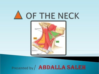
Triangles of the neck
- 2. OVERVIEW; The Ant. Region of the Neck is Divides into: Medial T. Lateral T. SCM R. SUBDIVISION: I. Medial II. Lateral Carotid Submandibular Submental Scapulotracheal (Omotracheal) Omotrapezius (occipital) Supraclavicular Infrahyoid R. Suprahyoid R. By the body of Hyoid bone By Inf. Belly of Omohyoid muscle
- 3. Suprahyoid region: • Boundaries: Inf. border of the mandible, Sup. Ant. border of SCM muscle, Lat. Line drown by the Hyoid bone, Inf. • Coverings: Skin, Superficial fascia, 2nd fascia, Sheath ,formed by 2nd fascia, gives two layers attached to the inf. border of the Mandible. • Floor: Mylohyoid and Hyoglossus muscles. Submental triangle • Boundaries: Chin, Sup. Body of the hyoid bone, Inf. Ant. Bellies if both digastric muscle, Lat. • Contents: Submental lymph node + beginning of AJV.
- 4. Submandibular triangle •Boundaries: Inf. Border of the Mandible, Sup. Both bellies of Digastric muscle, Ant. & Post. •Contents: The gland, The lymph, fat, fascial A&V •Structures: •Hypoglossal NHypoglossal N.. runs on hyoglossus muscle deep to the gland. •Lingual ALingual A.. ,from ECA, enters deep to the Post. border of Hyoglossus muscle. Maybe found for ligation in Pirgoff’s triangle.
- 5. Infrahyoid region: •Boundaries: Sternal notch, Inf. Ant. Border of SCM muscle, Lat. Line of the body of Hyoid bone, Sup. •Coverings: Skin, Subcutaneous tissue, Platysma, Proper fascia (superficial, deep, parietal, visceral) & the Infrahyoid Ms. •Contents: Branches of transverse cutaneous N. •Floor: Sternohyoid and sternothyroid muscles. Contents: Organs of the neck (Thyroid gland, Trachea, Larynx, Pharynx & Esophagus). Omotracheal triangle: • Boundaries: Medline of the Neck, Ant. Ant. Border of SCM muscle, Inf. Sup. Belly of Digastric muscle, Sup.
- 6. Carotid triangle •Boundaries: Post. Belly of Digastric muscle, Sup. Sup. Belly of Omohyoid muscle, Inf. Ant. Border of SCM muscle, Post. •Coverings: Skin, Fascia, Carotid sheath, Proper Fascia(Superficial), Platysma. •Contents: Carotid sheath (CCA+E,I CAs+IJV) Int+Ext. Laryngeal, Hypoglossal & descending branch, Accessory & Vagus Nerves. •Structures: CCA lies medial & deep to IJV. Left CCA comes from arch of Aorta. Right CCA is one of the terminal branches of right Brachiocephalic A. CCA gives NONO branches.
- 7. Structures: Descending branch of hypoglossal N. and Ansa Cervicalis embedded in the wall of Carotid sheath. Organs of Neck + Inf. Thyroid A. + recurrent laryngeal N. lie MedialMedial To CCA. Scalenus Ant. muscle + sympathetic trunk + prevertebral region lie LateralLateral . CCA lie opposite to Ant. Tubercle of C6 transverse process. ECA lies Anteromedial to ICA. Branches of ECA (Sup. Thyroid, Lingual, Fascial, Occipital, Ascending Pharyngeal, Post. Auricular, Maxillary, Superficial Temporal) . ICA has no branches, It enters the base of the skull through Carotid Canal Pharynx lies medial to it. IJV drains from Brain, Neck and Face and enters Jagular foramen and Carotid sheath. Units with Subclavian V. to form Brachiocephalic V. Vagus N. enters the Carotid sheath vertically and lie between IJV & ICA then between IJV & CCA. (meningeal, auricular, pharyngeal, sup.&recurrent laryngeal, cardiac) Right recurrent Laryngeal N. hooks around 1st part of Subclavian A. Left recurrent Laryngeal N. hooks around Arch of the Aorta.
- 8. Lateral triangle: •Boundaries: Clavicle, Inf. Post. Border of SCM muscle, Ant. Ant. Border of Trapezius muscle, Post. Omotrapezius triangle •Boundaries: SCM muscle, Ant. Trapezius muscle, Post. Inf. Belly of Omohyoid muscle, Inf. Fascias of the Neck:Fascias of the Neck: 11stst , 2, 2ndnd , 5, 5thth .. Supraclavicular triangle •Boundaries: Clavicle, Inf. SCM muscle, Med. Inf. Belly of Omohyoid muscle, Sup. Fascias of the Neck:Fascias of the Neck: 11stst , 2, 2ndnd , 3, 3rdrd , 5, 5thth ..
- 9. Supraclavicular triangle Structures: Subclavian V. lies Ant. & Inf. to the Clavicle. Subclavian A. lies Post. & Sup. To the Clavicle. Brachial plexus lies Post. To the Clavicle. Right Subclavian A. arises from Brachiocephalic A. Left Subclavian A. arises from arch of Aorta. Scalenus Ant. Passes Ant. To the A. and divides it into three parts: 11stst part:part: Subclavian A. Med. border of the muscle. Branches: Thyrocervical trunk + int. thoracic A. 22ndnd part:part: Behind the muscle. Branches: Costocervical trunk Sup. Intercostal A. 1st & 2nd ICSs. 33rdrd part:part: Lat. Border of the muscle Post. Triangle Lat. Border of 1st rib. No branches. Axillary A. Deep cervical A. deep Ms. of Neck.