
Recommended
More Related Content
What's hot
What's hot (20)
Viewers also liked
Viewers also liked (20)
Similar to Pulp protection
Similar to Pulp protection (20)
Recently uploaded
Recently uploaded (20)
Pulp protection
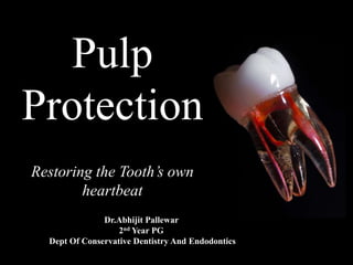
- 1. Pulp Protection Restoring the Tooth’s own heartbeat Dr.Abhijit Pallewar 2nd Year PG Dept Of Conservative Dentistry And Endodontics
- 2. CONTENTS • Introduction • Pulp –dentin complex • Pulp nerve supply • Pulp response • Pulp irritants • Protection in shallow and moderate cavities
- 3. • Pulp protection in deep cavities • indirect pulp capping • Direct pulp capping • Materials used for pulp protection • New materials and methods to protect pulp • References • conclusion
- 4. INTRODUCTION • The restoration and maintenance of dental health through adequate restorative treatment in order to protect pulp function is the main purpose of restorative dentistry. • The dental pulp is a soft connective tissue of mesenchymal origin present within the pulp chamber and root canals of teeth. • It is not considered an external tissue, yet its exposure to external stimuli is unceasing due to several factors that make the pulp extremely sensitive to environment outside.
- 5. • Protection of dentin-pulp complex is an important factor in pulp vitality during operative procedures. • This involves the avoidance of thermal stimuli caused by operative procedures, toxicity of restorative materials and bacteria penetration.
- 6. PULP-DENTIN COMPLEX • The dentin and pulp must be considered as one organ because of their intimate relationship between cellular tissue within dentin and peripheral pulp tissue. • As long as dentin is covered peripherally by enamel on coronal surfaces and cementum on radicular surfaces , dental pulp will remain healthy for life unless apical blood supply is disrupted by excessive orthodontic forces or severe impact of trauma.
- 7. • Most pathological pulp conditions begin with removal of these barriers via caries, fractures or abrasion. • Structure and response of dentin to injury are largely functions of the odontoblasts and other cells in pulp but these cells are dependent on dentin for their protection and their state of differentiation. • Normal form and function of one cannot be maintained without other.
- 8. • Hence its obvious response of pulp to any restorative material will be influenced by its surrounding dentin . • Therefore it is apparent that substances easily permeate dentin permit thermal, osmotic, and chemical insults to act on the pulpal constituents. • This involves stimulation of odontoblast in intial stages which may proceed to inflammation finally which often lead to tissue destruction
- 9. • A classical work done by pashley et al ,using radioactive I in dog teeth , which clearly demonstrated that once dentin tubules are opened, molecules can permeate to pulp easily .
- 11. PULP NERVE SUPPLY • Dental pulp has an abundant supply of both sensory and autonomic nerves. • Majority of the nerves are sensory. • The trigeminal ganglion supplies sensory innervations to the pulp via maxillary and mandibular nerves. • Nerve fibres enter the teeth via apical foramen and arborize coronally to form plexus of rakshow in subodondoblastic region of pulp. • Coronal dentin is more densely innervated than radicular dentin .
- 13. Classification Of Nerves TYPE OF FIBER FUNCTION Aα Motor, proprioception Aβ Pressure , touch Aγ motor Aδ Pain , temperature , touch B Preganglionic , autonomic C Pain , postganglionic sympathetic
- 14. PULP RESPONSE TO IRRITANTS • What happens when permeating substances reach the pulp chamber ?
- 15. Vicious cycle of pulpal inflammation that begins with irritation of pulp , leads to a localised response and may progress to a lesion increasing severity and eventually irreversible pulpitis
- 16. We have to protect it from any type of irritant
- 17. PULP IRRITANTS • Bacterial irritants • Trauma • Iatrogenic: i. during tooth preparation ii. Orthodontic movement of tooth iii. Periodontal and periapical curettage iv. use of chemicals v. idiopathic
- 18. Pulpal irritants A) Bacterial irritants (Most common cause for pulpal irritation) B) Traumatic 1-Caries Tooth fracture Luxation Avulsion Parafunctional habits like bruxism 2- Periodontal pocket and abscess 1-Acute trauma 2- Chronic Trauma
- 19. C) Iatrogenic: 1. During cavity preparation a) Heat production during cutting procedures: Pulp temperature 11°C Destructive reaction Pulpal temperature is critical and must not exceed normal values in dental restorative procedures. Clinical research has shown irreversible damage to pulp tissues at levels of 60% at 5.5°C and 100% at 11°C..
- 20. Excessive heat generation leads to change in dentin color due to vascular stasis and hemorrage in the subodontoblastic vascular plexus present in the pulp
- 21. b) Pressure exerted: Pressure of hand or rotary instruments Nuclear aspiration of odontoblasts or nerve endings from pulp tissues into the dentinal tubules Disturb odontoblasts metabolism leading to their complete degeneration and disintegration. c) Remaining Dentin Thickness (RDT)
- 22. 2. Orthodontic movement 3. Periodontal and periapical curettage
- 23. 4. Use of chemicals Temporary & permanent fillings, bases, liners, and use of alcohol that leads to pulpal injury due to its cytotoxicity, acidity, heat formed and marginal leakage Chemical irritants applied to dentin can result in damage and disorganization in the subadjacent pulp
- 25. Because of these various irritants pulp needs protection…
- 27. • Direct pulp capping • Indirect pulp capping Conventional methods
- 28. Direct pulp capping is placing a biocompatible material over the exposed pulp to maintain vitality and promote healing. Direct Pulp Capping WHY? 1) To maintain the vitality of the remaining pulp tissue 2) To prevent root canal treatment 3) To help conserve tooth structure
- 29. Indications Recent small mechanical exposure of pulp during (< 24 hours): a) Tooth preparation b) Traumatic injury. No or minimal bleeding at the exposure site.
- 30. Contraindications Wide pulp exposure Pre-operative history of Spontaneous pain Presence of bleeding at exposure site Radiograph doesn’t show any pulp pathology
- 31. Clinical Procedure 3.When vital & healthy pulp is exposed, check fresh bleeding 2. Isolate the tooth with rubber dam 1. Administer local anesthesia 4. Clean the area with saline solution 5. Dry it with a cotton pellet 6. Apply calcium hydroxide (preferably Dycal) over the exposed area
- 32. 7. Give interim restoration such as zinc oxide eugenol for 6 to 8 weeks b) If not pulpotomy or pulpectomy is requested a) Remove the cement to inspect the exposure site. If secondary dentin formation takes place over the exposed site restore the tooth permanently with protective cement base and restorative material.
- 33. In indirect pulp capping, all caries are removed except the ones that lie adjacent to the pulp. Caries near the pulp is left in place to prevent pulp exposure and preparation is enclosed with a biocompatible material. Indirect Pulp Capping
- 34. Indications 1. Deep carious lesion near the pulp tissue but not involving it 2. No mobility of tooth 3. No history of spontaneous toothache 4. No tenderness to percussion 5. No radiographic evidence of pulp pathology 6. No root resorption or radicular disease should be present radiographically. Root resorption
- 35. Clinical Procedure It’s the same procedure as the direct pulp capping except that the pulp is not exposed. A thin layer of dentin and some amount of caries is left to avoid exposure. Placement of calcuim hydroxide and zinc oxide eugenol dressing after excavation of soft caries
- 36. Factors affecting Pulp Capping success 1) Age of the patient: Due to vascularity of the pulp, young patients have greater potential for success than older ones Young patient Old patient 2) Type of exposure: Mechanically done pulpal exposure has better prognosis than exposure caused by caries, due to less pulpal inflammation and deleterious effect of bacterial toxins on the pulp
- 37. 3) Size of the exposure: In large exposures, it is difficult to control the hemorrhage and tissue seepage. Small pinpoint exposures are easy to manage and have a greater potential for success 4) History of pain: If previously pain has not occurred in the tooth, the potential for success is more
- 38. Conventional Materials Materials used for Pulp Protection Recent Materials Varnish Base Sealer Liner 1) Zinc oxide eugenol liners 2) Calcium hydroxide 3) Flowable composites 4) Glass ionomers 1) Zinc Oxide Eugenol 2) Zinc phosphate cement 3) Polycarboxylate cement 4) Glass ionomer cement
- 39. Ether or chloroform Organic copalResin gum Solvent evaporates Definition: It is an organic copal or resin gum suspended in solutions of ether or chloroform. When we put it on the tooth surface the organic solvent evaporates leaving a protective film Two coats of varnish should be applied using a small cotton pellet to ensure sufficient wetting of cavity walls VARNISH
- 40. Indications To seal the dentinal tubules Dentinal tubules Open Dentinal tubules Sealing dentinal tubules with varnish Dentinal tubules blocked by varnish 2. Protects the tooth from chemical irritants from cements reducing postoperative pain 3. Reduces microleakage around restorations 1. Prevents discoloration of tooth with an amalgam restoration by preventing migration of ions into the dentin
- 41. Under Composite Resin Varnishes dissolve in the monomer of the resin & also interfere with their polymerization of resins With Glass Ionomer Restorations It interferes the bonding of tooth to these cements Contraindications
- 42. Sealer Indications • To seal dentinal tubules • To treat dentin hypersensitivity. An adhesive sealer is commonly used under indirect restorations. For application, cotton tip applicator is used to apply sealer on all areas of exposed dentin.
- 43. • Liners can be classified as : Thin film liners( 1-50µm) a. solution liners ( varnish:2-5 µm) b. suspension liners ( zinc oxide / calcium hydroxide 20-25 µm) Thick film liners ( 200 -1000 µm) a. GIC ( type III) LINERS
- 44. Commercially available calcium hydroxide liners are DYCAL ( dentsply ) and single paste systems like CALCIMOL LC ( Voco) and Septocal LC ( Septodont)
- 45. 1. Zinc oxide eugenol liners • Used to alleviate pain from mild-to- moderate inflammation of pulp. In low concentration it acts as obtundant In high concentration it acts as chemical irritant Contraindication: It inhibits polymerization Should not be used under bonding agents & composite restorations
- 46. 2- Calcium hydroxide Most common agent considered as the “gold standard” of direct pulp capping materials against which new materials should be tested Advantages: 1. Causes dentin mineralization by activating the enzyme ATPase 2. Stimulates reparative dentin formation 3. Biocompatible 4. High pH (12.5) neutralizes acidity of silicate and zinc phosphate cements Disadvantages: 1. Low strength 2. High solubility Dissolves rapidlyUsed over small areas requiring pulp protection / Applying glass ionomer or zinc phosphate base to prevent its dissolution.
- 48. • The eluted Ca ions increase the proliferation of human dental pulp cells in a dose-dependent manner (Clapham 1995, Takita et al. 2006). • In addition, Ca ions specifically modulate osteopontin and bone morphogenetic protein-2 levels during pulp calcification (Rashid et al. 2003), • The release of Ca enhances the activity of pyrophosphatase, which helps to maintain dentine mineralization and the formation of a dentine bridge (Estrela & Holland 2003).
- 49. 3- Flowable composites Composites with a lower amount of filler more fluid consistency, less strength and lower modulus
- 50. 4- Glass ionomers Renewable source of fluoride under restorations Reduce the incidence of caries Fluoride Glass ionomer cements (GIC): Bond to tooth structure Act as a thermal barrier Ability to bond in a moist environment Easy to use. Anticariogenic.
- 51. Light-cured resin-modified glass ionomers (RMGIs) Provide good adhesion to both tooth structure and restorative materials High strength Flexible (low modulus of elasticity) Dual-setting reaction: 1) Light-activated, methacrylate crosslinking reaction 2) Slower, delayed, acid-base reaction Which gives RMGIs an additional period of maximum flexibility to absorb stress from the adjacent shrinking composite.
- 52. BASES
- 53. Classification of bases Protective bases Sedative bases Insulating bases They protect the pulp before restoration is placed They help in calming the pulp which has been irritated by mechanical, chemical or other means They protect the tooth from thermal shock. Bases should have sufficient strength so that they can withstand forces of mastication and condensation of permanent restorations.
- 54. Excellent sealing quality. Bacteriostatic in nature. Anodyne effect. Reduces the thermal conductivity of metallic restorations Blocks undercuts in the preparation wall in case of cast restorations. Chemically bonds to tooth Antibacterial properties Fluoride release Anticariogenic property Chemical bond to tooth Well tolerated by the pulp. Materials used as bases Zinc oxide eugenol Zinc phosphate cement Polycarboylate cement Glass ionomer cement
- 55. Pulp Protection according with depth of tooth preparation
- 56. Recent Materials used for Pulp Protection
- 57. Biodentin Biodentine is a calcium-silicate based material. Advantages: Biocompatible so no pulp inflammatory responses Can be used wherever dentin is damaged Outstanding sealing properties Used as base or liner under composite restorations Adequate compressive and flexural strength Creates faster dentin bridges Better properties than glass ionomer and calcium hydroxide Radio opacity for following up
- 58. • Biodentine (Septodont, France) is a new calcium silicate–based restorative cement with dentin-like mechanical properties, which can be used as a dentin substitute on crowns and roots similar to how MTA is used . • It has a positive effect on vital pulp cells and stimulates tertiary dentin formation.. • In direct contact with vital pulp tissue, it also promotes formation of reparative dentin. • Biodentine consists of a powder and liquid. • The powder mainly contains tricalcium and dicalcium silicate (the principal component of Portland cement, as well as calcium carbonate , Zirconium dioxide (ZrO2) serves as contrast medium.
- 59. • The liquid consists of calcium chloride, which is used as a setting accelerator and water-reducing agent in aqueous solution with an admixture of polycarboxylate (a superplasticizing agent) • Biodentine may be successfully used as a posterior restoration material for up to 6 months after direct pulp capping. • After validation of pulp health, it may be partially removed to place a permanent composite material Response of Human Dental Pulp Capped with Biodentine and Mineral Trioxide Aggregate - Alicja Nowicka JOE2013:39
- 61. (a & b) Pre-operative photograph showing in 11 with pulp exposure (c) Preoperative radiograph (d and e) A 3mm layer of Biodentine located over the uncovered pulp (f) Immediate post-operative radiograph showing 3mm barrier of Biodentine (g) Post-operative radiograph after 18 months showing a well-formed radio- opaque barrier (h) Post-operative recall photograph after 18 months Clinical Procedure:
- 62. Mineral Trioxide Aggregate (MTA) 1) Characteristics: Non-toxic material Low or no solubility Stimulate reparative dentin development by a normal defending process of an early pulpal wound healing (evidence was the presence of odontoblast like cells) Minimal inflammation at early healing stage 2) Composition: a. Tricalcium silicate b. Tricalcium aluminate c. Tricalcium oxide d. Silicate oxide
- 63. 3) Manipulation: Mixed with sterile water in a 3:1 powder to liquid ratio Setting time: MTA sets in 5 minutes 4) How does MTA work? Tricalcium oxide Tissue fluids Calcium hydroxide Hard tissue formation
- 64. 5) Clinical procedure a) Radiograph before performing the operative procedure b) A Photograph that shows the uncovered pulp tissue c) Photograph showing settlement of MTA above the pulp tissue d) Radiograph after restoring the tooth permenantly e) Six months follow up radiograph
- 65. Why is MTA better than Calcium Hydroxide? MTA Calcium hydroxide VS. 1. Rapid cell growth promotion in vitro 2. Greater ability to maintain the integrity of pulp tissue 3. Thicker and rapidly formed dentinal bridge 4. Less hyperemia 5. Lower level of necrosis
- 66. • Histological evaluations of exposed pulp tissue from animals capped with MTA have shown the formation of a thicker dentinal bridge, with low inflammatory response, hyperemia and pulpal necrosis compared to calcium hydroxide cement. • Calcium hydroxide does not adhere to dentine and lacks the ability to seal. • Tunnel defects in dentine bridges under calcium hydroxide dressings can act as pathways for microleakage (Cox et al. 1985). This material also has a tendency to dissolve over time (Schuurs et al. 2000). • MTA appears to induce the formation of a dentin bridge at a faster rate than calcium hydroxide Response of Human Dental Pulp Capped with MTA and Calcium Hydroxide Powder- MLR Accorinte et al Operative Dentistry 2008:33
- 67. • The ability of MTA to induce the formation of a dentine bridge may be due to its excellent sealing ability (Torabinejad et al or biocompatibility (Kettering &Torabinejad1995). • MTA can induce cytokine release from bone cells (Koh et al.1995)and can allow the attachment of osteoblasts in the form of a monolayer. Response of Human Dental Pulp Capped with MTA and Calcium Hydroxide Powder- MLR Accorinte et al Operative Dentistry 2008:33
- 68. • Calcium hydroxide powder has a pH ranging from 11 to 13, while MTA and calcium hydroxide cement possess a pH of approximately 10. • The higher the pH of the material, the thicker the mummified zone, the more complete the dentin bridge formation and the higher the inflammatory infiltrate. • Some question the superiority of calcium hydroxide, because of its degradation over time, tunnel defects through dentinal bridges under it and poor sealing properties • MTA has sufficient compressive strength to allow condensing of amalgam and a negligible solubility (Torabinejad et al. 1995). Response of Human Dental Pulp Capped with MTA and Calcium Hydroxide Powder- MLR Accorinte et al Operative Dentistry 2008:33
- 70. Thercal 2) Composition: Tricalcium silicate particles in a hydrophilic monomer that provides significant calcium release making it a uniquely stable and durable material as a liner or base. 3) Mechanism: Calcium release stimulates hydroxyapatite and secondary dentin bridge formation 4) Indications: Any pulpal exposures (carious exposures, mechanical exposures or traumatic exposures ) 1) Characteristics: TheraCal is a light cured, resin modified calcium silicate filled liner designed for use in direct and indirect pulp capping, as a protective base/liner under composites, amalgams, cements, and other base materials.
- 72. Why is Thercal better than MTA & Calcium Hydroxide? MTA Calcium hydroxideThercal VS. Higher calcium releasing ability Lower solubility than either MTA or Calcium Hydroxide due to the capability of TheraCal to be cured to a depth of 1.7 mm which avoids the risk of dissolution.
- 73. • Thera-Cal proved to be an ion-leaching material able to release calcium and hydroxyl ions for a period of at least 28 days, and it released significantly more calcium than either ProRoot MTA or Dycal throughout the test period. • The findings of this study suggest that the resin portion of TheraCal (comprising hydrophobic and hydrophilic monomers) is able to promote/sustain Ca and OH ion release within the wet surgical site (on the tooth pulp and/or dentine) and could favour the interaction of the formulation with the hydrophilic tooth dentine. Chemical–physical properties of TheraCal, a novel light-curable MTA-like material for pulp capping- M. G. Gandolfi IEJ 2012:45
- 74. • Most recent studies report long term success rates close to 90% for laser assisted pulp capping , compared to a success rate of about 60 % with traditional methods. • The use of the Er,Cr:YSGG laser allows cavity preparation to be completed with only one instrument in contrast to alternate use of high and low speed rotary instruments and other laser wavelengths ( CO2, Nd:YAG and diode lasers) which cannot be used for ablation of hard tissue. Lasers in pulp capping Erbium chromium laser in pulp capping treatment – dr.giovanni J Oral laser application 2006;6
- 75. • CO2 and erbium lasers are more superficial in their interaction with tissue than the diode and Nd: YAG wavelengths which penetrate more deeply and have a greater capacity for scattering . • Coagulating effect of laser guarantees a dry operating area with no bleeding and creation of zone of necrosis that is more superficial compared to chemical pulp capping agent. • Only erbium laser limits a pressure increase in dental cavity thus avoiding the risk of pushing either mechanically or manually infected dentinal chips into the pulp tissue during caries removal. Erbium chromium laser in pulp capping treatment – dr.giovanni J Oral laser application 2006;6
- 80. Caster Oil Bean (COB) Cement Histological sections comparing the rate of regeneration between calcium hydroxide and COB indicating that the regeneration is faster with COB The castor oil bean (COB) (Ricinus communis) is a polyester formed by an amino radical which was initially developed as a biomaterial for bone repair and regeneration after local bone injury. Advantages: Confers bactericidal effect Has biocompatibility with living tissues. It has great potential to facilitate tissue healing Excellent structural properties, Low cost Good physicochemical properties
- 81. Dental pulp engineering and regeneration
- 82. • Although the concept of engineering a whole tooth offers exciting potential and has been shown to be potentially feasible in controlled in vivo animal models, but significant clinical challenges remain. • Issues surrounding the control of tooth shape, size, availability of dental epithelium, growth, and eruption of an engineering bio-tooth have yet to be resolved • Investigations are currently in progress to develop methods to engineer a whole tooth and dental pulps. Review :Dental pulp stem cells: what, where, how?- ALASTAIR J. SLOAN International Journal of Paediatric Dentistry 2009; 19
- 83. • These range from tissue recombination studies to more complex scaffold-based engineering strategies, assembly of different bioengineered components, novel cell pellet engineering, chimeric tooth engineering, and gene-manipulated tooth regeneration. • Regeneration of the dental tissues provides an attractive alternative to more traditional restorative approaches because the diseased tissue is replaced by natural tissue, which forms an integral part of the tooth. • The development of such approaches, however, requires precise regulation of the regenerative events if they are to be effective. Review :Dental pulp stem cells: what, where, how?- ALASTAIR J. SLOAN International Journal of Paediatric Dentistry 2009; 19
- 84. Clarity on the biology of caries, comprehension of technological advances and conviction about enhanced restorative products has initiated pulp preservation that indeed is a benefit to the clinician and the patient. Science is a mystery that we won’t ever stop trying to reveal its secrets so what’s the next material we’ll discover?
- 85. References • Sturdevant’s Art and science of operative dentistry 5th edition • Ingle’s endodontics 6th edition • Philips science of dental materials – 5th south asia edition • Text book of operative dentistry – vimal k sikri 4th edition • Quoted Articles • Internet sources
Editor's Notes
- A) classical theory or direct neural stimulation theory – which proposed that stimuli applied to dentin caused direct stimulation of nerves in dentin. B) Modified theory or trasduction theory or odontoblast receptor theory _ proposed that stimuli applied to odontoblastic process would be trasmitted along the odontoblast and passed to sensory nerves via some sort of synapse. But this theory is not popular because there is no such neurotransmitter present in gap junction between odontoblast to facilitate synapse or synaptic transmission. C) Hydrodynamic theory – proposed that fluid movement within tubules transmits stimuli to highly sensitive pulpal nerves.
- A-δ fibres amounts approx. 90% of A fibers and these fibres in the dental pulp are involved in transmitting fast pain usually percived as sharp, piercing pain , while C fibers transmit slow pain described as dull, aching pain. There are 2 types of sensory nerve fibers : A and C where A is myelinated and C is unmyelinated.
- There may be a broad spectrum of pulp reaction from no inflammation to abscess formation which may depend upon concentration of injurious substances in the pulp. As exposed dentin may permit substances to permeate events associated with inflammation in pulp may not reach to higher levels as long as rate of Pulpal blood flow is normal , the microcirculation is very efficient in removing substances diffusing across dentin to pulp chamber. A reprative process may take place by forming tertiary dentin or reparative dentin by remaining odontoblast and newly formed odontoblast from undifferentiated mesenchymal cell from cell rich zone of pulp and it helps seal off the area of injury causing resolution of inflammation and remove dead cells. Since blood is confined to vasculature it comprises only 7% of total pulpal volume and blood volume of pulp is replaced 5 to 14 times each minute.
- If the pulpal blood flow is reduced There ll b as rise in interstitial fluid conc. Of substances permeated across dentin. Incresed conc of injurious substances may degranulate mast cells , release histamnine or activate plasma proteins . These effects would initate inflammation and these inflammatory mediators cause arteriolar vasodilation , elevated capillary hydrostatic pressure Increased leakage of plasma proteins and increased pulp tissue pressure. These events may lead to collapse of local venules and which may ultimately reduce or cut of blood flow to pulp which in turn may increase higher Interstitial conc of irritants … which finally may cause necrosis of pulp
- There are various factors should be considered during cavity preparation or tooth preparation like : Heat Pressure Remaining dentin thickness Revolution per minute (RPM) of the bur: As RPM increases, heat production increases. Speed must not exceed 3,000 rpm. Pressure: It is directly proportional to heat generation. Surface area of contact: The more the contact between the tooth structure and revolving tool, the more is the heat generation There are
- Remaining dentin thickness is dentin present between floor of tooth preparartion and pulp chamber. Normally dentin thickness in human teeth is approx. 3 mm thick Dentin permeability increases with decreasing RDT As dentin thickness decreases the pulpal response increases. RDT of 2 mm or more effectively precludes restorative damage to pulp At RDT of 0.75 mm effects of bacterial invasion are seen. When RDT is 0.25 mm , odontoblastic cell death is seen.
- Varnish—A solution of natural gum, synthetic resins, or resins dissolved in a volatile solvent, such as acetone, ether, or chloroform. Tooth varnishes are not used under composites because solvent in varnish could react with or soften resin component in the composite adversely affecting polymerization. And in turn free monomer of resin could dissolve varnsih film rendering it ineffective. Textbook of operative dentistry by sturdvent no longer recommends use of tooth varnishes. Instead sealers are recommended under nonbonded restorations and bonding systems for bonded restorations.
- Non bonded restorations should be sealed with gluma desensitizer before amalgam placement.
- According to skinners Cavity liner—Thin layer of cement, such as a calcium hydroxide suspension in an aqueous or resin carrier (after evaporation), used for protection of the pulp; certain glass ionomer cements that are used as an intermediate layer between tooth structure and composite restorative material are also considered liners. Purpose of cavity liners : To serve as physical barrier to ingress of bacterial products or bacteria To provide a therapeutic effect such as antibacterial , anticariogenic or pulpal anodyne To provide barrier for protection of pulp from residual reactants diffusing out of restoration To prevent oral fluids that may penetrate leaky restorations from reaching the pulp through dentin.
- Calcium hydroxide based liners : Suspensions of calcium hydroxide in an organic liquid such as methyl ketone or ethyl alcohol or in aqueous solution of methyl cellulose. Light cure calcium hydroxide liners is liner which consists of urethane dimethacrylate resin matrix in which calcium hydroxide and barium sulfate are incorporated and also consist of hydroxyl methacryalte and a light activator –initiator system Fluid materials that can adapt more readily to all aspects of a tooth preparation Used to create a uniform, even surface that aids in adaptation of more viscous filling materials (amalgams, composites) Do not have sufficient thickness, hardness and strength not used alone in deep preparations Indications : Protect pulp from chemical irritants by sealing ability Stimulate formation of reparative dentin.
- Calcium hydroxide based liners : Suspensions of calcium hydroxide in an organic liquid such as methyl ketone or ethyl alcohol or in aqueous solution of methyl cellulose. Light cure calcium hydroxide liners is liner which consists of urethane dimethacrylate resin matrix in which calcium hydroxide and barium sulfate are incorporated and also consist of hydroxyl methacryalte and a light activator –initiator system Its is available in form of simple non settable powder which can be mixed with aqueous or viscous vehicles and is used as intracanal medicament. Paste form of calcium hydroxide is a dual paste system : it contains base in one paste and catalyst in other . For ex:- dycal which is most popular paste is form of calcium hydroxide Composition: base paste: calcium tungstate, calcium phosphate , and zinc oxide in glycol salicylate. Catalyst paste: zinc oxide and zinc stearate in toluene sulfonamide. Both the paste are dispensed in equal amounts and mixed to uniform color and setting results from the formation of amorphous calcium disalicylate. Setting time : is less than 10 secs
- The eluted Ca ions increase the proliferation of human dental pulp cells in a dose-dependent manner (Clapham 1995, Takita et al. 2006). In addition, Ca ions specifically modulate osteopontin and bone morphogenetic protein-2 levels during pulp calcification (Rashid et al. 2003), and the release of Ca enhances the activity of pyrophosphatase, which helps to maintain dentine mineralization and the formation of a dentine bridge (Estrela & Holland 2003).
- Sandwich technique—Process of placing glass ionomer cement as an intermediate layer between the tooth structure and a resin-based composite; this restoration design benefits from the adhesive quality and fluoride-releasing ability of glass ionomer cement and the aesthetic quality and durability of resin-based composite
- According to skinners Base—Layer of insulating, sometimes medicated, cement, placed in the deep portion of the preparation to protect pulpal tissue from thermal and chemical injury.
- MTA is a bioactive, biocompatible, antibacterial material with unique stability and high sealing ability. However, MTA is reportedly difficult to use because of its long setting time, poor handling properties, high material costs, and the discoloration potential of dental tissue.
- Intial setting time: 5-6 mins Final setting time : 2 hours 45 mins
- Histological evaluations of exposed pulp tissue from animals capped with MTA have shown the formation of a thicker dentinal bridge, with low inflammatory response, hyperemia and pulpal necrosis compared to calcium hydroxide cement. Calcium hydroxide does not adhere to dentine and lacks the ability to seal. Tunnel defects in dentine bridges under calcium hydroxide dressings can act as pathways for microleakage (Cox et al. 1985). This material also has a tendency to dissolve over time (Schuurs et al. 2000). MTA appears to induce the formation of a dentin bridge at a faster rate than calcium hydroxide The ability of MTA to induce the formationof a dentine bridge may be due to its excellent sealing ability (Torabinejad et al. 1993, 1994, Bates et al. 1996, Fischer et al.1998,Wu et al.1998) or biocompatibility (Kettering &Torabinejad1995, Torabinejad et al.1997,1998, Holland et al. 1999, Mitchell et al. 1999, Keiser et al. 2000). MTA can induce cytokine release from bone cells (Koh et al.1995,1997,1998) and can allow the attachment of osteoblasts in the form of a monolayer. calcium hydroxide powder and cement, a more alkaline environment is known to favor the further differentiation of fibroblasts into odontoblasts, inducing calcified dentin bridge formation Calcium hydroxide powder has a pH ranging from 11 to 13, while MTA and calcium hydroxide cement possess a pH of approximately 10.14,24 The higher the pH of the material, the thicker the mummified zone, the more complete the dentin bridge formation and the higher the inflammatory infiltrate. it has been reported that Ca(OH)2 does not adhere to dentin and dissolves over time, and dentin bridges adjacent to the material may contain multiple tunnel defects Subsequent to pulp capping with this conventional alkaline agent, the adjacent pulp tissue is usually completely deranged and distorted, forming a zone of obliteration. A weaker chemical e!ect on the subjacent, more apical tissue results in a zone of coagulation necrosis. This layer causes sufficient stimulation to the vital pulp tissue to respond (Stanley 1989). Some question the superiority of calcium hydroxide, because of its degradation over time, tunnel defects through dentinal bridges under it and poor sealing properties Mta has sufficient compressive strength to allow condensing of amalgam and a negligible solubility (Torabinejad et al. 1995).
- ProRoot MTA (Dentsply, Johnson City, TN, USA) is a bioactive (Gandolfi et al. 2009, 2010a,b,c, 2011b,c, Taddei et al. 2009), biocompatible (Torabinejad & Parirokh 2010) and self-setting hydrophilic calcium silicate cement (Gandolfi et al. 2008, Parirokh & Torabinejad 2010a) now successfully used for direct pulp capping (Tuna & Olmez 2008, Parirokh & Torabinejad 2010b). MTA is more effective and better than calcium hydroxide materials, as it has an enhanced interaction with dental pulp tissue (Takita et al. 2006) with limited pulp tissue necrosis (less caustic effect) shortly after its application and less pulp inflammation (Moghaddame-Jafari et al. 2005). MTA facilitated the proliferation/differentiation of human dental pulp cells (Takita et al. 2006, Sawicki et al. 2008) and exhibited calcified tissue–conductive activity with the ability to stimulate more/faster complete dentine bridge formation and new hard tissue formation (Moghaddame-Jafari et al. 2005, Bogen et al. 2008, Okiji & Yoshiba 2009).
- TheraCal (Bisco Inc, Schamburg, IL, USA) is a new light-cured resin-modified calcium silicate-filled base/ liner material designed with direct and indirect pulp capping containing approximately 45% wt mineral material (type III Portland cement), 10% wt radiopaque component, 5% wt hydrophilic thickening agent (fumed silica) and approximately 45% resin (Suh et al. 2008). The patent stated that the resin consists of a hydrophobic component (comprising hydrophobic monomers) such as urethane dimethacrylate (UDMA), bisphenol A-glycidyl methacrylate (BisGMA), triethylene glycol dimethacrylate (TriEDMA or TEGDMA) and a hydrophilic component (containing hydrophilic monomers) such as hydroxyethyl methacrylate (HEMA) and polyethylene glycol dimethacrylate (PEGDMA) (Suh et al. 2008). TheraCal has good sealing capabilities (Suh et al. 2008) and was well-tolerated by immortalized odontoblast cells (Hebling et al. 2009).
- TheraCal is a new light-curable pulp capping material able to release calcium ions and create an environmental pH close to physiological pH after 7 days. Its ability to polymerize to a depth of 1.7 mm may avoid the risk of untimely dissolution. The ability of TheraCal to provide free calcium ions could favour the formation of apatite and induce the differentiation of odontoblasts with the formation of new dentine. TheraCal is a resin-modified MTAlike material and a class 2 material ‘in which the setting reaction of the polymerizable component is light-activated’ (ISO 9917 part 2 clause 4.1.) Thera-Cal proved to be an ion-leaching material able to release calcium and hydroxyl ions for a period of at least 28 days, and it released significantly more calcium than either ProRoot MTA or Dycal throughout the test period. Some calcium ion release from Dycal occurred during the 28-day experimental period, in agreement with other studies (Shubich et al. 1978, Tamburic et al. 1993), but ProRoot MTA released The findings of this study suggest that the resin portion of TheraCal (comprising hydrophobic and hydrophilic monomers) is able to promote/sustain Ca and OH ion release within the wet surgical site (on the tooth pulp and/or dentine) and could favour the interaction of the formulation with the hydrophilic tooth dentine. The results of the water absorption test showed that the hydrophilic resin in TheraCal formulation allows some water absorption that is likely responsible for the initiation of the hydration reaction of the Portland cement particles with subsequent formation of portlandite or calcium hydroxide. The occurrence of similar chemical–physical events in a light-curable MTA-based material containing an amphiphilic resin was recently reported
- The erbium, chromium-doped yttrium, scandium, gallium and garnet (Er,Cr:YSGG) CO2 and erbium lasers are more superficial in their interaction with tissue than the diode and Nd: YAG wavelengths which penetrate more deeply and have a greater capacity for scattering . Coagulating effect of laser guarantees a dry operating area with no bleeding and creation of zone of necrosis that is more superficial compared to chemical pulp capping agent. Only erbium laser limits a pressure increase in dental cavity thus avoiding the risk of pushing either mechanically or manually infected dentinal chips into the pulp tissue during caries removal.
- Erbium lasers compared to other lasers limits temperature increase in pulp chamber. On SEM evaluation the obliteration of dentinal tubules creating a limited area of melting that protects the underlying pulp tissue could be observed . Use of erbium laser is also important for its selective ablation of caries , minimally invasive preparation and possibility of reducing use of local anesthesia.