

Recommended
More Related Content
What's hot
What's hot (20)
Similar to Cestodes (The Taeenid tapeworms)
Similar to Cestodes (The Taeenid tapeworms) (20)
More from med zar
More from med zar (16)
Recently uploaded
Recently uploaded (20)
Cestodes (The Taeenid tapeworms)
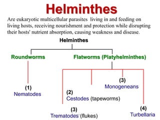
- 1. (4) Turbellaria Helminthes Are eukaryotic multicellular parasites living in and feeding on living hosts, receiving nourishment and protection while disrupting their hosts' nutrient absorption, causing weakness and disease. (2) Cestodes (tapeworms) Roundworms (3) Trematodes (flukes) Flatworms (Platyhelminthes) (1) Nematodes Helminthes (3) Monogeneans Souzan Eassa 1
- 2. Cestodes (The Taeenid tapeworms) Dr. Souzan Eassa Department of Microbiology Souzan Eassa 2
- 3. The taeenid tapeworms known to cause zoonotic infections in humans are: 1. Taenia solium, pork tapeworm/ Hook tapeworm. - Humans are the definitive hosts - Pigs are the intermediate hosts. - Causing cysticercosis 2. Taenia saginata, beef tapeworm/ Hookless tapeworm. - Humans are the definitive hosts. - Cattels and humans are the intermediate hosts. - Intestinal taeniasis Souzan Eassa 3
- 4. Differences between T. solium and T. saginata T. solium T. saginata Size Body length 2-4 meters up to 10 meters (70' worms have been reported) Scolex 4 suckers, rostellum & hooklets 4 suckers Mature proglottid Ovary 3 lobes 2 lobes Testes 150-200 300- 400 Gravid proglottid: Uterine branches 7-12 15-30 Life expectancy - 25 years or more 25 years or more Souzan Eassa 4
- 5. T. saginata T.solium D.H Human Human I.H Cattle Swine and Human Habitation Small intestine (Human) Tissue like brain, eye, muscle, liver and lungs etc. ( in Cattle) Small intestine (Human) Tissue like brain, eye, muscle, liver and lungs etc. (in Swine and humans) Infective stage Cysticercus bovis larvae (Humans) Egg (Cattle) Cysticercus Cellulosae larvae (Humans) Egg (Swine and humans) Disease Taeniasis (Humans) Cysticercosis (Cattle and humans) Taeniasis (Humans) Cysticercosis (Swine and humans) Differences between T. solium and T. saginata Souzan Eassa 5
- 6. Egg, The eggs of Taenia saginata and T. solium are identical. The eggs are spherical, diameter 31 to 43 µm, with a thick radially striated brown shell (embryophore). Inside each shell is an embryonated oncosphere with 6 hooklets. Souzan Eassa 6
- 7. Life cycle of T. solium 1. final host: man. 2. Intermediate host: pig (or man). 3. Infective stage: cysticercus and egg. 4. Infective mode: eating raw bean-pork. 5. Site of inhabitation: adult in small intestine; cysticercus in tissues. 6. Infective mode of cysticercosis: endogenous, exogenous auto-infection and foreign source. 7. Life span: more than 25 years; cysticercus can survives 5-6 years in human body. Souzan Eassa 7
- 8. Life cycle of T. saginata 1. final host: man. 2. Intermediate host: Cattle. 3. Infective stage: cysticercus. 4. Infective mode: eating raw beef. 5. Site of inhabitation: adult in small intestine. 7. Life span: more than 25 years. Souzan Eassa 8
- 10. Life cycle of T. solium and T. saginata Infective eggs (from human feces) are ingested by the intermediate host (cow or pig). The onchosphere hatches and penetrates the intestinal mucosa of the intermediate host. Onchosphere delivered to various parts of body via the circulatory or lymphatic systems. Most localize & encyst in muscle. Infective stage - the encysted larva, called a cysticercus develops within 2 months. Human infections take place when uncooked or undercooked meat containing larvae is ingested.Souzan Eassa 10
- 11. Pathogenesis and Clinical Manifestations 1. Taeniasis: It is caused by the adult residing in small intestine of the man. The patient is usually no obvious symptom, only complaining passing proglottides. Some time the adult irritates the small intestine causing: Deprivation of nutrition Disfunction and discomforts of the intestine, such as abdominal pain, anorexia, chronic indigestion, emaciation, eosinophilia and etc. vomiting or diarrhea. Allergic reactions Appendicitis. Obstructions of the intestineSouzan Eassa 11
- 12. 2. Auto-infection, due to T.solium egg only; Pathogenic factor: cysticercus cellulosae). Symptoms vary with site (the tissues and organs involved) and intensity of infection (number of the cysticerci). Clinical aspects: headache, dizziness, epilepsy, blurred vision, subcutaneous nodule etc. Cysticercosis is divided into three types. Pathogenesis and Clinical Manifestations Souzan Eassa 12
- 13. The subcutaneous nodules are usually found in head, limbs, neck, abdomen and back. (1) Subcutaneous type: Souzan Eassa 13
- 14. The cysticercus is usually found in the vitreous body or subretina. Visual disturbance often occurs. (2) Ocular type: Souzan Eassa 14
- 15. (3) Brain type: The symptoms are related to the site of infection. The patients may manifest headache, nausea, vomiting, epilepsy, paralysis, weakness in limbs, diplopia, dizziness, mental disorder. Epilepsy is the most frequent symptoms of brain cysticercosis. Souzan Eassa 15
- 16. Brain type Souzan Eassa 16
- 17. • cysticercus in the tongue Souzan Eassa 17
- 18. Diagnosis • Taeniasis Anal swab: to find egg at perianal region Finding of gravid proglottids or eggs at the perianal region by cellophane tape method. Fecal exam: to find segment (species identification). • Cysticercosis Biopsy (subcutaneous nodule). X-ray/CT/MRI: cerebral cysticercosis. • Serological tests • Molecular Tests Souzan Eassa 18
- 19. Treatment and Prevention Treatment of Taeniasis: Praziquantel. Treatment of cysticercosis: Surgical removal is required for ocular and superficial cysticercoses. (1) Health education. (2) Avoid eating raw beef and pork. Souzan Eassa 19