Assessment of immunomolecular_expression_and_prognostic_role_of_tlr7_among_pa...
P1-01-17_poster
1. Integrative Pathology: Analysis of Cellular Multiplex Technology to Detect Proteomic, Genomic and DNA Data from Fine Needle Aspiration Biopsy Specimens
Mittendorf EA1, Dogan B1, Morgan R2,4, Chargin A2,4, Wu Y1, Cornett-Risher S1, Green L 3, Zhang P3, Shults K2
1The University of Texas MD Anderson Cancer Center, 2Penfold-Patterson Research Institute, 3MTSU Biostatistics, 4IncellDx
BACKGROUND
An integrative system capable of detecting proteomic, genomic and DNA content from
cell isolates obtained by fine needle aspiration (FNA) biopsy may offer distinct
advantages in diagnosing breast cancer and monitoring response to therapy. Cellular
Multiplex™ (figure 1) is such a system. An initial pilot study evaluating this technology
established a series of variables that could separate normal from cancerous elements
using cells obtained from an FNA performed on excised tumors and reduction
mammoplasty specimens. In order for the technology to be clinically relevant, it must
perform robustly on intact tumors. The current study was therefore undertaken to
validate Cellular Multiplex™ on cells obtained by FNA performed on intact tumor at the
time of diagnosis.
METHODS
Patients undergoing lumpectomy requiring either needle or 125I seed
localization were identified. FNA was performed on intact tumor (A
samples) at the time of radiographic localization prior to lumpectomy
and repeated on the excised tumor (B samples). Cells obtained by FNA
were placed in a proprietary fixative (incellFP, IncellDx, Menlo Park, CA)
then hybridized and stained to detect multiple mRNA and protein targets
(OncoBreast 3Dx®, IncellDx, Menlo Park, CA) along with DNA content
(see Table 1) Estrogen receptor (ER), progesterone receptor (PR), and
HER2 were included in the panel of targets and compared to the routine
clinical pathology report where ER, PR and HER2 were evaluated using
immunohistochemistry. Cell morphology was assessed by mean
corpuscular volume. Samples were analyzed using a 3 laser EC800
(Sony Biotechnology, San Jose, CA). The quality control of the system
contains known bead targets as well as mixtures of peripheral blood
mononuclear cells (PBMC), MCF7 and SK-BR-3 cultured breast cancer
cells to ensure the daily performance is within acceptable limits.
The study is designed to enroll 50 patients and here we report an
interim analysis of the first 21 cases. Data regarding ER, PR and HER2
expression generated from the analyzed samples were paired with ER,
PR and HER2 data generated by IHC performed for clinical care (Table
2). The fused dataset was cleaned and validated after which SAS code
was used to create the statistics package. The variables measured or
derived were compared for the both A and B samples. The paired
Student T-test and Mann-Whitney nonparametric test were used to
determine difference using p-values. In most cases, these tests agree.
In the few cases where they disagreed, a test for normality
(Kolomogorov-Smirnov) was used to determine which test is most
appropriate.
CONCLUSIONS
•Results obtained by IHC and Cellular
Multiplex match with high precision.
•In cases where at least 100 epithelial cells are
obtained, the measured/derived parameters
match when comparing the two sample
sources.
•This interim data suggests that Cellular
Multiplex may be used as a diagnostic tool.
Other applications to include following
response to neoadjuvant therapy warrant
investigation.
Table 1. mRNA and protein targets identified by identified by
Cellular Multiplex™
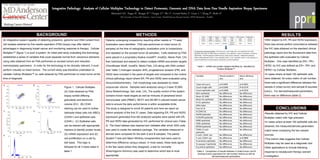
Figure 1. Cellular Mulitplex.
(A) Cells obtained by FNA
biopsy exhibit differing
granularity and electronic
volume (EV). (B) CD45
staining can be used to further
delineate these cells into WBCs
(CD45+) and epithelial cells
(CD45-). (C) Epithelial cells
can be stained with appropriate
markers to identify protein levels,
(D) mRNA expression and (E)
cell proliferation on a cell by
bell basis. This logic is
followed for all 3 tubes listed in
table 1.
A. B.
C. D.
E.
RESULTS
•With respect to ER, PR and HER2 expression,
there was almost perfect concordance between
the IHC data obtained on the standard clinical
pathology report and the fluorescent data from
the epithelial cells evaluated by Cellular
Multiplex. One case identified as ER+, PR+,
HER2- by IHC was defined as ER+, PR+ and
HER2+ by Cellular Multiplex.
•In cases where at least 100 epithelial cells
were obtained, for every metric of cell number,
there was a significant difference between the
sample A (intact tumor) and sample B (excised
tumor). For derived/measured parameters,
there was no difference (table 2).
Table 2. Comparison of results between A (intact tumor) and B
(excised tumor) specimens for cell number metrics as well as
derived/measured parameters.