PNEUMONIA
•
166 likes•39,024 views

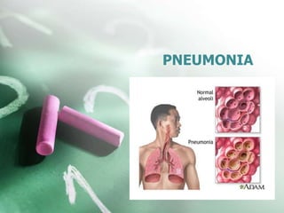
Pneumonia is an inflammation of the lung parenchyma caused by various microorganisms, including bacteria, mycobacteria, fungi, and viruses. Pneumonitis is a more general term that describes the inflammatory process in the lung tissue that may predispose and Pneumonia is an inflammation of the lung parenchyma that is caused by a microbial agent. place the patient at risk for microbial invasion. Pneumonia is classified into four: community-acquired pneumonia (CAP) and hospital-acquired pneumonia (HAP), pneumonia in the immunocompromised host, and aspiration pneumonia.

Recommended
More Related Content
What's hot
What's hot (20)
Similar to PNEUMONIA
Similar to PNEUMONIA (20)
More from ANILKUMAR BR
More from ANILKUMAR BR (20)
Recently uploaded
Recently uploaded (20)
PNEUMONIA
- 1. PNEUMONIA
- 2. • Pneumonia is an inflammation of the lung parenchyma caused by various microorganisms, including bacteria, mycobacteria, fungi, and viruses. • Pneumonitis is a more general term that describes the inflammatory process in the lung tissue that may predispose and place the patient at risk for microbial invasion.
- 3. Classifications • Pneumonia is classified into four: community-acquired pneumonia (CAP) and hospital-acquired pneumonia (HAP), pneumonia in the immunocompromised host, and aspiration pneumonia.
- 4. Community-Acquired Pneumonia ( CAP ) • CAP occurs either in the community setting or within the first 48 hours after hospitalization. • The causative agents for CAP that needs hospitalization include streptococcus pneumoniae, H. influenza, Legionella, and Pseudomonas aeruginosa. • Only in 50% of the cases does the specific etiologic agent become identified. • Pneumonia is the most common cause of CAP in people younger than 60 years of age.
- 5. Hospital-Acquired Pneumonia (HAP) • HAP is also called nosocomial pneumonia and is defined as the onset of pneumonia symptoms more than 48 hours after admission in patients with no evidence of infection at the time of admission. • HAP is the most lethal nosocomial infection and the leading cause of death in patients with such infections.
- 6. Hospital-Acquired Pneumonia (HAP) • Common microorganisms that are responsible for HAP include Enterobacter species, Escherichia coli, influenza, Klebsiella species, Proteus, Serratia marcescens, S. aureus, and S. pneumonia. • The usual presentation of HAP is a new pulmonary infiltrate on chest x-ray combined with evidence of infection.
- 7. Pneumonia in the Immunocompromised Host • Pneumonia in immunocompromised hosts includes Pneumocystis pneumonia, fungal pneumonias and Mycobacterium tuberculosis. • Patients who are immunocompromised commonly develop pneumonia from organisms of low virulence. • Pneumonia in immunocompromised hosts may be caused by the organisms also observe in HAP and CAP.
- 8. Aspiration Pneumonia • Aspiration pneumonia refers to the pulmonary consequences resulting from entry of endogenous or exogenous substances into the lower airway. • The most common form of aspiration pneumonia is a bacterial infection from aspiration of bacteria that normally reside in the upper airways. • Aspiration pneumonia may occur in the community or hospital setting. • Common pathogens are S. pneumonia, H.influenza, and S. aureus.
- 9. RISK FACTORS FOR PNEUMONIA 1. Conditions that produce mucus or bronchial obstruction and interfere with normal lung drainage (e.g., cancer, cigarette smoking, COPD) 2. Immunosuppressed patients and those with a low neutrophil count (neutropenic) 3. Smoking; cigarette smoke disrupts both mucociliary and macrophage activity Prolonged immobility and shallow breathing pattern.
- 10. 1. Depressed cough reflex (due to medications, a debilitated state, or weak respiratory muscles); 2. aspiration of foreign material into the lungs during a period of unconsciousness (head injury, anesthesia, depressed level of consciousness), or abnormal swallowing mechanism 3. Nothing-by-mouth (NPO) status; placement of nasogastric, orogastric, or endotracheal tube 4. Antibiotic therapy (in very ill people, the oropharynx is likely to be colonized by gram- negative bacteria)
- 11. 1. Alcohol intoxication (because alcohol suppresses the body’s reflexes, may be associated with aspiration, and decreases white cell mobilization and tracheobronchial ciliary motion) 2. General anesthetic, sedative, or opioid preparations that promote respiratory depression, which causes a shallow breathing pattern and predisposes to the pooling of bronchial secretions and potential development of pneumonia
- 12. Clinical Manifestations • Pneumonia varies in its signs and symptoms depending on the organism and the patient’s underlying disease. • However, regardless of the type of pneumonia (CAP, HAP, immunocompromised host aspiration), a specific type of pneumonia cannot be diagnosed by clinical manifestations alone.
- 13. • For example, the patient with streptococcal (pneumococcal) pneumonia usually has a sudden onset of shaking chills, rapidly rising fever (38.5°to 40.5°C [101°to 105°F]), and pleuritic chest pain that is aggravated by deep breathing and coughing.
- 14. • Advanced age, because of possible depressed cough and glottic reflexes and nutritional depletion Respiratory therapy with improperly cleaned equipment
- 15. Tachypnea SOB Pleuritic chest pain (Deep breathing and coughing aggravate the pain in the chest). Use of accessory muscles in respiration Loss of appetite and fatigue Cyanosed lips and nail beds Purulent sputum. The sputum becomes purulent because of the infection in the lung parenchyma which produced sputum-filled with pus.
- 17. Prevention • It is better to prevent the occurrence of pneumonia instead of treating the disease itself. Here are several ways that can help prevent pneumonia. • Pneumococcal vaccine. This vaccine can prevent pneumonia in healthy patients with an efficiency of 65% to 85%. • Staff education. To help prevent HAP, the CDC (2004) encouraged staff education and involvement in infection prevention.
- 18. Prevention • Infection and microbiologic surveillance. It is important to carefully observe the infection so that there could be an appropriate application of prevention techniques. • Modifying host risk for infection. The infection should never be allowed to descend on any host, so the risk must be decreased before it can affect one.
- 19. Complications associated with Pneumonia • Pneumonia has several complications if left untreated or the interventions are inappropriate. These are the following complications that may develop in patients with pneumonia.
- 20. Complications associated with Pneumonia 1. SHOCK AND RESPIRATORY FAILURE (Severe complications of pneumonia include hypotension and shock and respiratory failure (especially with gram-negative bacterial disease in elderly patients).
- 21. Complications associated with Pneumonia 2. ATELECTASIS AND PLEURAL EFFUSION (Atelectasis (from obstruction of a bronchus by accumulated secretions) may occur at any stage of acute pneumonia)
- 22. SUPERINFECTION • Superinfection may occur with the administration of very large doses of antibiotics, such a penicillin, or with combinations of antibiotics.
- 23. ASESSMENT & DIAGNOSTIC • Assessment and diagnosis of pneumonia must be accurate since there are a lot of respiratory problems that have similar manifestations. • The following are assessments and diagnostic tests that could determine pneumonia.
- 24. ASESSMENT & DIAGNOSTIC 1. History taking. The diagnosis of pneumonia is made through history taking, particularly a recent respiratory tract infection. 2. Physical examination. Mainly, the number of breaths per minute and breath sounds is assessed during physical examination
- 25. ASESSMENT & DIAGNOSTIC 1. Chest x-ray studies 2. Blood culture (bloodstream invasion, called bacteremia, occurs frequently) and 3. Fiberoptic bronchoscopy. May be both diagnostic (qualitative cultures) and therapeutic (Reexpansion of lung segment).
- 26. • ABGs/pulse oximetry. Abnormalities may be present, depending on extent of lung involvement and underlying lung disease. • Bronchoscopy : Bronchoscopy is often used in patients with acute severe infection, patients with chronic or refractory infection, or immunocompromised patients when a diagnosis cannot be made from an expectorated or induced specimen.
- 27. Sputum examination. • The sputum sample is obtained by having the patient. 1. Rinse the mouth with water to minimize contamination by normal oral flora. 2. Breathe deeply several times. 3. Cough deeply and 4. Expectorate the raised sputum into a sterile container
- 28. The sputum sample is obtained by having the patient.
- 29. Medical management • The treatment of pneumonia includes administration of the appropriate antibiotic as determined by the results of the Gram stain.
- 30. The management of pneumonia centers is a step- by-step process that zeroes on the treatment of the infection through identification of the causative agent. 1. Blood culture. Blood culture is performed for identification of the causal pathogen and prompt administration of antibiotics in patients in whom CAP is strongly suspected. 2. Administration of macrolides. Macrolides are recommended for people with drug- resistant S. pneumoniae.
- 31. • Hydration is an important part of the regimen because fever and tachypnea may result in insensible fluid losses. • Administration of antipyretics. Antipyretics are used to treat fever and headache. • Administration of antitussives. Antitussives are used for treatment of the associated cough. • Bed rest. Complete rest is prescribed until signs of infection are diminished.
- 32. • Oxygen administration. Oxygen can be given if hypoxemia develops. • Pulse oximetry. Pulse oximetry is used to determine the need for oxygen and to evaluate the effectiveness of the therapy. • Aggressive respiratory measures. Other measures include administration of high concentrations of oxygen, endotracheal intubation, and mechanical ventilation.
- 34. Nursing Assessment Nursing assessment is critical in detecting pneumonia. • Assess respiratory symptoms. Symptoms of fever, chills, or night sweats in a patient should be reported immediately to the nurse as these can be signs of bacterial pneumonia. • Assess clinical manifestations. Respiratory assessment should further identify clinical manifestations such as pleuritic pain, bradycardia, tachypnea, and fatigue, use of accessory muscles for breathing, coughing, and purulent sputum.
- 35. • Physical assessment. Assess the changes in temperature and pulse; amount, odor, and color of secretions; frequency and severity of cough; degree of tachypnea or shortness of breath; and changes in the chest x-ray findings. • Assessment in elderly patients. Assess elderly patients for altered mental status, dehydration, unusual behavior, excessive fatigue, and concomitant heart failure.
- 36. The nurse should monitor the following: 1. Changes in temperature and pulse 2. 2. Amount, odor, and color of secretions 3. 3. Frequency and severity of cough 4. 4. Degree of tachypnea or shortness of breath 5. 5. Changes in physical assessment findings (primarily assessed by inspecting and auscultating the chest)
- 37. The nurse should monitor the following: 1. Changes in the chest x-ray findings 2. In addition, it is important to assess the elderly patient for unusual behavior, altered mental status, dehydration, excessive fatigue, and concomitant heart failure.
- 38. Nursing Management 1. Maintain a patent airway and adequate oxygenation. 2. Obtain sputum specimens as needed. 3. Use suction if the patient can’t produce a specimen. 4. Perform chest physiotherapy. 5. Provide a high calorie, high protein diet of soft foods.
- 39. Nursing Management 1. To prevent aspiration during nasogastric tube feedings, check the position of tube, and administer feedings slowly. 2. Provide a quiet, calm environment, with frequent rest periods. 3. Explain the importance of respiratory exercise such as spirometry, deep breathing, effective coughing, and chest physical therapy etc. 4. Monitor the patient’s ABG levels, especially if he’s hypoxic. 5. Assess the patient’s respiratory status, auscultate breath sounds at least every 4 hours
- 40. Nursing Diagnosis Ineffective airway clearance related to copious tracheobronchial secretions 2. Activity intolerance related to impaired respiratory function 3. Risk for deficient fluid volume related to fever and dyspnea 4.
- 41. 1. Imbalanced nutrition: less than body requirements 2. 5. Deficient knowledge about the treatment regimen and preventive health measure
- 42. Nursing Interventions 1. Improve airway patency. 2. Rest to conserve energy. 3. Maintenance of proper fluid volume. 4. Maintenance of adequate nutrition. 5. Understanding of treatment protocol and preventive measures. 6. Absence of complications.