
Recommended
More Related Content
What's hot
What's hot (20)
Similar to Residual ridge resorption
Similar to Residual ridge resorption (20)
Recently uploaded
Recently uploaded (20)
Residual ridge resorption
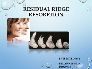
- 1. RESIDUAL RIDGE RESORPTION PRESENTED BY : DR. ANNESHA K. KONWAR
- 2. CONTENTS: DEFINITIONS CLASSIFICATIONS CONSEQUENCES MECHANISM PATHOGENESIS ETIOLOGY TREATMENT & MANAGEMENT SUMMARY AND CONCLUSION
- 3. DEFINITIONS : • “Bone - a highly vascularized, living, constantly changing, mineralized connective tissue”. [Gray’s anatomy-40th edition] • “Alveolar process -- that part of the maxilla and mandible that forms and supports the sockets of the teeth”. [Orban’s]
- 4. • “Alveolar bone is the bony portion of the maxilla and the mandible in which roots of the teeth are held by fibers of periodontal ligament”. [GPT-8]
- 5. • “Residual alveolar ridge is the portion of the alveolar ridge and its soft tissue covering which remains following the removal of or loss of teeth. [GPT-8]
- 6. • The residual bony architecture of the maxilla and mandible undergoes a life-long catabolic remodelling. • The rate of reduction in size of the residual ridge is maximum in the first 3-6 months and then gradually tapers off. • However, bone resorption activity continues throughout life at a slower rate, resulting in loss of varying amount of jaw structure, ultimately leaving the patient a ‘dental cripple’.
- 7. The mechanical aspect of bone remodeling is usually associated with Wolff’s law of bone transformation which states that “Every Change In Change In The Form And Function Of Bone , Or Of Their Function Alone,is
- 9. According to Atwood : (JPD 1971 vol.26) • Order 1 : pre-extraction • Order 2 : post-extraction • Order 3 : high, well rounded • Order 4 : knife-edge • Order 5 : low, well rounded • Order 6 : depressed
- 10. SIEBART’S CLASSIFICATION • Class I : defects – faciolingual loss of tissue width with normal ridge height. • Class ii : defects- loss of ridge height with normal ridge width • Class iii : defects-combination of loss in both the directions
- 11. According to Wical and Swoope : Class I : upto one third of the original vertical height lost. Class ii : from one third to two thirds of the vertical height lost. Class iii : two third or more of the mandibular height lost.
- 12. According to Niel’s classification Class 1 : 0.5” space exists between mylohyoid ridge and floor of the mouth Class 2 : less than 0.5” space Class 3 : mylohyoid muscle at the same level as mylohyoid ridge
- 13. According to Misch’s classification Bone Density D1 dense cortical bone D2 thick dense to porous cortical bone on crest and cortical trabecular bone within D3 thin porous cortical bone on crest and fine trabecular bone within D4 fine trabecular bone D5 immature, non mineralized bone
- 14. According to the American College of Prosthodontists : Mcgarry et al, J prosthodont 8(1):27-39, 1999 Based on bone height (mandible only) Type I : residual bone height of 21 mm or greater measured at the least vertical height of the mandible. Type II : residual bone height of 16 - 20 mm measured at least vertical height of the mandible. Type III : residual alveolar bone height of 11 - 15 mm measured at the least vertical height of the mandible. Type IV : residual vertical bone height of 10 mm or less measured at the least vertical height of the mandible.
- 15. BONE CELLS 1) Osteoblasts : • Derived from osteoprogenitor cells • Periosteum serves as important reservoir . • Bone resorbing factors that act via the osteoblast are parathyroid hormone, vitamin D3, interlukin -1 and tumor necrosis factor.
- 16. 2)OSTEOCYTES Nerve cells Sense the change in environment and send signals that affect response of other cells involved in bone remodelling Maintains balance between resorption and remodelling Bone that forms more rapidly shows more osteocytes.
- 17. 3) OSTEOCLASTS - Bone destructing cells.
- 19. The organic components of the intercellular substance are removed by proteolytic action of the osteoclasts Then, the Ca salts (inorganic) are dissolved by a chelating action of the osteoclasts. As resorption takes place, the osteocytes released may revert to osteoblasts or become osteoclasts, depending on the physiologic and pathologic demands. MECHANISM OF BONE RESORPTION Histologically, bone apposition and resorption take place in close approximation, making possible the bone balance of shape and size.
- 20. SEQUENCE OF RESORPTIVE EVENTS Attachment of osteoclasts to mineralized surface of bone Creation of a ruffled border and a sealed acidic environment through action of the proton pump Dissolution of the Hydroxyapatite Fall in pH to 2.5-3 in the osteoclast resorption space Digestion of the organic components of the matrix by proteolytic enzymes
- 21. BONE RESORPTION FACTORS LOCAL SYSTEMIC -Endotoxins from dental plaque -Osteoclast activating factor(OAF) -Prostaglandins -Human gingival bone resorption factor -Trauma due to ill fitting dentures which leads to increased or decreased vascularity and changes in oxygen tension. -Correct amount of circulating estrogen, thyroxine, growth hormone, calcium, phosphorus, -vitamin D , -Osteoporosis - Hypophosphetemia - Parathormone - Calcitonin
- 22. BONE RESORPTION AND CA HOMEOSTASIS: The only sources of Ca for the body are •Diet •Bone reservoir. Ca homeostasis is maintained by controlling Ca obtained from these 2 sources. This can occur by altering internal absorption mechanisms (income) or tubular reabsorption (recycling) or by liberation of Ca from the skeleton via resorption (savings). There is a reciprocal relationship between Ca concentration and bone resorption to maintain Ca homeostasis. As the level of serum calcium develops, resorption is stimulated and factors that would inhibit resorption are depressed.
- 23. Skeletal depletion of calcium occurs as a result of stimulation of parathyroid gland and the alveolar bone is the first to be affected. This is due to the function of parathyroid hormone in maintaining the blood calcium level by mobilizing it from bones by osteoclastic activity. Simultaneously , there is an increased renal excretion of phosphate, which disturbs the blood calcium:phosphorous ratio by raising the blood calcium level. This results in mobilization of phosphates from bones by osteoclastic activity. •Under these conditions , alveolar bone becomes susceptible to diseases like osteoporosis.
- 24. In equilibrium the two antagonistic actions (of osteoblasts and osteoclasts) are in balance. In growth, although resorption is constantly taking place in the remodeling of bones as they grow, increased osteoblastic activity more than makes up for the bone destruction. Whereas in osteoporosis, osteoblasts are hypoactive, and, in the resorption related to hyperparathyroidism, increased osteoblastic activity is unable to keep up with the increased osteoclastic activity. The normal equilibrium may be upset and pathologic bone loss may occur if either bone resorption is increased or bone formation is decreased, or if both occur.
- 25. Since bone metabolism is dependent on cell metabolism, anything that influences cell metabolism of osteoblasts and osteoclasts is important. The thyroid hormone affects the rate of metabolism of cells in general and hence the activity of both, the osteoblasts and osteoclasts. Parathyroid hormone influences the excretion of phosphorous in the kidney and also directly influences osteoclasts.
- 26. •The degree of absorption of Ca, P and proteins determines the amount of building blocks available for the growth and maintenance of bone. •Vit C aids in bone matrix formation. •Vit D acts through its influence on the rate of absorption of calcium in the intestines and on the citric acid content of bone. •Various members of Vit B complex are necessary for bone cell metabolism.
- 27. According to Reifenstein, In the young person, there is a relative predominance of anabolic hormones (estrogen and testosterone) over the anti anabolic hormones( cortisone and hydrocortisone) resulting in continued growth of skeleton. As people get older, the anabolic hormones are so reduced that the antianabolic hormones are in relative excess with the result that bone resorption may take place faster than bone formation and that bone mass may be reduced.
- 28. OSTEOPOROSIS Osteoporosis is defined by the WHO as bone mineral density (BMD) greater than 2.5 standard deviations below that of the young adult BMD. Osteoporosis is common in aging individuals, especially post menopausal women when the estrogenic blood level is low. In elderly men and women, osteoporosis is caused by a variety of factors such as calcium loss, calcium deficiency, hormonal deficiency, change in protein nutrition and decreased physical activity.
- 29. osteoporosis Primary (no known cause) Type 1 Post menopausal Type 2 Age related secondary Traceable etiology
- 31. PATHOPHYSIOLOGY • The most popular theory of how osteoporosis occur in females is based on the central role of oestrogen in bone remodelling.
- 32. Decreased oestrogen levels leads to increased pro-inflammatory cytokine levels like IL1 and TNF leading to increased osteoclast formation and hence increased bone loss. Oestrogen acts through two receptors: oestrogen receptor a (ERa) and ERb, ERa appears to be the primary mediator of the actions of oestrogen on the skeleton. Another line of action is the decreased antagonistic action of oestrogen on parathyroid leads to more parathormone secretion and consequently increased bone resorption.
- 33. One •loss and/or mobility of teeth Two •edentulism, Three •excessive residual ridge resorption Four •dentures which require repeated revision or remakes PROSTHODONTIC IMPLICATIONS
- 34. PATHOGENESIS OF RRR Immediately following the extraction (order II), any sharp edges remaining are rounded off by external osteoclastic resorption leaving a high well rounded ridge (order III). As resorption continues from the labial and lingual aspects ,the crest of the ridge becomes increasingly narrow, ultimately becoming knife edged (order iv). As the process continues, the knife-edge becomes shorter and eventually disappears leaving a low well-rounded or flat ridge (order v). Eventually this too resorbs, leaving a depressed ridge (order VI).
- 35. PHYSIOLOGY V/S PATHOLOGY??? • Some clinicians feel that RRR is not a disease but a normal physiological process. • However there is wide variation in the rate of RRR in different individuals- depending on multiple factors. • The need to elucidate these major differences warrants labeling this process a “ disease” or “pathology”
- 36. • The mechanism of the reduction of the mandibular residual ridge actually represents a modified version of the enlow’s “V” principle, showing external resorption accompanied by endosteal deposition. Principles of bone remodeling. By Donald H. Enlow
- 37. PATHOLOGY OF RRR Gross Microscopic
- 38. The careful superimposition of portions of tracings of lateral cephalograms clearly shows the gross reduction of bone in size and shape that occurs on the labial, crestal, and lingual aspects of the residual ridge.
- 39. IN DRY SPECIMENS *External cortical surface of maxilla and mandible are uniformly smooth & crestal area of residual ridge shows porosities and imperfections. *Bones with more severe RRR display gross porosities of medullary bone on the crest of ridge.
- 40. • Panoramic radiograph showing severe RRR in both maxilla and mandible in contrast to dentulous area that support three mandibular teeth.
- 41. MICROSCOPIC PATHOLOGY Osteoclastic activity occurs on the external surface of crest of ridges . Scalloped margins of howships lacunae sometimes contain visible osteoclasts. Frequently the scalloped external surface seems inactive without bone resorbing cells.
- 42. MEASUREMENT OF RRR 1. Serial examination of diagnostic casts. 2. Lateral cephalometric radiographs • Most accurate • Measures RRR over a period of time. 3. Panoramic radiographs.
- 43. ORIGINAL BONE HEIGHT = THREE TIMES THE DISTANCE FROM INFERIOR BORDER OF MANDIBLE TO THE LOWER EDGE OF MENTAL FORAMEN. {Kenneth E. Wical and Charles C. Swoope. Studies of residual ridge resorption. Uses panaromic radiographs for evaluation and classification of mandibular resorption. JPD;1974;32;7}
- 44. •To date, it appears that RRR world-wide, occurs in males and females, young and old, sickness and in health, with and without dentures and is unrelated to the primary reason for the extraction of the teeth (Caries / periodontal disease). Rate of RRR is variable -between persons. -within the same person at different times. -within the same person at different sites. EPIDEMIOLOGY OF RRR
- 45. AMOUNT AND RATE OF BONE RESORPTION • According to Boucher, During the first year after tooth extraction, the reduction in residual ridge height in the midsagittal plane is 2-3 mm for maxilla 4-5 mm for mandible Annual rate of reduction in height 0.1-0.2 mm for mandible 4 times less in the maxilla
- 46. DIRECTION OF BONE RESORPTION This progressive change of the edentulous mandible and maxilla makes many patients appear prognathic. Maxilla resorbs upward and inward to become progressively smaller The mandible inclines outward and becomes progressively wider.
- 48. Thus RRR is centripetal in maxilla and centrifugal in mandible.
- 49. ETIOLOGY OF RRR : According to Atwood : RRR is a multifactorial biomechanical disease caused by a combination of : Some clinical factors related to rate of resorption of residual ridges JPD vol 12,issue 3, pages 441-450
- 50. ANATOMIC FACTORS • It is postulated that RRR varies with the quantity and quality of the bone of residual ridges.. The more bone there is, the more rrr will ultimately be. But this cannot be considered a good prognostic factor, because in some cases large ridges resorb rapidly and some knife-edge ridges may remain with little change for long periods of time. RRR α Anatomic factors
- 51. • We should always try to evaluate the present status of the residual ridge to determine what has gone on before. • If a ridge has existed as high and well rounded (order iii) for several years, it will likely to continue to do so. • But if a ridge has gone from an order ii to order iv in just two years it will probably continue to resorb rapidly.
- 52. MECHANICAL FACTORS • Disuse atrophy vs. Abuse resorption. • Duration of functional forces do not exceed 15 mins. /Day (brewer) • 3500 – 4200 lbs loading force by 1500 empty swallows. RRR ≈ force
- 53. • Projected maxillary denture bearing area – 4.2 in.2 , mandibular denture bearing area – 2.3 in.2 ( ratio 1.8:1) • Thus biting force of 50 lbs translates to 12lb/in2 on maxilla and 21lb/in2 on mandible Rrr ≈ 1 Damping effect • Damping effect or energy absorption by • Viscoelastic mucoperiosteum • Bone
- 54. RRR ≈ Anatomic factors + Bone resorption factors Bone formation factors + Force factors Damping effect factors + 1 Time
- 55. METABOLIC FACTORS • RRR varies directly with certain systemic or localized bone resorptive factors and inversely with certain bone formation factors. RRR BONE RESORPTION FACTORS BONE FORMATION FACTORS
- 56. Excessive stress resulting from artificial environment. Abuse of tissues from lack of rest- Bone is moldable. It can tolerate masticatory forces within the limits of physiologic tolerance. But exceeding that it causes damaging forces which will result in resorption of the alveolar bone. PROSTHETIC FACTORS
- 57. Long continued use of ill fitting dentures: May be due to long use, loss of bone, Incorrect occlusion, Incorrect jaw relation. Lack of freeway space due to increased vertical dimension of occlusion: Freeway space is present in the teeth in the physiologic rest position. It is normally around 2mm. At times, due to lack of freeway space the bone resorbs because of increased vertical height in an attempt to create the space.
- 58. Incorrect Centric relation record If the Centric relation is not recorded properly, the mandibular teeth will not occlude properly with those on the maxillary arch. This proper occlusion is essential to the health of bony support. Otherwise, during eccentric movement, it causes pressure on bone due to failure of denture stability. Hence resorption of base occurs. Error in relating maxilla to the cranial landmarks (orientation relation): The plane of the maxilla should be oriented to the facial reference line (Camper’s plane or ala tragus line). If not, may cause instability of denture leading to resorption.
- 59. If occlusal corrections are not done: • These errors which may be caused due to processing techniques if not corrected causes premature contacts resulting in increased stress. • Selective grinding should be done to minimize lateral stress and resulting tissue trauma.
- 60. CONSEQUENCES OF RRR : • Apparent loss of sulcus width and depth. • Displacement of muscle attachment close to the ridge. • Loss of vertical dimension of occlusion. • Reduction of the lower face height. • Increase in relative prognathia
- 61. • Changes in inter alveolar relationship. • Morphological changes of the alveolar bone such as sharp, spiny uneven residual ridges. • Location of mental foramina close to the ridge crest.
- 63. Prosthetic manageme nt Systemic evaluatio n. Tissue treatmen t therapy Pre prosthe tic surgery Diet
- 64. Pre-prosthetic surgery It aims at providing a good healthy surface for the insertion of the dentures. It includes all the surgical procedures by virtue of which an ideal smooth, healthy U shaped ridge , without any unfavourable undercuts or bony growths and with sufficient vestibular depth is achieved.
- 65. It includes the following surgical procedures: •Ridge correction. •Ridge extension/vestibuloplasty. •Ridge augmentation •Surgical correction of maxillomandibular relation.
- 66. Ridge Corrective surgery Soft tissue deformities •Labial frenectomy. •Lingual frenectomy. •High buccal frenal attachments. •Hyperplasia of soft tissues.
- 67. Bony deformities •Sharp irregular ridge. •Alveoloplasty. •Alveolectomy. •Excision of tori and genial tubercles.
- 68. RIDGE EXTENSION SURGERY/VESTIBULOPLASTY: •Labial. •Lingual. •High mental foramen. •Zygomaticoplasty. •Tuberoplasty.
- 69. RIDGE AUGMENTATION It is aimed at : •Increase in the ridge height and width providing a large denture bearing area , •Protection of neuro vascular bundles •Restoration of proper maxillomandibular arch relationship.
- 70. Impression techniques. Denture base selection. Teeth selection and arrangement Implant supported prosthesis. Prosthetic manageme nt
- 71. 1) IMPRESSION TECHNIQUE The main aim of the impression procedure is to gain maximum area of coverage. For e.g., in mandibular ridge, obtaining a fairly long retromylohyoid flange helps to achieve a better border seal and retention. Selection of proper trays and the correct impression procedure is very essential for an accurate impression.
- 72. Selective pressure technique This technique is most widely advocated to manage RRR. It makes it possible to confine the forces acting on the denture to the stress bearing areas . This helps in better withstanding the mechanical forces induced by denture wearing. Miller used mouth-temperature waxes instead of tissue conditioners.
- 73. Mucodynamic technique It is intended to integrate the changes in the shape of the vestibules when functional movements are made. A highly viscous thermoplastic reversible impression material is placed in the custom tray, then carefully adapted to the residual ridge and held with light and uniform pressure while the functional movements are made. As soon as the entire surface is smooth and the buccal and lingual borders are molded to the outer circumference without any folds, the impression is complete.
- 74. 2) Selection of denture base For degenerative ridge patients there are three types of denture bases: •Methyl methacrylate resin denture bases •Cast metal bases •Processed resilient , lined denture bases
- 75. 3) TEETH SELECTION AND ARRANGEMENT Teeth can be selected acc. to their form and size. The following requirements have to be met during teeth arrangement: •Stability of occlusion in centric relation. •Balanced occlusion for eccentric contacts. •Unlocking of the cusps mesio distally to accommodate the settling of denture bases.
- 76. Control of horizontal force by buccolingual cusp height reduction acc. to residual ridge shape and inter arch space. Functional balance by favorable tooth to ridge crest position. Cutting and shearing efficiency. Anterior clearance of teeth during mastication. Minimal occlusal stop areas for reduced pressure during function. Teeth should be placed in neutral zone to create co ordination between the primary and secondary masticatory organs.
- 77. Relative to each other, the maxillary and mandibular residual ridges are known to be in a favorable position for normal arrangement of posterior teeth if the connecting line between the midridge line of the max. and mand. residual ridges are at an angle of more than 80 degrees. An angle less than 80 degrees necessitates a cross bite or reverse occlusion arrangement of posterior teeth. A prognathic mandible necessitates the arrangement of anterior teeth in a reverse occlusion.
- 79. •Non anatomic teeth have known to cause fewer denture sore spots and lesser ridge resorption. •Anatomic posterior teeth cause more denture soreness and ridge resorption •Few studies state that anatomic posterior occlusion favors lower dentures and non anatomic posterior teeth favor upper denture.
- 80. 4) IMPLANT SUPPORTED PROSTHESIS The various problems associated with RRR and stability of removable soft tissue borne dentures have aroused interest in dental implantology to provide stable mechanical support to the dental prosthesis. This is because of the following advantages offered by implant supported prosthesis: Maintenance of alveolar bone. Maintenance of occlusal vertical dimension. Height of alveolar bone is found to be maintained as long as the implant remains healthy.
- 81. Regained proprioception. Increased stability, retention and phonetics. Maintenance of structure and function of muscles of mastication and facial expression. Immune to caries. Increased trabeculation and density of bone.
- 82. •Overall volume of bone is maintained. •Efficiency to take up stress and strain. •There is 20 fold decrease in the loss of structure with implants when compared with resorption that occurs with removable prosthesis. •Preventive implant is given following extraction to retard ridge resorption.
- 83. IMMEDIATE DENTURES: Some authors claim that extraction followed by immediate dentures reduces the ridge resorption.
- 84. OVERDENTURES Tooth supported over dentures help in improved stress distribution there by maintaining the integrity of residual ridge. The occlusal and parafunctional stresses are distributed through the abutment teeth. A study was conducted with overdentures supported by canines and it was seen that, the bone loss was 0.6mm where as 5mm in conventional complete dentures.
- 85. THE ADVANTAGES OF THE OVERDENTURE OVER THE CONVENTIONAL DENTURE ARE: 1. The denture bearing mucosa of the residual ridges are spared abuse. 2. Maintenance of the alveolar bone. 3. Sensory feedback. 4. Tactile sensitivity discrimination. 5. Masticatory performance. 6. Reduction of psychological trauma.
- 86. JOURNAL REVIEW
- 87. • MARKOVIC D ET AL., “ CURRENT KNOWLEDGE ON RESORPTION OF THE EDENTULOUS ALVEOLAR RIDGE” (1967) He proved that there is direct relation between the pressure under the mobile denture and the residual ridge resorption level on the other side. It seems that the frontal areas of the edentulous are more susceptible to resorption. Orthopantomography is the most acceptable method in level measuring of the RRR.
- 88. • MORRIS ET AL HAVE CONDUCTED STUDIES ON THE RESIDUAL RIDGE RESORPTION IN FIVE YEARS AFTER IMPLANT PLACEMENT The rate of resorption decreases significantly from that seen in conventional mandibular dentures • -XIE Q, ET AL., “ORAL STATUS & PROSTHETIC FACTORS RELATED TO RESIDUAL RIDGE RESORPTION IN ELDERLY SUBJECTS” -1997 He concluded that the local factors related to RRR more often in the maxilla than in the mandible,thus suggesting that severe resorption in the mandible is influenced more by systemic factors
- 89. • WYATT CC “THE EFFECT OF PROSTHODONTIC TREATMENT ON ALVEOLAR BONE LOSS” Concluded that an implant-supported fixed Prosthesis to restore missing teeth in partially or completely edentulous jaws seems to be the best means of preserving residual alveolar bone. • KLEMETTI E ET AL., “ FLUORIDATED DRINKING WATER,OESTROGEN THERAPY AND RESIDUAL RIDGE RESORPTION.” J ORAL REHABIL 1997 JAN:24(1):47-51 Artificially fluoridated water may also reduce the degree of residual ridge resorption
- 90. • AINAMO A ET AL., “ ASSOCIATION OF RESIDUAL RIDGE RESORPTION WITH SYSTEMIC FACTORS IN HOME-LIVING ELDERLY SUBJECTS” -1997 Concluded that asthma due to corticosteroid treatment is to be considered a risk indicator for severe resorption of the edentulous mandible, alcohol intake in the elderly may be related to a lesser degree of resorption of the edentulous maxilla.
- 91. PROSTHODONTIC MANAGEMENT OF A SIEBERT CLASS III DEFECT IN MANDIBULAR ANTERIOR REGION WITH A MODIFIED ANDREW’S BRIDGE . MANJUSHA PALEPU, DEVIPRASAD NOOJI, PRANAV MODY, SUHAS
- 94. SUMMARY AND CONCLUSION • The ultimate aim of a successful prosthesis is stability in function and excellent esthetics. • The expectations of edentulous patients are highly variable therefore the outcome of patient treatment varies significantly. • Patients should be educated regarding the type and extent of treatment that is ideal for them, the prognosis of the treatment outcomes with various types of removable or fixed prostheses and the alternatives that are available.
- 95. •Ortman HR: Factors of bone resorption of the residual ridge. J Prosthet Dent 1962;12,3:429-440. •Wendt DC: The degenerative denture ridge – Care and treatment. J Prosthet Dent 1974;32,5:477-492. REFERENCES
- 96. •Winkler S : Essentials of complete denture prosthodontics. 2nd edition,2000. •Boucher CO : Prosthodontic treatment for edentulous patients. 12th edition,2004. •Misch CE : Contemporary implant dentistry. 2nd edition,1999.
- 97. THANK YOU