Maddox rod and double maddox rod
•
41 likes•12,880 views

Measurement of Different Types of deviations with Maddox Rod and Double Maddox Test

Recommended
More Related Content
What's hot
What's hot (20)
Similar to Maddox rod and double maddox rod
Similar to Maddox rod and double maddox rod (20)
Recently uploaded
Recently uploaded (20)
Maddox rod and double maddox rod
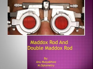
- 1. Maddox Rod And Double Maddox Rod By: Anu Musyakhwo M.Optometry
- 2. 1. Maddox Rod (Introduction, Objective, Equipment required, Set-up, Procedures, Recording, Interpretation,, Disadvantages) Distance Horizontal Deviation Distance Vertical Deviation Near horizontal and Vertical Deviation 2. Double Maddox Rod (Introduction, Set-up, Procedures, Recording, Interpretation,, Disadvantages) Distance and near cyclodeviation 3. References
- 3. a red or white lens consisting of a series of parallel cylinders that converts a point source of light into a streak of light or a line image perpendicular to the axis of parallel cylinders a dissimilar image test in which a single target is artificially made dissimilar in both eyes 1. Maddox Rod
- 4. Principle: Here, a point source of light is seen as spotlight with one eye and a red line with other eye, before which the maddox rod is placed. Therefore, the test is based on the principle of ‘Diplopic Projection’. position of the red line/streak in relation to the image of the light source seen by fellow eye indicates the presence and the amount of deviation of the eye subjectively
- 5. results of the test may be influenced by a head position, so the test should be performed in the habitual position since use of a phoropter will limit the patient’s ability to adopt a habitually abnormal head position, the measurement of vertical deviations using maddox rod is best performed using a trial frame or a hand-held maddox rod in free space
- 6. this test is performed to assess the type and size of a latent or manifest horizontal, vertical or torsional strabismus for distance and near fixation subjectively
- 7. Equipment required Maddox rod Source of spotlight Prism bar or loose prisms Trial frame Set up patient should wear his/her optimal refractive correction patient should be explained about the test and the procedures of the test room light should be dimmed and make sure that other source of light does not disturb the patient’s view of the streak and target spotlight
- 8. Distance Horizontal Deviation 1) place the maddox rod in front of the right eye making sure the grooves in the maddox rod are absolutely horizontal so that the patient sees vertical streak 2) spotlight is provided at distance of 6 meter using projector chart or Snellen Yellow light 3) ask the patient to see on the spotlight 4) ask the patient whether s/he sees both the vertical streak and spotlight a) if s/he finds difficulty in recognizing or seeing the streak demonstrate the patient by alternately covering the eyes b) if s/he sees only streak or spot, suppression may be present. To confirm the presence of suppression, maddox rod is transferred to the left eye and try again. If the patient has suppression, the spotlight or the streak cannot be seen again and follow up test should be performed
- 9. 5) With the maddox rod on right eye (see Fig. 3): a) if the patient sees streak passing through or overlapping the spotlight s/he is orthophoric i.e. patient does not have any deviation b) if the patient sees streak and spot light separated; streak on right side and spot on the left side which are uncrossed images, the patient has esodeviation c) if the patient sees crossed images that is the streak on the left side and the spotlight on the right side, the patient has exodeviation Fig. 3 :Positions of the streak and the spotlight in different types of deviations when maddox rod is placed before the right eye. (green dot represents the white/yellow spotlight)
- 10. 6)Measurement of deviations is done by placing loose prisms or prism bar in front of the either eye according to the type of deviation of eyes(see Fig. 3): a) to determine the amount of esodeviation, base out prism is placed in front of any eye until the patient sees the streak passing through the spot light a) to determine the amount of exodevaition, base in prism is placed in front of any eye until the patient sees the streak passing through the spotlight
- 11. Distance Vertical Deviation 1. rotate the maddox rod so that the grooves are absolutely vertical in orientation 2. ask the patient to see the spotlight 3. ask the patient if the streak is above or below the spotlight 4. With the maddox rod on the right eye (see Fig. 3): a. if the streak passes through the spotlight then the patient has no vertical deviation b. if the streak is above the spotlight then, the patient has right hypodeviation or left hyperdevaition. It is possible to specify vertical deviations with respect to other eye. Thus, this type of deviation can also be called as left over right. c. if the streak is below the spotlight then, the patient has right hyperdeviation or left hypodeviation. This type of deviation can also be called as right over left.
- 12. 5. To determine the amount of vertical deviations base up or base down prisms are used (see Fig. 3): a. if the deviation is left over right, base down prism is placed before the left eye (or base up prism before the right eye) until the streak passes through the spotlight b. if the deviation is right over left, base down prism is placed before the right eye (or base up prism before the left eye) until the streak passes through the spotlight
- 13. the horizontal and vertical deviations at near can be similarly measured the penlight (used as spotlight) is shone at nose bridge between the eyes at about 40 cm the near horizontal deviation measurements with maddox rod are considered unreliable in young patients due to the lack of a good accommodative stimulus in presbyopes, near deviation should be measured with the addition. The measurement technique is exactly the same as measurement of deviation at distance
- 14. Recording a. ortho is recorded for absence of horizontal, vertical as well as cyclodeviation or in other words for absence of any kind of deviation b. record the horizontal deviation and amount of deviation in prism diopters (▲) as: 6▲E for 6 prism diopter esophoria 4▲X for 4 prism diopter exophoria c. record the vertical deviation and amount of deviation in prism diopters (▲) as: 6▲R/L, or 6▲Rhyper, or 6▲Lhypo for 6 prism diopter for right over left. 4▲L/R, or 4▲Lhyper, or 4▲Rhypo for 4prism diopter for left over right.
- 15. Interpretation Most of the people with true normal binocular vision have some amount of latent deviation (heterophoria). In children and young adults the mean distance heterophoria is 1▲ exophoria ±1▲ and mean near heterophoria is 3▲exophoria ±3▲ (Scheiman and Wick 2002). In older adults, exophoria upto 6▲is common (Evans 2002). The vertical phoria of about 0.50 ▲ only is considered normal in both children and adults. In some patients even this amount of vertical phoria can cause symptoms. The amount of deviation with optimal correction got from maddox rod test should be compared with the cover test. If there is no significant change in refractive error after subjective refraction, the deviation got with maddox rod test should be similar to cover test. If there is change in refractive correction there occurs changes in horizontal deviation determined using the cover test and subjective assessment. For instance: if the optimal refractive correction shows an increase in plus or decrease in minus power from patient’s previous glass prescription then increase in exodeviation or decrease in esodeviation is expected and vice versa.
- 16. Disadvantages a) a spotlight represents a poor stimulus for accommodation. Some clinicians consider that this limits the usefulness of maddox rod to the measurement of vertical deviations which are assumed to be unaffected by accommodation changes a) cannot be performed under certain circumstances: sensory anomalies Suppression Abnormal Retinal Correspondence (This may mask the true size of deviation). c) cannot differentiate between tropias and phorias.
- 17. 2. Double Maddox Rod Test subjective test which uses two maddox rods, one before each eye used to determine cyclodeviation or torsion the test can be done in all the position of gazes for comparison traditionally, a red Maddox rod was placed before the right eye and a white Maddox rod before the left, but evidence suggests the different colors can cause fixation artifacts that do not occur if the same color is used bilaterally therefore, nowadays red maddox rods are preferred before both the eyes during the test
- 18. Set-up: place each maddox rod in the trial frame aligning the grooves of the rods at 90◦ making the red line/streaks seen by the patient horizontal (see Fig. 4). explain the patient about the test and procedures of the test dim the room light and make sure other light source does not disturb the view of the streaks for the patient Fig. 4: A patient wearing a trial frame in which a pair of maddox rods are placed, one before each eye
- 19. Procedures 1) use a source for spotlight at 33cm at the bridge of nose at the eye level for near measurement and for distance use a source of spotlight at 6 meter 2) ask the patient what the patient sees 3) ask the patient to show the orientation of the streaks using his/her arms 4) if patient feels difficult to compare the orientation of streaks, place the loose prism in the trail frame if the patient does not have any vertical or horizontal deviation, the streaks can be difficult to compare. Thus, loose prisms can be used to separate the images/streaks to let easy comparision or, if the patient has large horizontal or vertical deviation, prisms can be used to bring the streaks closer together to let the easy comparision
- 20. examiner can either use base-out, base-in, base-up or base-down prism depending on the deviation. For example: a patient with an large esotropia may need a base-out prism placed in front of deviated eye to bring the streak corresponding to this eye closer to other streak 5) ask the patient whether the streaks are parallel to each other •if the two streaks are parallel to each other, patient does not have cyclodeviation •if the patient sees the streaks not parallel to each other, rotate the maddox rod or ask the patient to rotate the maddox rod with help of dial in trial frame to make the lines parallel
- 21. Recording When recording the result, remember to list the name of the test as well as the direction and degrees of the torsion. Degrees taken from 90◦ (When maddox rod is vertical in trial frame) 15◦ excyclo (When maddox rod is before either of the eye rotated 15◦ outward/temporally from 90◦) Note: You are recording the position of the eye not the image.
- 22. Interpretation 1) Starting from 90◦; rotation of grooves of the maddox rod towards the nose (nasally) measures the incyclodeviation and towards the lateral side (temporally) measures the excyclodeviation. 2) The amount of cyclodeviation is measured in degrees, shown by the scale in the trial frame. For example: if the axis or grooves of maddox rod is rotated at 100◦, the patient has 10◦ excyclodeviation but if the axis of maddox rod is rotated at 80◦, the patient has 10◦ incyclodeviation. Along with the results gathered from other tests, you can use this information to consider the action of each muscle and also to indicate which muscle is likely to be affected. In most of the cases double maddox rod test helps in detection of the defect of the oblique muscles. For patients with unilateral superior oblique weakness the excyclophoria is generally between 1 and 7 degrees, whereas in bilateral superior oblique weakness the excyclophoria is typically greater than 10 degrees.
- 23. Disadvantages a) patients with congenital/longstanding cyclodeviation may not report torsion as it is subjective test. (Fundus Photography should therefore be performed to determine the presence or absence of cyclodeviation in such cases) b) cannot be performed under certain circumstances: sensory anomalies suppression abnormal Retinal Correspondence
- 24. References 1. David B. Elliot, Butterworth Heinamann Elsevier. Clinical Procedures in Primary Eye Care, Third Edition, 2007. Maddox Rod. 2. Mitchell Scheiman, Bruce Wick. Clinical Management of Binocular Vision, Fourth Edition, 2015. Dissimilar Image Tests. 3. Keneth W. Wright. Handbook of Pediatric Strabismus and Amblyopia, 2006. The Ocular Motor Examination. Sensory Aspects of Strabismus, 204. 4. Sandeep. Pediatric Ophthalmology and Strabismus. 2007-2008. Dissimilar Image Tests. 5. Md. Azizul Islam. Ispahana Islamia Eye Institute and Hospital. Maddox Rod Test. Slideshare 6. Simons, Kurt. Brown, Mary H. Dissociation artifacts in double Maddox rod cyclodeviation testing. Ophthalmology 101(12):1897- 901 · January 1995.