

Recommended
More Related Content
What's hot
What's hot (20)
Similar to Laparoscopy
Similar to Laparoscopy (20)
More from Arya Anish
More from Arya Anish (20)
Recently uploaded
Recently uploaded (20)
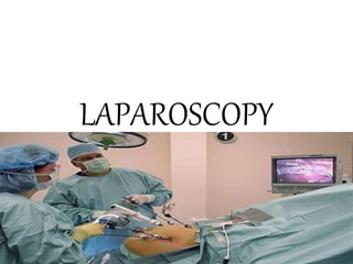
Laparoscopy
- 2. LAPAROSCOPY • Also called ‘key hole surgery’ • It can be therapeutic as well as diagnostic • It is called operative laparoscopy if it is done therapeutically • Telescope with fibre optic cable introduced through a port is used to visualise the abdominal and pelvic contents • Operating instruments are introduced through separate ports
- 3. INDICATIONS DIAGNOSTIC • Infertility • Acute/chronic pelvic pain • Ectopic pregnancy • Endometriosis OPERATIVE • Sterilisation • Ectopic pregnancy • Hysterectomy • Tubal anastomoses
- 4. ADVANTAGES • Less blood loss • Less post operative pain • Shorter hospital stay • Avoids large incision • Early return to normal activity • Minimal risk of incisional hernia
- 5. EQUIPMENTS • Equipments consist of an imaging system,an insufflating system and specialised surgical instruments
- 6. IMAGING SYSTEM Consist of laparoscope,light source and fibroptic cord and a camera unit.
- 7. LAPAROSCOPE • It is a telescope usually a 10mm one • Can be 5mm or 3mm • Commonly used is a 0 degree angle telescope.30 degree telescope allows better visualisation but requires careful orientation
- 8. LIGHT SOURCE AND FIBREOPTIC CORD • LIGHT SOURCE • Light is introduced through the laparoscope with fibreoptic cable powered by a light source • A high intensity light source like halogen or preferably xenon is used.Xenon is more powerful
- 10. • CAMERA UNIT • The camera unit consists of camera head,cable,camera control and TV monitor • The image seen through eye piece of a laparoscope is converted to electric signals by a charge coupled device(CCD) in camera head • The electric signals are then processed by camera control facility which is connected to TV monitor
- 11. camera
- 12. • RECORDING EQUIPMENT • Recording is done by a video recorder or DVD recorder • It may be useful in medicolegal procedures
- 14. INSUFFLATING SYSTEM • This system allows gas to fill abdominopelvic cavity for better visualisation • Gas used is carbondioxide as it is rapidly absorbed by blood • In patients with CVS risk factors gasless laproscopy is done where a mechanical lifting arm is attached to a fanlike retractor along peritoneal surface of abdomen thereby obliviating need for gas distension
- 15. VERESS NEEDLE • It is used to create pneumoperitoneum • It is spring loaded to prevent visceral injury • Once peritoneal cavity is entered blunt tip projects out • Insufflators provide carbondioxide to create pneumoperitoneum with continuous monitoring of volume,flow rate and intra abdominal pressure.A safety device is there to ensure maximum pressure is not exceeded
- 16. • Other methods to create pneumoperitoneum includes direct trocar insertion and open laproscopy
- 17. INSTRUMENTS
- 18. TROCARS • The 10mm trocar is usually used for intraumbilical entry to accommodate telescope • The ancillary ports are 5mm trocars through which operating instruments are introduced • Trocar and canula is inserted and trocar removed.Then telescope or ancillary instrument is introduced through cannula
- 19. trocar
- 20. ANCILLARY INSTRUMENTS • They are different instruments essential in laproscopic surgery like scissors,forceps,probes,etc
- 22. • General anaesthesia is preferred for diagnostic as well as operative laproscopy • After induction of anaesthesia patient is placed in low lithotomy position with legs supported in stirrups • The arms are positioned at patient’s side by adduction and pronation • Bladder is catherised
- 24. PROCEDURE • 1.ENTERING ABDOMINAL CAVITY o Veress needle and umbilical entry Umbilical site is used for entry.A small incision is made infraumbilically with scalpel and abdominal wall lifted away manually or with instruments.Patient’s position is normal (never Trendelenberg position)
- 25. Veress or trocar needle is introduced.Shaft of needle is held by fingers and introduced into abdominal cavity. A rubin cannula is inserted for uterine manipulation and chemoperfusion If hysterectomy is planned,uterine manipulator is inserted
- 26. • Alternatively,an open entry method can be used.In this method,rectus sheath is pulled up with Allis clamps through skin incision and incised.Then trocar is inserted directly • Carbondioxide insufflation is started at rate of 1L/min.Flow rate can be increased to maintain intra abdminal pressure at 10-12mm Hg once intra abdominal gas has been confirmed by percussion
- 27. CORRECT PLACEMENT • Correct placement can be assessed by several methods Hanging drop method-a drop of saline will be placed in top of veress needle which will be sucked in by negative intraabdominal pressure. Syringe test-attaching a syringe to veress needle ad watching the column of saline descend the barrel
- 28. INSERTION OF TROCAR • Once pneumoperitoneum is sufficient(3-5L) head down tilt allows good visualisation of pelvis. • A 10mm trocar can be inserted at lower border of umbilicus • Once insertion is complete,trocar is withdrawn and a laparoscope is introduced through sleeve
- 30. SECONDARY TROCAR PLACEMENT • Secondary trocars for introduction of operating instruments • Usual points are Lateral ports-5cm above pubic symphysis and 8cm lateral to midline Suprapubic port-5-6 cm above pubic symphysis
- 32. • Pelvic viscera is visualised and operative procedures performed • Gas is allowed to escape after completing of procedure
- 33. COMPLICATIONS
- 34. • At needle or trocar entry injury to vessels-inferior epigastric,aorta,vena cava Injury to bowel or other organs • Pneumoperitoneum Subcutaneous emphysema • Laproscopic surgery Injury to vessel,viscera Injury to bowel,bladder,ureter