Ptosis workup
•
52 likes•12,677 views

To know about various type of ptosis. Ptosis measurement procedure

Recommended
More Related Content
What's hot
What's hot (20)
Similar to Ptosis workup
Similar to Ptosis workup (20)
More from Azizul Islam
More from Azizul Islam (20)
Recently uploaded
Recently uploaded (20)
Ptosis workup
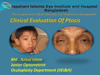
- 1. Clinical Evaluation Of Ptosis Md . Azizul Islam Junior Optometrist Oculoplasty Department (IIEI&H) IIEI&H
- 2. DEFINITION Abnormal drooping of upper eyelids is called ptosis. Normally upper eyelid covers 2mm of cornea. Therefore in ptosis it covers more than 2mm. IIEI&H
- 3. CLASSIFICATION OF PTOSIS A. Congenital B. Acquired I. Neurogenic II. Myogenic III. Mechanical IV. Traumatic C. Pseudotosis IIEI&H
- 4. Congenital ptosis It is associated with congenital weakness (maldevelopment) of the levator palpebral superior (LPS) muscle. 1.Simple congenital ptosis : Not associated with any other anomaly. congenital ptosis with associated weakness of superior rectus muscle. 2.Blepharophimosis syndrome: which comprises congenital ptosis, blepharophimosis, telecanthus and epicanthus inversus . 3.Congenital synkinetic ptosis : (Marcus Gunn jaw winking pheno menon). Its the condition of misdirection 3rd nerve , retruction of the upper lid with various ocular movements. IIEI&H
- 5. Acqured Ptosis • Third nerve palsy 1. Neurogenic • Third nerve misdirection • Horner syndrome • Myasthenia gravis • Ocular myopathies • Simple congenital 2.Myogenic • Blepharophimosis syndrome IIEI&H
- 6. 3.Mechanical ptosis Due to excessive weight on the upper lid lid tumours, multiple chalazion . lid oedema. 4.Traumatic ptosis: Trauma to levator muscle, post surgies. IIEI&H
- 7. Mechanical/Traumatic ptosis Dermatochalasis Large tumours Severe lid oedema due to Tr. Tr.Anterior orbital lesions IIEI&H
- 8. PSEUDOPTOSIS Pseudoptosis is the appearance of ptosis in the absence of levator abnormality. Exclude pseudoptosis (simulated ptosis) on inspection. Its common causes are: Microphthalmos, Anophthalmos, Endophthalmos Phthisis bulbi. Double elevator palsy Blepharospasm Contralateral proptosis Due to phthisis Bulbi Due to Contralateral proptosis IIEI&H
- 9. HISTORY Ptosis Age of onset Duration One/both eye Diurnal variability Associated history : Diplopia Fatigability Variable Muscle weakness Vision IIEI&H
- 10. Association with Jaw movements Abnormal ocular movements Abnormal head posture History of Trauma or previous surgery Poisoning Use of steroid drops Any reaction with anesthesia Bleeding tendency Previous photographs may prove to be of great help. Is there a family history of ptosis or of other muscle weakness? IIEI&H
- 11. This is a test for determining corneal reflex, if the eyes are in alignment, You shine a light at the eyes and observe where the light reflex is located in reference to the pupil. Its may Central , Eccentric , In ward , Out ward. HB / CL Reflex Measurments of Ptosis IIEI&H
- 12. Upper lid crease • Distance between lid margin and lid crease in down-gaze. • Normal-females 10 mm; males 8 mm. • Absence in congenital ptosis indicates poor levarator function. Distance between lash line and skin fold in primary position of gaze. Pretarsal show crease fold IIEI&H
- 13. Measurements Of MRD Margin-reflex distance (MRD). MRD1: distance between upper lid margin and CLR. N: 4-4.5 mm MRD2: distance between lower lid margin and CLR. N: 5-5.5 mm Mild ptosis (2 mm ) Moderate ptosis (3 mm) Severe ptosis (4 mm or more) (MRD) Normal MRD IIEI&H
- 14. Measurements LPSA Levator Palpebral Supirior Action(LPSA) • Place thumb against brow to stop frontalis • Patient look down • Then look up • Measure with a ruler • Results: – >15mm: normal – 12-14 mm: good – 5-11 mm: fair – <4 mm: poor IIEI&H
- 15. Measurements PFH Palpebral fissure height(PFH) • Distance between upper and lower lid margin • Normal: – Women: 8-12 mm – Men: 7-10 mm N.B: Upper lid margin rests about 2mm below upper limbus and Lower lid margin 1mm above lower Limbus. So we determind amount of unilateral ptosis by PFH. IIEI&H
- 16. Bell’s phenomenon Upward rotation of globe on lid closure Good Poor - risk of postoperative corneal exposure IIEI&H
- 17. Marcus Gunn jaw winking pheno- menon (MJWP). Its the condition of misdirection 3rd nerve , retruction of the upper lid with various ocular movements IIEI&H
- 18. Ocular Motility: Importance in myogenic ptosis, To R/O 3rd nerve palsy presence of strabismus, especially vertical strabismus entails that it be corrected prior to the correction of the ptosis. Visual acuity Best-corrected visual acuity should be assessed to record any amblyopia if present, especially in cases of congenital ptosis. Pupillary Examination: To diagnosis Horner’s syndrome Involvement in a case of third nerve palsy IIEI&H
- 19. Documentation of ptosis mesurements HB –Reflex :Central , Eccentric , In ward , Out ward. Lid Crease :10 mm / 8 mm. MRD : mild 2 mm , moderate 3 mm, severe 4 mm. LPSA : 15 mm normal,8-12 mm good,5-11 mm fair, <4 mm poor. PFH : 8-10, 7-9 mm Bell’s phenomenon : Good, Poor. Ocular motility : full, restricted. MGJWP : + (ve), - (ve) . Corneal sensitivity : Good, fair. IIEI&H
- 22. TREATMENTS Non-surgical – Rehabilitative crutch glasses Surgical - Definitive Treatment Decision making When to operate Which procedure concern is cosmetic any age. concern is amblyopia early surgery. squint has to be operated first. Blepharophimosis, telecanthus, epicanthus operated first Depends on levator function + associated anomaly IIEI&H
- 23. Pre - oparative Ptosis Post oparative ptosis Results of ptosis Sx IIEI&H
- 24. References IIEI&H Congenital Ptosis". MEDgle. Retrieved 2008-10-20. Adult Ptosis at eMedicine "Eye Ptosis Congenital". Retrieved 2010-06-14. Finsterer, J (2003). "Ptosis: causes, presentation, and management". Aesthetic Plastic Surgery. Pic: Google + Me.
- 25. IIEI&H IIEI&H