Basics of bladder tumors
•Download as PPTX, PDF•
24 likes•1,594 views

Presentation on pathology of tumors of Urinary bladder

Recommended
More Related Content
What's hot
What's hot (20)
Similar to Basics of bladder tumors
Similar to Basics of bladder tumors (20)
Recently uploaded
Recently uploaded (20)
Basics of bladder tumors
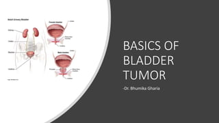
- 1. BASICS OF BLADDER TUMOR -Dr. Bhumika Gharia
- 2. Localization of bladder female & male
- 4. UROTHELIUM • Normal 6-7 layer thick • On distension 2-3 cells
- 5. Normal bladder histology Inner & outer longitudinal, central circular
- 6. CONGENITAL ANOMALIES- exstrophy of bladder vesicoureteral reflux urachal anomalies diverticula Lithiasis Endometriosis Amyloidosis
- 7. CYSTITIS • ACUTE • Hunner’s cystitis • Eosinophilic cystitis • Emphysematous cystitis (Cl. perfringens) • CHRONIC • Malakoplakia • Polypoid cystitis (confuse with papillary urothelial Ca, h/o indwelling catheter)
- 9. Cystitis glandularis & cystitis cystica
- 10. Adenocarcinoma Chronic inflammation form brunn’s island cystitis glandularis & cystitica Trigone area
- 11. SQUAMOUS METAPLASIA & NEPHROGENIC ADENOMA
- 12. TUMORS OF THE URINARY BLADDER Urothelial (transitional) tumours Exophytic papilloma Inverted papilloma Papillary urothelial neoplasms of low malignant potential Low-grade & high-grade papillary urothelial cancers Carcinoma in situ (CIS, or flat non invasive urothelial carcinoma) Mixed carcinoma Adenocarcinoma Small cell carcinoma sarcomas
- 13. Neoplasms of Urinary bladder Epithelial neoplasms •Benign neoplasms •Papilloma •PUNLMP •Malignant neoplasms •Urothelial Carcinoma •Squamous cell carcinoma •Adenocarcinoma •Metastases •Small cell or neuroendocrine Ca •Carcinoid •melanoma Non epithelial neoplasms •Benign neoplasms •Leiomyoma •Paraganglioma •Fibroma •Plasmacytoma •Hemagioma •Solitary fibrous tumor •Neurofibroma •lipoma •Malignant neoplasms •Rhabdomyosarcoma •Leiomyosarcoma •Lymphoma •Osteosarcoma •Angiosarcoma •Malignant fibrous histiocytoma
- 15. UROTHELIAL TUMORS • Precursor lesions • -non invasive papillary tumor • -flat non invasive urothelial carcinoma
- 17. Genetic alterations • Low grade papillary, non inavsive gain of function mutation in FGFR3 • Activating mutation in HRAS oncogene (frequently seen) • Chromosome 9 deletions • High grade loss of function mutation in TP53 & RB tumor suppressor genes
- 18. Molecular genetics Loss of heterozygosity of chromosome 9 Loss of heterozygosity of 3p, 5q and 17p – invasive tumors Other markers – loss of heterozygosity in chromosomes 4,8 and 11 Overexpression of p53- correlates with prognosis Associated high levels of MDM2 C-erbB-2 increased expression as grade of tumor increases Overexpression of bcl-2- inversely correlated with tumor stage Cyclin D1- corelats with tumor grade & stage Genes- FHIT (tumor suppresssor gene at 3p14.2) & INK4A (chromosome 9p21)
- 19. Biopsy Ideally- include underlying muscle In addition to main tumor- three other sites (one lateral to each ureteral orifice & upper posterior wall) biopsies separately submitted- recommended Biopsy report Grade Configuration Depth of penetration Presence of muscle Lymphatic invasion Blood vessel invasion Changes in adjacent mucosa if present
- 20. cytology
- 21. Grading of urothelial tumours WHO/ ISUP grades • Urothelial papilloma • Urothelial neoplasm of low malignant potential • Papillary urothelial carcinoma, low grade • Papillary urothelial carcinoma, high grade WHO grades • Urothelial papilloma • Urothelial neoplasm of low malignant potential • Papillary urothelial carcinoma, grade 1 • Papillary urothelial carcinoma, grade 2 • Papillary urothelial carcinoma, grade 3
- 22. Benign tumors Inverted papilloma Villous adenoma Condyloma acuminatum Squamous papilloma Paraganglioma Solitary fibrous tumor Others • Mucin-secreting “cystadenoma” of possible Mullerian origin • Leiomyoma • Hemangioma • Arteriovenous malformation • Lymphangioma • Chondroma • Granular cell tumor • Schwannoma • Neurofibromatosis • Angiomyolipoma • Clear cell myomelanocytic tumor
- 23. Exophytic papilloma Delicate structure superficially attached to mucosa by a stalk
- 25. Inverted papilloma • Benign lesion • Inter anastomosing cords of cytologically bland urothelium extend into lamina propria
- 26. PUNLMP Papillary urothelial neoplasm with low malignant potential • > papilloma in size • Thicker urothelium
- 27. IHC CK 7 & CK 20 positivity CK 8 & CK 18- interface between tumor & stroma. Higher the grade less the chance of expression Thrombomodulin & uroplakin • Thrombomodulin : sensitivity > specificity • Uroplakin: specificity > sensitivity • CEA, Cathepsin B, CA19-9, CD 15, survivin, androgen receptors • Laminin- detect early stromal invasion
- 28. LET’S SEE • NOT CROSSING Basement membrane – Low grade papillary Uca – High grade papillary Uca – Cis • CROSSING Basement membrane • invasive
- 30. Low grade papillary urothelial carcinomas • Mild nuclear atypia • Scattered hyperchromatic nuclei • Infrequent mitotic figures
- 31. High grade papillary urothelial cancers • Dyscohesive cells • Large hyperchromatic nuclei • Highly anaplastic cells • Atypical mitotic figures
- 33. Normal wall Tis-Carcinoma in situ High grade lesions with pronounced cytological atypia,but lacking papillary configurtn
- 34. Tis ≠ Superficial carcinoma Superficial carcinoma= used by urologists, for tumors not invading muscularis propria
- 35. Denuding cystitis • Cystoscopic appearance • Tumor cells of CIS spread along basement membrane & lift up normal transitional cells- pagetoid growth pattern • Cause of false negative biopsies • Repeat biopsies, if high clinical suspicion.
- 36. IHC – in situ vs reactive atypia Panel: CK20, cadherin, p53, CD44 CK20, cadherin, p53 +ve in CIS CD44 +ve in invasive
- 39. cystoscopy
- 42. IHC markers- CK7,CK20 & uroplakin
- 44. Nested variant Urothelial cancer with deceptively bland cytology
- 49. Squamous cell carcinoma • More in schistosomiasis endemic areas • Mixed urothelial with areas of SCC more frequent • Most are invasive, fungating or infiltrative & ulcerative
- 50. • Gross- large ulcerated mass • Micro- always invades muscle • -very Poor prognosis • Othe variants • Basaloid squamous cell carcinoma • Verrucous carcinoma • Warty carcinoma
- 51. Cross-section through the bladder, uterus, and vagina with squamous cell carcinoma of the bladder infiltrating through the bladder wall into the vaginal wall.
- 52. Adenocarcinoma of bladder • Similar to GIT adenoca • Arise from urachal remnant
- 53. Adenocarcinoma Bladder adenocarcinoma Clear cell type (mesonephric,mesonephroid) Signet ring cell Hepatoid adenocarcinoma Chronic inflammation from brunn’s island cystitis glandularis & cystitica adenocarcinoma Trigone area
- 54. Adenocarcinoma of bladder Grossly- fungating mass, ulcerating mucosa
- 55. Microscopicaly- mucinous (MC) non mucinous adenocarcinoma of bladder
- 56. IHC • Β catenin –membranous (cytoplasmic) positivity • (helps differentiate with colorectal adenoCa) • CK7 positve
- 59. IHC NSE, chromogranin & / synaptophysin – reactivity Low molecular weight keratin (Cam5.2) – dot like perinuclear pattern CD 44 – absent (in contrast to TCC)
- 60. CLEAR CELL ADENOCARCINOMA OF BLADDER hobnail pattern & glycogen rich cells
- 61. Nephrogenic adenoma Vs clear cell Ca Pax-8 - nephrogenic MIB-1, p53 strongly expressed in clear cell
- 62. SARCOMATOID CARCINOMA Sarcomatoid (spindle/metaplastic) Ca-high grade neoplasm of bladder Malignant epithelial (transitional,glandular,squamous,undifferentiated type) coexists with areas having sarcoma like appearance (spindle cell,osteoclast giant cell, rhabdomyosarcoma, chondrosarcoma, liposarcoms,MFH)
- 66. Inflammatory myofibroblastic tumor ALK 1 staining positive Panel: Pancytokeratin,CAM5.2, SMA, desmin, ALK positive CD34, S100, CD 117 negative
- 67. Leiomyosarcoma of bladder Located in bladder dome
- 73. OTHER SARCOMAS
- 74. METASTATIC TUMORS Most from Breast & malignant melanoma
- 75. Breast Mets vs Uca plasmacytoid variant Clinical history Panel- ER,PR, GCDFP, UP III, TM , CD 138
- 77. Direct extension- Prostate cancer into bladder can also be from large bowel & cervix
- 78. Transitional vs prostate Ca Panel: CK 34-β-12, CK7, p53, PSA, PSAP and Leu7 CK 34-β-12, CK7, p53- TCC PSA, PSAP and Leu7 – prostate Ca
- 79. • Representative panel of immunohistochemical markers in most cases of prostate adenocarcinoma. Positive immunoreactivity for prostate-specific antigen (A), prostate-specific membrane antigen (B), prostate acid phosphatase (C), P501s (D), NKX3.1 (E), and α-methylacyl coenzyme A racemase (F). Negative immunoreactivity for CK34βE12 (G), p63 (H), thrombomodulin (I), S100P (J), and GATA binding protein 3 (K).
- 81. • STAGE Ta: Non invasive, papillary • STAGE Tis: Carcinoma in situ
- 82. • STAGE T1: lamina propria invasion
- 83. • STAGE T2: Muscularis propria invasion
- 84. TREATMENT • Grade I & II • Without muscle invasion • Initially transurethral resection • Sometimes supplemented with intravesical chemotherapy or radiation therapy (if multiple/recurrent) • Intravesical BCG immune therapy also given
- 85. STAGE 3
- 89. TREATMENT • Grade III & IV • With muscle invasion • Radical cystectomy (with/without pripr radio/chemo therapy) • Radical cystectomy –in males = bladder+prostate+seminal vesicles+adjacent perivesical tissues • Radical cystectomy –in females = bladder+uterus+tubes+ovaries+anterior vagina+urethra • With enbloc pelvic lymph nodes dissection
- 90. Take home message Bladder is simple, lets not complicate it.!! • Epithelial • Urothelial • adenoCarcinoma • squamous • Mesenchymal • Leiomyosarcoma • Rhabdomyosarcoma • IHC always helps- CK7,CK20, HMWCK, Uroplakin, thrombomodulin Bladder tumors
- 91. References Surgical pathology book by Ackerman Dabb’s immunohistochemistry book Robbins & cotran pathology book From archives of AFIP: Neoplasms of the urinary bladder:radiologic-pathologic correlation: Fade F. Wong-You-Chong et al Google
Editor's Notes
- In females-bladder rests on uterus,vagina In males it is in contact with prostate and colon
- Lateral wall- urothelial Base- adenocarcinoma Trigone area – squamous, it can also be in lateral wall Bladder neck – carcinoid tumors
- Transitional= cause can change shape from cuboidal to flattened
- Layer of cell- superficial- umbrella like, intermediate- cuboidal and basal layer Bladder neoplasms can arise from any layer- epithelial or non epithelial=submucosal layer
- Exstrophy= adenoCa of bladder , Diverticula = urothelial Ca,
- Hunner’s cystitis seen in females, very common condition- lower perineal pain, urinary frequency Malakoplakia- seen at trigone area as multiple nodules, seen mostly in immunocompromised pts
- Cystoscopy appearance = frond like- can confuse with papillary
- Here there is focal proliferation of basal cells which form buds then nodules which are called von brunn nests or islands, central cystic area with accumulation of mucin CK 7 positivity
- Can confuse with clear cell carcinoma of baldder- hob nail pattern in both, put ki 67- high index in Ca IHC can help- put Pax 2 & pax 8 markers if from kidney
- Chemical like beta napthylamines, drugs like phenacetin, cyclophosphamide Smoking cigarettes not pipes or cigars
- Variety of markers are being tried on urine – bladder tumor antigen, FDPs, telomerase, nuclear matrix protein 22 Chromosomal 9 abnormality study
- Exophytic benign growth- benign epithlium
- If confuse with URCa – put Ki 67, p53, CK20 all positive in Cancer
- New addition to WHO Low grade small solitary neoplasm, that neither invades nor metastasizes Distinction from low grade can be difficult & subjeective
- sEen in lamina propria,May be confused with brunn nest of cystitis glandularis and underdiagnosed This tumor has aggressive behaviour
- Resembles ovarian serous papillary Ca, but it has aggressive clinical course
- Occur in chronic bladder infection & irritation. Also called bilharzial bladder carcinoma More at trigone areas & lateral walls & in bladder diverticula
- Cytoscopy- large ulcerated infiltrating mass
- Primary – urachal or non urachal Secondary (metastases)
- Primary Adenocarcinoma- favors base of bladder or urachal regiom- lies in midline of dome of bladder Easily identified on USG- soft tissue mass with heterogenous areas & calcified
- <0.5% prevalence most common in lateral bladder wall, central necrosis & cystic changes
- Sheets of small cells with round hyperchromatic nuclei,sparse cytoplasm & mitoses & necrosis
- Non infiltrating smooth muscle tumor lacking mitotic activity, cellular atypia & necrosis
- Proliferated spindle cells in myxoid background
- Pts present with hematuria & obstruction Can behave aggressively
- Arise from primitive muscle cells, can occue anywhere in body except bone Around age 10 years May be assoc wd congen anomalies like neurofibromatosis, nephroblastomas, etc
- Diffusely infiltrating lesion Gelatinous Majority –embryonal type some alveolar type
- Malignant spindle cells with myxoid edematous stroma Highly cellular zobe- cambium layer beneath urothelium is seen
- GCDFP=gross cystic disease fluid protein
- Lets wait for urine cytology to help diagnose bladder tumors early