Facemask , oral and nasal airways
•Download as PPTX, PDF•
274 likes•61,835 views

facemask and nasopharyngeal airway

Recommended
More Related Content
What's hot
What's hot (20)
Similar to Facemask , oral and nasal airways
Similar to Facemask , oral and nasal airways (20)
More from DR SHADAB KAMAL
More from DR SHADAB KAMAL (13)
Recently uploaded
Recently uploaded (20)
Facemask , oral and nasal airways
- 1. FACEMASKS, ORAL AND NASAL AIRWAYS DR. SHADAB
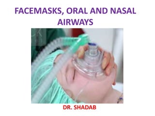
- 2. INTRODUCTION • Facemask is an interference device between the patient’s upper airway and the breathing system. • It allows gas administration to the patient from the breathing system without introducing any apparatus into the patient's mouth. • They may be made up of antistatic material to prevent ignition of anaesthetic gases.
- 3. DESCRIPTION Face masks may be made up of: -Black Rubber, -Clear Plastic, -Elastomeric Materia or Disposable Plastic or -Combination of these.
- 4. PARTS OF FACEMASK Body ( Shell or Dome) • Constitutes the main part of the mask • Transparent facemask allows observation for vomitus, secretions , blood, lip color, and exhaled moisture and is better accepted by a conscious patient .
- 5. Seal (Rim, Flap, Edge) It comes in contact with the face. Pad (cushion) type seal Inflated with air or filled with a material that will conform to the face when pressure is applied. Flap type seal Flexible extension of the body that conforms to the contour of the face. It is pressed onto the face to create a seal.
- 6. Connector (Orifice, Collar, Mount) • Present at the opposite side from the seal. • Has a thickened fitting with a 22-mm internal diameter. • A ring with hooks may be placed around the connector to allow a mask strap to be attached.
- 7. Face mask-types 1) Anatomical mask Black rubber Latex Plastic 2) Ambu transparent mask 3) Rendell-Baker- Soucek mask
- 8. ANATOMICAL BLACK RUBBER FACE MASK • Also called as Connell mask. • Body is made of rubber that can be widened or narrowed to fit the face • Available in sizes 0, 1, 2, 3 & 4.
- 9. Snuggy Silicone and black rubber Facemask Size Use 0 Infant 1 Small Child 2 Child 3 Small Adult 4 Adult 5 Large Adult • It is autoclavable soft contoured cuff snug facemasks • its provides patient’s comfort as well as makes for easy cleaning. • The one piece construction and silicone rubber also allows for clear view of presence of vomit, patient’s lip color as well as other oral secretions.
- 10. EcoMask™ • It is a non-PVC single-use anatomical facemasks. • It has clearer version of the traditional, reusable rubber face mask - Non-slip rings for a better grip and tighter seal - Anatomical shape, soft seal for a comfortable fit - Transparent shell for excellent visibility - available in a range of seven sizes with distinct colour-coding for easy selection.
- 11. Trimar Mask It is similar to Connell mask. It has shallower body & less dead space Trimar Mask
- 12. AMBU TRANSPARENT MASK Made of transparent plastic with inflatable cuff for a seal and has thumb rest built into the body
- 13. RENDELL-BAKER-SOUCEK MASK • Designed for the pediatric patients, • Has a triangular body. • Usually available in sizes 00, 0, 1 & 2 • Has a low dead space i.e. 4ml in size 1 & 8ml in size 2 • Adequately fits the child’s face & no special seal is needed. • Some of these masks are scented and may have a pacifier
- 14. RBS MASKS-(A) CLEAR PLASTIC VERSION,(B) WITH PACIFIER,©BLACK RUBBER VERSION.
- 15. SCENTED MASKS • ROUND TYPE SILICONE PAEDIATRIC MASKS uses scents to camouflage the odors of inhalational agents
- 16. Laerdal Peadiatric Mask It is made of silicon rubber It has an inward curving circular face seal It can be boiled or autoclaved It has been found to be better for ventilation of newborn & paediatric patients
- 17. Endoscopic Masks • An endoscopic mask is designed to allow mask ventilation while an endoscope is being used • Port/diaphragm in the mask body allows a fiberscope to be inserted into the nose or mouth. • A tracheal tube previously loaded over the fiberscope can then be advanced, if desired.
- 18. Hudson’s Face Mask It is made of clear plastic & is transparent On sides there are air entrainment holes It is variable performance mask (i.e FiO2 delivered is O2 flow dependant)
- 19. •It is under-the-chin design for excellent fit on a wide variety of face sizes. •Clear, soft vinyl for patient comfort and visual patient assessment. •Adjustable nose clip assures comfortable fit. •Complete with 7 feet oxygen supply tubing. Method O2 flow (l/min) Estimated FiO2(%) Face mask 1 24 2 28 3 32 4 36 5 40 6-7 50 7-8 60 Facemask with reservoir 6 60 7 70 8 80 9 90 10 95
- 20. TECHNIQUES OF USE • Smallest mask that is appropriate is the most desirable as it will cause: -least increase in dead space -easiest to hold -less likely to result in pressure on the eyes.
- 21. One-hand Method • Mask is holded by dominant hand and placed over the bridge of the nose and on the chin • The thumb and index finger of the dominant hand are placed on the mask body and these finger slightly push downwards to hold the mask to the face and prevent leak. • The remaining three fingers are placed on bony part of the mandible to pull the mandible up into the mask • Use the free hand to squeeze the reservoir bag
- 22. The middle and ring finger is placed on the mandibular ridge to pull the jaw backward and extend the neck. Little finger is placed under the angle of the jaw and pull jaw upward Digital pressure in region of sub mental triangle is avoided as it can push the tongue up to the roof & increases obstruction.
- 23. Two-handed Method • Mask is placed over the bridge of the nose and on the chin • index finger and thumb distal phalanges of both hand is placed along the ridges of the mask • The remaining fingers are placed on the mandible • Mandible is pulled up into the mask to perform a jaw-thrust and chin-lift maneuver • The assistance squeeze the reservoir bag
- 24. A: Holding the mask with two hands. Esmarch-Heiberg maneuver, involves dorsiflexion at the atlanto-occipital joint and protrusion of the mandible anteriorly by exerting a forward thrust on the rami. B: The anesthesiologist's chin on the mask elbow can help to create a better seal between the mask and the patient's face.
- 25. Mask Straps • A mask strap is used to hold the mask firmly on the face. • Care needs to be paid when using a mask strap because obstruction is more likely to go unrecognized than when the mask is being held by the anesthesia provider's hand. • A typical mask strap consists of thin strips arranged in a circle with two or four projections. • The head rests in the circle, and the straps attach around the mask connector. The straps at the jaw may tend to pull the jaw posteriorly. • Crossing the two lower straps under the chin may result in a better fit and counteract the pull from the upper straps so that there is less tendency for the mask to creep up above the bridge of the nose.
- 26. Straps should be no tighter than necessary to achieve a seal in order to avoid pressure damage from the mask or the straps. They should be released periodically and the mask moved slightly. Gauze sponges placed between the straps and the skin will help to protect the face from excessive pressure. Another risk of using a mask strap is that it will take longer to remove the mask if vomiting or regurgitation occurs.
- 27. Complications Skin Problems: • Dermatitis in allergic pts. If rubber is a component of a face mask, a serious reaction can occur in the patient with latex allergy. • Chemical or gas used for sterilization of reusable masks can cause allergy. • Pressure necrosis under the face mask . Nerve Injury: • Pressure injury to underlying nerves. • Forward jaw displacement may result in stretching nerve injury. User Fatigue: cramped hands and tired muscles and limits the user's ability to perform other tasks specially in obese and patient with larger head
- 28. Eye Injury: corneal abrasion may be caused by a face mask inadvertently placed on an open eye • Pressure on the medial angles of the eyes and supraorbital margins may result in eyelid edema, chemosis of the conjunctiva, pressure on the supraorbital or supratrochlear nerve, corneal injury, and temporary blindness from central retinal artery occlusion Gastric Inflation of stomach ( So Inspiratory pressure should be kept<20 cm H2O) Foreign Body Aspiration: occur in case of endoscopic mask Postoperative Jaw Pain.
- 30. Pharyngeal Airway • Most common cause of airway obstruction is due to fall back of tongue and epiglottis into the posterior pharynx due to relaxed muscles in the floor of the mouth and pharynx supporting the tongue.
- 31. AIM OF OROPHARYNGEAL AIRWAY • To lift the tongue and epiglottis away from the posterior pharyngeal wall and • prevent them from obstructing the space above the larynx .
- 32. OROPHARYNGEAL AIRWAY • May be made of elastomeric material, metal, or plastic • Curved tube used to provide free passage of air between the mouth and pharynx of an unconscious person • FLANGE at the buccal end to prevent Oro-Pharyngeal Axis from moving deeper into the mouth and a means to fix the airway in place
- 33. • BITE PORTION is short firm and straight portion that fits between the teeth or gums, prevents patient biting and obstructing the air channel • CURVED PORTION corresponds to the curvature of the tongue and oropharynx for their effective separation, • The pharyngeal end rests between the posterior wall of the pharynx and the base of the tongue, by exerting pressure along the base of the tongue, also pulls the epiglottis forward.
- 34. USES • Helps to maintain an open airway in unconscious patient. • Prevents a patient from biting and occluding an oral endotracheal tube. • Protect the tongue from biting. • Facilitate oropharyngeal suctioning. • Provides a pathway for inserting devices into the esophagus or pharynx .
- 35. SPECIFIC AIRWAYS WATERS AIRWAY • made of metal • an oval flange, a straight bite block section, an anatomically curved pharyngeal section • holes at the side near the distal end • Discarded due to its propensity to damage teeth and soft tissue and inability to see any foreign material lodged within it.
- 36. GUEDEL’s AIRWAY • single use • integrated bite block, colour coded • smooth bevelled tip for easy insertion and minimising the risk of trauma • available in 9 size depnding on distance between the center of the incisors and the angle of the jaw.
- 37. • #000, 00, 0, 1- 6 with length of 30 to 110 mm. • size #000 and 00 are for premature and full term new born babies respectively. Size 0, 1 and 2 are for children and rest are for adults
- 38. BERMAN • has a center support and channels along each side that allow a suction catheter or ETT to slide into the pharyngeal space. WILLIAMS AIRWAY INTUBATOR • proximal half is cylindrical, while the distal half is open on its lingual surface. • designed for blind orotracheal intubations
- 39. PATIL-SYRACUSE ENDOSCOPIC AIRWAY • made from aluminum • designed to aid fiberoptic intubation • has lateral channels and a central groove on the lingual surface to allow a fiberscope with a tracheal tube to pass • slit at the distal end allows the fiberscope to be manipulated in the AP direction.
- 40. OVASSAPIAN FIBEROPTIC INTUBATING AIRWAY • Designed to deliver a fiberscope as close to the larynx as possible • at the buccal end are two vertical sidewalls and between them is a pair of guide walls that curve toward each other. • accommodate a tracheal tube up to 9.0mm .
- 41. BERMAN INTUBATING/PHARYNGEAL AIRWAY • Tubular along its entire length • Open on one side so that it can be split and removed from around a tracheal tube • used as an oral airway or as an aid to fiberoptic or blind orotracheal intubation.
- 42. Technique of Insertion • Pharyngeal and laryngeal reflexes should be depressed. • correct size estimated by holding the airway next to the patient's mouth. The tip should rest cephalad to the angle of the mandible. • lubricate the airway • the jaw is opened with the left hand • the airway is inserted with its concave side facing the upper lip
- 43. • When the junction of the bite portion and the curved section is near the incisors, the airway is rotated 180° and slipped behind the tongue into the final position.
- 44. NASOPHARYNGEAL AIRWAY • Resembles a shortened tracheal tube with a flange at the outer end to prevent it from completely passing into the nare. • Flange is movable in some models. • When fully inserted, the pharyngeal end should be below the base of the tongue but above the epiglottis
- 45. USES • Used during and after pharyngeal surgery • To apply continuous positive airway pressure (CPAP) • To facilitate suctioning and as a guide for nasogastric tube. • as a guide for a fiberscope and to maintain ventilation during fiberoptic endoscopy • To dilate the nasal passages in preparation for nasotracheal intubation • Used in dental surgery • can be fitted with a tracheal tube connector and used with an anesthesia breathing system
- 46. • • Better tolerated than an oral airway • Preferable if the patient's teeth are loose or there is trauma or pathology of the oral cavity • used when the mouth cannot be opened for introducing an oral airway. Advantages over Oropharyngeal Airway
- 47. LINDER NASOPHARYNGEAL AIRWAY • made of plastic with a large flange • distal end lacks bevel • supplied with an introducer, which has a balloon on its tip that can be inflated and deflated by attaching a syringe to the one -way valve at the other end .
- 48. CUFFED NASOPHARYNGEAL AIRWAY • similar to a short cuffed tracheal tube • inserted through the nose into the pharynx, the cuff is inflated, and then is pulled back until resistance is felt. BINASAL AIRWAY • consists of two nasal airways joined together by an adaptor for attachment to the breathing system • used to administer anaesthesia or to provide CPAP to babies
- 49. Insertion • Diameter of the nasal airway should be the same as needed for a tracheal tube (0.5 to 1 mm smaller than for an oral tracheal tube) • Lubricated thoroughly along its entire length. • Inspect each nostril for size, patency, and the presence of polyps • Use vasoconstrictor drops before insertion
- 50. Correct method :NPA is inserted perpendicularly, in line with the nasal passage Incorrect method : The airway is being pushed toward the roof of the nose and into the turbinates Airway is held with the bevel against the septum and gently advanced posteriorly while being rotated back and forth. If resistance is encountered during insertion, the other nostril or a smaller size airway should be used.
- 51. Contraindications Of Nasopharyngeal Airway • Anticoagulation; • Basilar skull fracture; • Pathology(polyp), or deformity of the nose or nasopharynx; • Bleeding disorder or a history of nose bleeds requiring medical treatment. There is no evidence that nasal airways cause significant bacteremia
- 52. COMPLICATIONS Airway Obstruction • The tip of an airway can press the epiglottis or tongue against the posterior pharyngeal wall and cover the laryngeal aperture. • With a nasopharyngeal airway, neck movement in rotation or anteroposteriorly may result in the lumen becoming obstructed. The use of a fenestrated airway may overcome this problem. • The nasopharyngeal airway lumen may be compressed inside the nose tip can press the epiglottis or tongue against the posterior pharyngeal wall Trauma Injury to the nose and posterior pharynx may cause epistaxis . Central Nervous System trauma can occur if the patient has basilar skull fracture and if the nasal airway enters the anterior cranial fossa.
- 53. Tissue Edema Ulceration and Necrosis of the nose or tongue can occur. Dental Damage Laryngospasm and Coughing Retention, Aspiration, or Swallowing Allergic Reaction to Latex Gastric Distention
- 54. Thank You…