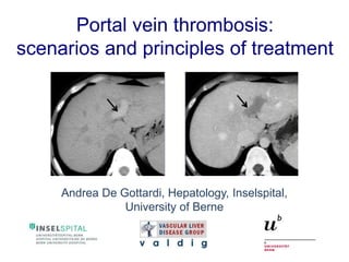
Portal vein thrombosis: scenarios and principles of treatment
•
29 likes•11,524 views

Portal vein thrombosis: scenarios and principles of treatment

Recommended
More Related Content
What's hot
What's hot (20)
Viewers also liked
Viewers also liked (20)
Similar to Portal vein thrombosis: scenarios and principles of treatment
Similar to Portal vein thrombosis: scenarios and principles of treatment (20)
Recently uploaded
Recently uploaded (20)
Portal vein thrombosis: scenarios and principles of treatment
- 1. Portal vein thrombosis: scenarios and principles of treatment Andrea De Gottardi, Hepatology, Inselspital, University of Berne
- 2. First things first venous thrombus formation →endothelial dysfunction or injury →hemodynamic changes →hypercoagulability (Rudolf Virchow 1821 – 1902)
- 3. Outline The scenarios of portal vein thrombosis →Anatomical brush-up →Acute (with or without cirrhosis) →Chronic →Local factors →General factors →Localisation →Extension →Degree of obstruction →Complications →Principles of treatment
- 4. Clinical case: history and examination 43 year-old man presenting with diffuse abdominal pain and vomiting for 2 days one episode of diarrhea, chills, but no fever his daughter had fever, nausea and vomiting reduced general conditions, T 37.3°C, BP 130/85 mmHg, HR 99/min diffuse abdominal tenderness on palpation, reduced peristaltic sounds
- 5. Clinical case: laboratory Haematology white blood cells 14.4 G/L hemoglobin 144 G/L thrombocytes 82 G/L Biochemistry C reactive protein 143 mg/L creatinine 66 uM bilirubin 27 uM glucose 6.9 mM ALT 17 U/L alcaline phosphatase 65 U/L
- 6. Anatomy of the portal system Pressure gradient to hepatic veins: < 6 mmHg = normal ≥ 6 mmHg = portal hypertension ≥ 10 mmHg = clinically relevant PHT Blood flow: 750 – 1500 mL/min Velocity: 10-15 cm/s spleno-mesenteric confluence
- 10. Acute PVT Definition • Sudden formation of a thrombus within the portal vein • Can involve a variable portion of the mesenteric veins and/or the splenic vein • Occlusion can be complete or it can be partial
- 11. Acute PVT Degree and extension of occlusion short partial long partial long complete short complete
- 12. Acute PVT Symptoms • no symptoms • abdominal or lumbar pain of sudden onset or progressing over a few days • abdomen might be moderately distended by ileus, but without any other features of intestinal obstruction • ascites, bloody diarrhea (suggesting infarction) • acute septic PVT = acute pylephlebitis provided there is no extension of the thrombus to mesenteric venous arches, all manifestations of acute PVT are completely reversible Condat, Nat Clin Pract Gastroenterol Hepatol, 2006 Hoekstra, Neth J Med, 2009 Primignani, Dig Liver Dis, 2009
- 13. Acute PVT Laboratory • marked systemic inflammatory response • liver function is preserved (compensatory increase of hepatic arterial blood flow) • acidosis and renal or respiratory dysfunction are also suggestive of intestinal infarction Condat, Nat Clin Pract Gastroenterol Hepatol, 2006 Hoekstra, Neth J Med, 2009 Primignani, Dig Liver Dis, 2009
- 14. Acute PVT Imaging Abdominal ultrasound: - hyperechoic intraportal material - absence of portal flow, no Doppler signal CT-scan: - contrast enhanced (portal phase, evaluation of the extension of the thrombosis)
- 15. Acute PVT General factors Primignani, Dig Liver Dis, 2010. Plessier, Hepatology, 2010. Leebeek, Neth J med, 2012 FACTOR PREVALENCE % JAK2 V617F mutated MPD 17-53 Anti-phospholipid syndrome 1-11 Paroxysmal nocturnal hemoglobinuria 0-9 Hyperhomocystinemia 11-15 Factor V Leiden 3-9 Prothrombin mutation G20210A 2-22 Protein C deficiency 1-9 Protein S deficiency 0-7 Oral contraceptive 0-4 Pregnancy and post-partum 7-44
- 16. James, Nature, 2005 JAK2 V617F mutation
- 17. FACTOR PREVALENCE % Abdominal inflammatory lesions 7-34 - diverticulitis, appendicitis, pancreatitis - inflammatory bowel disease - CMV infection/hepatitis - abdominal abcess Abdominal surgery/trauma 3-45 - splenectomy, gastrectomy, cholecystectomy, LT Abdominal cancer Acute PVT Local factors Primignani, Dig Liver Dis, 2010. Plessier, Hepatology, 2010. Leebeek, Neth J med, 2012
- 18. Chronic PVT Definition the obstructed portal vein is replaced by a network of hepatopetal collateral veins connecting the patent portion of the vein upstream from the thrombus to the patent portion downstream
- 19. Cavernous transformation Zhang, World J Gastroenterol, 2011 Cavernoma may be identified as soon as 15-30 days after the onset with abdominal symptoms
- 20. Chronic PVT Consequences With occlusion of the trunk of the portal vein, antral, duodenal and biliary veins are markedly enlarged. This enlargement can produce compression and deformation of large bile ducts = portal cholangiopathy or portal biliopathy. Complete occlusion of the portal vein trunk is virtually always associated with portal hypertension and the development of portosystemic collaterals.
- 21. Portal cholangiopathy Llop, Gut, 2011 Symptomatic portal cholangiopathy develops in 19% after 5 years
- 22. Chronic PVT Complications Portal hypertension - formation of esophageal or gastric varices - the occurrence of ascites or encephalopathy in patients with chronic PVT is uncommon Portal cholangiopathy - it mimics the bead-like appearance of primary sclerosing cholangitis - It is much more commonly seen on biliary tract imaging than with clinical or laboratory features of biliary disease. - A tumor-like cavernoma can be confused with carcinoma of the main bile duct.
- 23. PVT in cirrhosis PVT occurs rarely in early cirrhosis High prevalence in patients on transplant WL (5-26%) Severe liver disease ⇆ aggravates ⇆ PVT Decreased portal flow velocity (<10 cm/s) Coagulation inbalance and role of thrombocytopenia and hyperfibrinolysis Changes in portal endothelial cells Plessier, J Hepatol, 2012. Francoz, J Hepatol, 2012. Zocco, J Hepatol, 2009. Rijken, Throm Hemost, 2012
- 24. PVT in cirrhosis Virchow’s triad (1) La Mura, Gut, 2011 Endothelial dysfunction in the hepatic vascular bed
- 25. PVT in cirrhosis Virchow’s triad (2&3) Lisman, J Hepatol, 2010 Hepatic vascular resistance is increased and consequently portal flow velocity is decreased or even inversed (hepatofugal). Cirrhosis is characterized by a hemostatic rebalance.
- 26. Principles of treatment Goals of treatment ACUTE PVT • To recanalize the obstructed veins, which will prevent intestinal infarction and portal hypertension • To treat or avoid infection • Recanalisation of the portal vein virtually never occurs without treatment
- 27. Treatment options Anticoagulation for acute PVT Recanalization can be expected until the 6th/12th month of anticoagulation portal 39%, splenic 80%, mesenteric 73% (Condat, Hepatology, 2000. Plessier, Hepatology, 2010) Optimal duration of anticoagulation from 3 months to long-term (prothrombotic conditions) In patients with DVT, a lack of complete recanalization indicates a high risk of recurrence after cessation of anticoagulation therapy. Is this also true for patients with acute PVT? Bleeding under anticoagulation occurs in 9%, however without related mortality
- 28. Treatment options Anticoagulation for acute PVT Unfractionated heparin or low molecular weight heparin represent the mainstay of initial therapy. Vitamin K antagonists are generally used after recovery from the acute event. Very few data on DOACs. Thrombolysis • Transhepatic or transjugular route • Limited number of patients, but • Recanalisation rate similar to those achieved with anticoagulation • Major procedure-related bleeding in up to 50%
- 29. Treatment options Other treatment modalities for acute PVT Antibiotics are indicated with chills and positive blood cultures Surgical intestinal resection when clinical and radiological features indicate that a patient has (or may have) intestinal infarction Surgical thrombectomy Recanalisation is obtained in 30% of the patients and recurrence of PVT is frequent
- 32. Treatment options Aim: to prevent recurrence or extension of thrombosis Anticoagulation for chronic PVT Risk of GI bleeding and severity not increased (Condat, Gastroenterology, 2001, Kitchens, J Thromb Thrombolysis, 2007) Should be discussed in patients with a prothrombotic condition (De Franchis, Baveno V, J Hepatol, 2010)
- 33. Prevention of PVT in cirrhosis Dashed line: controls Continuous line: enoxaparin-treated patients Villa, Gastroenterology, 2012
- 34. TAKE HOME MESSAGES Recurrence or extension should be avoided by treating local or systemic factors Recanalisation should be the aim of anticoagulation or invasive procedures if indicated PVT should be correctly diagnosed
- 35. Further reading EASL Clinical Practice Guidelines: Vascular liver diseases 2015 J. Hepatol. In preparation AASLD Practice Guidelines: Vascular disorders of the liver Hepatology, 2010