SlideShare a Scribd company logo
1 of 18
Download to read offline
REVIEW
A Review: Pharmaceutical and Pharmacokinetic Aspect
of Nanocrystalline Suspensions
DHAVAL A. SHAH,1
SHARAD B. MURDANDE,2
RUTESH H. DAVE1
1
Arnold & Marie Schwartz College of Pharmacy and Health Sciences, Long Island University, Brooklyn, New York 11201
2
Drug Product Design, Pfizer Worldwide R&D, Groton, Connecticut 06340
Received 7 August 2015; revised 23 September 2015; accepted 25 September 2015
Published online in Wiley Online Library (wileyonlinelibrary.com). DOI 10.1002/jps.24694
ABSTRACT: Nanocrystals have emerged as a potential formulation strategy to eliminate the bioavailability-related problems by enhancing
the initial dissolution rate and moderately super-saturating the thermodynamic solubility. This review contains an in-depth knowledge of,
the processing method for formulation, an accurate quantitative assessment of the solubility and dissolution rates and their correlation to
observe pharmacokinetic data. Poor aqueous solubility is considered the major hurdle in the development of pharmaceutical compounds.
Because of a lack of understanding with regard to the change in the thermodynamic and kinetic properties (i.e., solubility and dissolution
rate) upon nanosizing, we critically reviewed the literatures for solubility determination to understand the significance and accuracy of the
implemented analytical method. In the latter part, we reviewed reports that have quantitatively studied the effect of the particle size and
the surface area change on the initial dissolution rate enhancement using alternative approaches besides the sink condition dissolution.
The lack of an apparent relationship between the dissolution rate enhancement and the observed bioavailability are discussed by reviewing
the reported in vivo data on animal models along with the particle size and food effect. The review will provide comprehensive information
to the pharmaceutical scientist in the area of nanoparticulate drug delivery. C 2015 Wiley Periodicals, Inc. and the American Pharmacists
Association J Pharm Sci
Keywords: nanocrystals; nanosuspensions; nanoparticles; solubility; dissolution; pharmacokinetics; food interactions; bioavailability;
particle size reduction
INTRODUCTION
Recent advances in synthetic, analytical, and purification
chemistry, along with the development of specialized tools
such as high-throughput screening, combinatorial chemistry,
and proteomics, have led to a sharp influx of discovery com-
pounds entering into development. Many of these compounds
are highly lipophilic, as the in vitro screening techniques place
considerable emphasis on the interaction of compounds with de-
fined molecular targets. In recent years, it has been estimated
that up to 70% of the new drugs discovered by the pharmaceu-
tical industry are poorly soluble or lipophilic compounds. Poor
aqueous solubility is one of the major hurdles in the develop-
ment of new compounds into oral dosage forms, as absorption
is limited by dissolution for these compounds.1
The well-known Biopharmaceutics Classification System
(BCS) is frequently used to categorize pharmaceutical com-
pounds. According to the BCS system, poorly soluble com-
pounds belong to Class II (low solubility, high permeability)
or Class IV (low solubility, low permeability). In another words,
we can also say that Class II and IV compounds provide more
opportunities for the development of newer technologies to
overcome the solubility- or dissolution-related issues based on
chemical and physical properties of the compounds. This per-
ception is widely used and well established within the pharma-
ceutical industry. However, using the BCS system for guidance
in formulation selection may sometimes oversimplify the com-
Correspondence to: Rutesh H. Dave (Telephone: +718-488-1660; Fax: +718-
780-4586; E-mail: Rutesh.Dave@liu.edu)
Journal of Pharmaceutical Sciences
C 2015 Wiley Periodicals, Inc. and the American Pharmacists Association
plex nature of drug dissolution, solubility, and permeability.
Poorly water-soluble compounds can possess such a low aque-
ous solubility that the dissolution rate, even from micronized
particle, is very slow. In this case, it is not possible to reach suf-
ficiently high drug concentrations in the gastrointestinal tract
for an effective flux across the epithelial membrane. Other fac-
tors, such as efflux transport or pre-systemic metabolism, can
also negatively influence oral bioavailability.
Therefore, it is recommended to classify compounds into
slightly different categories, as they can show dissolution
rate-limited, solubility-limited, or permeability-limited oral
bioavailability. Butler and Dressman2
designed the “Developa-
bility Classification System (DCS),” as another way to catego-
rize compounds in a more bio-relevant manner. This system dis-
tinguishes between dissolution rate-limited compounds (DCS
Class IIa) and solubility-limited compounds (DCS Class IIb).
In order to select the right formulation approach and to ad-
dress the compound-specific issues with a suitable formulation
type, it is imperative to first understand the bioavailability lim-
iting factors. Selection of the right formulation approach is one
of the key activities for formulators in the pharmaceutical in-
dustry. Key factors include the physicochemical properties of
active pharmaceutical ingredient (API), such as aqueous solu-
bility, the melting point temperature, and chemical stability. In
addition, the formulator needs information about the potency
of the compound and the desired route of administration to de-
termine the type of final dosage form as well as the required
drug load. All these factors can be considered in decision trees,
which are often used in the industry to guide the formulator.
However, there are some biopharmaceutical-relevant as-
pects that need more attention in order to avoid false nega-
tive results. In addition, it is also important to note that there
Shah, Murdande, and Dave, JOURNAL OF PHARMACEUTICAL SCIENCES 1
2 REVIEW
is no uniform approach that solves all the formulation-related
problems. Each technology has its own advantages and disad-
vantages. Depending on the formulator’s understanding of the
interplay between the physicochemical properties of the drug,
the special aspects of the various formulation options and the
required in vivo performance, the higher the chance that the op-
timal formulation approach will be chosen. This minimizes the
risk of late failures in the human clinical trials, for example, due
to insufficient or highly variable drug exposures. Compounds
showing dissolution rate limited bioavailability may be referred
to as DCS Class IIa compounds, but they represent only one
part of the BCS Class II compounds. The extent of the oral
bioavailability of such compounds directly correlates with their
dissolution rate in vitro. The fraction of the dose that dissolves
in the lumen is readily absorbed through the intestinal mem-
brane. Consequently, the bioavailability of such compounds can
be improved by any technique that increases the primarily the
dissolution rate. Various formulation approaches are known to
lead to increased dissolution rate and bioavailability, includ-
ing salt formation, the use of cocrystals, particle size reduction,
complexing with cyclodextrins,3
microemulsions,4
and solid dis-
persion technologies.5,6
The formulator has to select the optimal
formulation approach based on the properties of a specific drug
molecule. However, all these technologies have certain limita-
tions and cannot be used as universal formulation techniques
for all the poorly soluble compounds, especially those which
are insoluble in both aqueous as well as non-aqueous solvents.7
To prevent the removal of poorly soluble compounds from the
pharmaceutical pipeline, a broad-based technology is required
for drug molecules that are insoluble or poorly soluble in both
aqueous and non-aqueous solvents. This will have the tremen-
dous impact in discovery sciences and will improve the perfor-
mance of existing molecules suffering from formulation-related
issues.8
In the last two decades, after the introduction of Nano
crystal R
technology, particle-size reduction approaches have
grown to a commercial level. Several formulation ap-
proaches have been reported to formulate the nanoparticles,
such as nanocrystalline suspensions, Poly Lactic-co-Glycolic
acid(PLGA)based nanoparticles, nanosphears, and solid-lipid
nanoparticles. By the virtue of their large surface area (SA)
dc
dt = AD(Cs−C)
h
ln S
S0
= 2MY
DrRT hH = k
√
L/
√
V
Noyes–Whitney Equation Ostwald–Freundlich Prandtl Equation
dc/dt = Dissolution velocity S = Solubility at Temp T hH = Hydrodynamic boundary layer thickness
A = Surface area S0 = Solubility of infinite big particle k = Constant
D = Diffusion coefficient M = Molecular weight L = length of surface in flow direction
Cs = Saturation solubility D = Density V = relative velocity of flowing liquid
C = Drug concentration in U = Interfacial tension
Solution at time t R = Gas constant
h = Thickness of diffusion layer r = Radius
T = Temperature
to volume ratio, nanocrystals provide an alternative method
to formulate poorly soluble compounds. Nanosizing refers to
the reduction of the APIs’ particle size down to the sub-micron
range. Nanosuspensions are sub-micron colloidal dispersions of
discrete particles that have been stabilized using a surfactant
and a polymer or a mixture of both.9
Stabilized sub-micron
particles in nanosuspensions can be further processed into
standard dosage forms, such as tablets or capsules, which are
best suited for oral administration.
It has been studied and observed that the reduction in par-
ticle size in the micron or nano range have a positive impact on
the in vitro dissolution rate, which can be used to predict in vivo
enhancement in bioavailability for poorly soluble compounds.10
Compound-specific properties, such as high melting point, high
log P value and poor aqueous solubility, are required to consider
before the selection of this approach. Therefore, BCS Class II
and IV compounds would theoretically be good candidates for
the nanosizing approach, along with some exceptions, such as
fenofibrate (FBT) (low melting point).11
Drug nanocrystals ex-
hibit many advantages, including high efficiency of drug load-
ing, easy scale-up for manufacture, relatively low cost for prepa-
ration, and applicability to various administration routes, such
as oral, parenteral, ocular, and pulmonary delivery (Table 1).
All these advantages have led to successful promotion of drug
nanocrystals from experimental research to patients’ usage.
The availability of several products on the market shows the
therapeutic and commercial effectiveness of the approach.12
The pioneering work of many academics and industrial re-
searchers has laid the foundation for broad utilization and ac-
ceptance of this approach within the field of pharmaceutical
sciences.
By definition, nanosizing is particle-size reduction to 1 and
1000 nm. Because of their small size, these particles can vary
distinctly in their properties from micronized drug particles.
Similarly to other colloidal systems, drug nanocrystals tend to
reduce their energy state by forming larger agglomerates or
crystal growth, which is why they are often stabilized with sur-
factants, stabilizers, or with a mixture of both. Reduction of the
particle size to the nanometer range results in a substantial
increase in SA (A), thus, this factor alone will result in a faster
dissolution rate as described by Noyes–Whitney.13
In addition,
the Prandtl equation shows that the drug nanocrystals showed
decreased diffusional distance “h”. This further enhances the
dissolution rate. Finally, the concentration gradient (Cs − Cx) is
also of high importance. There are reports that drug nanocrys-
tals have shown increased saturation/thermodynamic solubil-
ity (Cs). This can be explained by the Ostwald–Freundlich
equation14
and by the Kelvin equation.15
It is still not clear to what extend the saturation solubility
can be increased solely as a function of particle size. Most prob-
ably the increased solubility of drug nanocrystals is a combined
effect of nanosized drug particles and solid-state effects caused
by the particle fractionation during the process. A number of
authors have reported improvement from a 10% increase in
Shah, Murdande, and Dave, JOURNAL OF PHARMACEUTICAL SCIENCES DOI 10.1002/jps.24694
REVIEW 3
Table 1. Advantages of Nanocrystals in Different Route of
Administration
Route Advantages
Oral r Increase bioavailability
r Decrease in fed/fast variations
r Increase rate of absorption; decrease in Tmax
and increase in Cmax
r Quick and easy to formulate
Parenteral r About 100% bioavailability can be achieved if
given as an IV formulation
r Targeting drug delivery
r Avoidance of organic solvent, surfactants, pH
extremes
Pulmonary r Used in nebulizer as a liquid solution or dry
powder
r A single drop can contain many nanoparticles
r Increase the concentration and or loading of
nanocrystalline dispersion
saturation solubility to several folds using different
approaches.16–20
Below are the established equations to de-
scribe nanocrystals and their physicochemical properties.
Advantages of nanocrystals over conventional and special
drug delivery systems:
1. Because of high surface enlargement factor in nanocrys-
tals, there is an increase in the dissolution rate as well
as a modest increase in saturation solubility as compared
with micronized particles.
2. With a size range in nanometers, it can be injected as a
IV to get 100% bioavailability.
3. Dose reduction and patient compliance.
4. Lessen or eliminate the food effect on bioavailability.
5. Targeted drug delivery either by transcellular or intra-
cellular uptake.
6. Molecule can be delivered via a required route with ease
in scale up.
This review focuses on the various established approaches
for the formulation of nanocrystals, the different published an-
alytical methods applied for thermodynamic solubility deter-
mination, assessment of dissolution properties and dissolution
rate enhancement upon nanosizing, the effect on pharmacoki-
netic (PK) properties such as bioavailability, the area under
curve (AUC), and the half-life due to size reduction as well as
future research opportunities.
FORMULATION APPROACHES FOR NANOCRYSTALS
Before the first top-down processes were developed (i.e., tech-
niques reducing the size of larger crystals by means of attrition
forces), nanosized drug particles were produced using a sim-
ple precipitation approach known as solvent–anti-solvent ad-
dition technique. It is also referred as one of the “bottom-up”
approaches. However, it is often difficult to control the particle
growth/crystal growth using this technique as well as to scale
up by maintaining all the parameters constant. Therefore, it
was suggested to perform the precipitation step in conjunction
with immediate lyophilization, or spray-drying, in order to re-
duce the risk of crystal growth.
Top-Down Approach
There are two basic approaches which are well established for
the formulation of nanocrystals:
1. Top down: Involves the mechanical reduction of the par-
ticle size by wet media milling or high-pressure homoge-
nization (HPH).
2. Bottom up: Involves the generation of nanosized particles
from dissolved molecules by means of precipitation.9
Top-down methods can be further divided in to two
approaches—homogenization and attrition wet media milling.
Attrition Wet Media Milling
This technology was developed at the Pharmaceutical Research
Division of Eastman Kodak (Sterling Winthrop, Inc.), which
was set-up as NanoSystems LLC and later acquired by Elan.
An active drug substance is dispersed with an aqueous solution
in which the stabilizers were pre-dissolved. As the surface of
nanocrystals is highly cohesive and has high surface energy,
it should be stabilized by a single or mixture of stabilizers.
Stabilizers can be ionic or stearic and can be used as a single
and/or in a combination of polymeric as well as surfactant sta-
bilizers. This solution is poured in the grinding chamber along
with spherical beads/balls while the beads are rotated at very
high speed. It is believed that because of the attrition between
molecules’ surface and surface of the beads, particle size reduc-
tion occurs; the beads/balls serving as a milling media. Beads
are available in various sizes and are of different materials,
but generally are made of glass, zirconium oxide, or polymeric
material. The type of material the beads are made of is a crit-
ical factor as they can interact with the active drug substance.
There is a fair chance that an impurity related to the material
of beads may contaminate the final product. Yttrium-stabilized
zirconium oxide is the most widely used type of bead by ma-
jor pharmaceutical companies because in most cases, it does
not interact with active drug substances. Although expensive,
these beads are the best alternative to avoiding impurities in
the final formulation.21
The size of the beads has a direct relationship with the de-
sired particle size range in the formulation of nanocrystals.22
The usual duration for conventional milling using overhead
stirring is somewhere between 3 and 12 h. Certainly, these pa-
rameters can change from molecule to molecule. Milling should
be stopped once the desired particle size range is achieved. The
rotational speed of the milling media is also a critical parame-
ter. With the too slow speed, the beads cannot rotate efficiently
and milling cannot be performed accurately, and with the too
fast speed, the evenly rotating balls may remain at the upper
surface of the media and milling does not take place. With a
systematic study by trial and error the formulator selects the
stabilizers, as well as other milling parameters and optimizes
them in order to achieve the desired particle size range and
stability. The final product characteristics can vary, depend-
ing on the amount of beads, the ratio of active drug substance
to the amount of beads, the ratio of concentrations of active
substance to the stabilizer, milling time, milling temperature
as well as milling duration.23
This method is simple, inexpen-
sive, and easily scalable. The only drawback associated with
this technology is the contamination related to the beading
DOI 10.1002/jps.24694 Shah, Murdande, and Dave, JOURNAL OF PHARMACEUTICAL SCIENCES
4 REVIEW
material. That aside, several products have successfully
reached the commercial level using this technology.
High-Pressure Homogenization
There are several established methods for the formulation of
nanocrystals using the homogenization approach. The microflu-
idization technology (Insoluble Drug Delivery-Particles IDD-
PTM
Technology), Dissocubes R
technology, and Nanopure R
technology are examples of the methods that fall under this
category. Microfluidizers are known as high shear fluid pro-
cessors that are unique in their ability to achieve monomodal
particle size reduction. It reduces particle size by a frontal colli-
sion of fluid streams under pressure of up to 1700 bar.24
At very
high pressure, collision and cavitation occur. The major draw-
back associated with this method is that it requires at least
50–75 cycles to achieve the desired nanometer size range. This
makes the method more tedious and relatively more time con-
suming as compared to milling. Dissocubes R
technology works
with piston gap homogenizer, which was developed by Muller
and his colleagues. In this method, a crude aqueous suspension
of active drug substance and stabilizer is forced through a tiny
hole, which can reach a pressure of up to 4000 bar. The width
of the homogenization gap is adjustable, which is typically in
the micrometer range.
Compared with wet media ball milling, there are fewer
chances to generate impurities with HPH. The negative aspects
of using this method are cavitation, which causes mechanical
wear, as well as noise, although fragmentation is a beneficial
effect associated with cavitation. The main source of impurity
comes from the wearing out of equipment parts. Almost all ma-
chine parts are made of stainless steel, which leads to a very low
impurity level when the nanosuspension is prepared using the
HPH. Krause and Muller25
carried out a comparative study and
observed a negligible amount of iron impurities in the nanosus-
pension formulated with 20 cycles at 1500 bar. Wear and tear
occurs only when very hard material is processed through the
piston gap. Using stainless steel material can also lead to wear
and tear as the new type of homogenization valves used today
are made of ceramic tips which are able to withstand the harsh
processing conditions.26
Homogenizers vary in size from a small
scale to large scale production.27
Many research studies have
reported minimal growth of microorganisms as a result of the
HPH process.28
These improve the shelf life of the nanosuspen-
sion and avoid the need for further studies that are required
if it is administered orally. However, it is not a rule of thumb,
the HPH is generally used for relatively soft material and bead
media mill is used for relatively harder or harsh material.
Combinative Approach
In order to proceed with both the top down technology (wet me-
dia milling, HPH) micronized powder is required as the starting
material, which leads to a long process time. In order to over-
come this drawback, a combinative formulation approach was
developed. The combinative approach was first developed and
introduced by Baxter Inc. as NanoedgeTM
technology. Today five
combinative methods have been successfully developed.
1. NanoedgeTM
—microprecipitation + HPH
2. H69—microprecipitation immediately followed by HPH
(minimization of time between two steps in order to pro-
duce even smaller crystals)
3. H42—drug pre-treatment by means of spray-drying fol-
lowed by standard HPH
4. H96—Freeze drying combination with HPH
5. CT—Media milling followed by HPH
In the microprecipitation stage, the drug is usually dissolved
in a suitable organic solvent that is miscible with water. The
drug solution is then added to an aqueous solution in which
stabilizers have been pre-dissolved. The drug solution is added
in a controlled manner to prevent inadequate crystal growth.
After the microprecipitation step, precipitates are converted
into more stable crystals in the nanometer size range with the
help of top down technologies (i.e., HPH, media milling, and
sonication). The amount of residual content in the final prod-
uct is the major concern while using a combinational approach
during scale up. The presence of organic solvent can alter the
physicochemical properties of the active drug substance.29
It
may also be responsible for the Ostwald’s ripening. To prevent
this from happening, an alternative method was developed by
Salazar et al.21
known as H 42 and H 96 technology. H42 uses
the spray drying of the microprecipitated solution that was de-
veloped with the bottom-up approach, and then followed by
HPH. In the case of H96, it employs the freeze drying of the mi-
croprecipitated solution, followed by a top-down approach. In-
deed, on the one hand, this method has more advantages than
any single step conventional method, but on the other hand,
any additional steps in the procedure require more careful and
more extensive research, and control of additional parameters,
which will increase the cost of the end product development. To
date, no product has been developed and marketed using this
technology, but research papers have been published for the
formulation or production of nanocrystals using the combina-
tive approach. Among these, the top-down approaches are more
convenient because of the ease of being able to govern the parti-
cle size range as well as the ease of scaling up. Because of these
benefits, several products have been successfully launched to
the commercial level.
Bottom-Up Approach
This method is also known as the precipitation approach.
Hydrosols30
and Nanomorph31
techniques are examples of the
bottom-up approach. The particles generated by Nanomorph
technology are amorphous in nature, which give an advantage
of both a higher supersaturation and a higher dissolution rate.
It is well known that amorphous systems are high energy sys-
tems; therefore, because of their high rate of crystallization, un-
controllable crystal growth occurs, which leads to a reduction in
solubility and eventually, reduction in bioavailability. Although
both technologies are scalable, they require the control of dif-
ferent parameters, such as temperature and the stoichiometry
of the solute, solvent, and the stabilizer.
UNDERSTANDING SOLUBILITY BEHAVIOR AND
METHODS OF DETERMINATION
Several research papers have discussed the impact of solubility
and particle size on the PK performance of nanocrystals. Some
of the literature has reported that the generation of surface cur-
vature and crystal defects on the particle surface have an enor-
mous impact on its solubility behavior. Another possible cause
might be the development of high energy surfaces through
Shah, Murdande, and Dave, JOURNAL OF PHARMACEUTICAL SCIENCES DOI 10.1002/jps.24694
REVIEW 5
attrition during particle size reduction. According to the litera-
ture, solubility may range from one-fold to several fold, based on
particle size.17–19
Bioavailability enhancement associated with
nanocrystalline API is attributed to an increase in the dissolu-
tion rate because of the enlargement of SA and some increase
in solubility based on particle size. This solubility enhance-
ment should be in fair agreement with what would be expected
based on the Ostwald–Freundlich equation. A change in solu-
bility is more significant when particle size is reduced to below
100 nm, which can also be described by the Ostwald–Freundlich
equation. Rapid dissolution associated with a nanoparticulate
system is clear evidence of a generation of transient super sat-
uration of a solution compared with the bulk solubility of a
stable crystal form. In the case of crystalline nanoparticles, the
degree of supersaturation is low compared with high energy
amorphous solids, as particle size has limited impact on satu-
ration solubility.
Determining accurate solubility is vital to characterize the
effectiveness of the formulation. There are several challenges
associated with the accurate determination of solubility of any
formulation, as it varies case by case. Accurate measurement
is significantly more complicated in the case of nanoparticulate
systems, as they have the tendency to remain in suspended
form in the solution after using conventional approaches. It is
almost impossible to visualize the presence of nanoparticles in
the filtrate with the naked eye. The intrinsic solubility of poorly
soluble compounds is extremely low in number; therefore, the
presence of a couple of undissolved particles can lead to a signif-
icant error in measurement. While reviewing the literature for
determining solubility by a separation-based method, we have
found the absence of a validated universal method for accurate
solubility determination. This makes it even more challenging
when dealing with particle size in the nanometer range, as com-
pared with the micronized or bulk particles. The following are
the general challenges associated with solubility determination
of the nanoparticulate system.
1. No standard method is available in the literature for sol-
ubility determination,
2. Difficulty in separation of the dissolved and undissolved
nanocrystals/particles because of smaller size.
3. Confirmation that equilibrium is attained or not.
4. Reproducibility of results.
5. Validation of method for accuracy.
In addition to the above challenges, one also has to consider
other process parameters which vary with the physicochemical
properties of the active drug substance for solubility determina-
tion. For instance, if the API is weakly acidic or basic, then the
pH of the solution plays an important role. It is difficult to deter-
mine whether or not equilibrium is attained in this particular
case. Several researches have published different approaches
for the solubility determination of nanocrystals. Although nu-
merous methods for the separation of dissolved and undissolved
nanoparticles have been reported in the literature, these are the
most common approaches used described in Figure 1. An aque-
ous solubility determination by separation-based approach is
widely accepted, has been used in industry and academia for
many decades, and is the most convenient way to determine
solubility. Typically, it is a two-step process: initially, an excess
amount of drug is dispersed in an aqueous or buffer solution.
The equilibrium is established by shaking or stirring the solu-
tion at a specific rpm for a specific time and temperature, at
which we want to determine the solubility. Usually the sam-
ples are withdrawn after 24 and 48 h. Samples were either cen-
trifuged or filtered from syringe filters. A sample analysis was
performed using the HPLC and UV. In the case of crystalline
nanoparticles, the research articles listed in the Table 2 have
reported the solubility determination data for nanosuspensions
and nanocrystals by utilizing a separation-based approach as
their primary method for solubility determination. The most
commonly reported approaches for solubility determination of
nanoparticles are by shake-flask method at a specific temper-
ature. Most of them have overestimated the thermodynamic
solubility associated with nanocrystals, which is why it is im-
portant to consider some additional factors during solubility
determination when particle size is reduced to nano from mi-
cron range. The selection of appropriate pore size filters with
respect to the particle size of crystals, and the selection of an
appropriate spectroscopic analytical method, is the key factor
that needs to be taken in to consideration for accurate solubility
determination of crystalline nanoparticles.
Bernard Van Eerdenbrugh reported that the UV spectra is a
reliable tool to determine the concentration of micronized par-
ticles, however, it is not a reliable tool for the determination
of the concentration of nanosuspensions, as it is overestimat-
ing the actual solubility data. With particles in the nanome-
ter range, they itself absorbs the UV light. Therefore, the ab-
sorbance data are the mixture of the dissolved and undissolved
nanosized particles.32
Today, the measurement of the dissolved
drug concentration using an in situ UV probe is the preferred
noninvasive method because of its sophistication in terms of
contingency and its ability to record data from the start of dis-
solution. However, absorption of light from particles is size de-
pendent and it is having a great influence on smaller particles.
With felodipine as a model drug, an observation has been made
that both nanoparticulates of felodipine and free felodipine in
the solution absorb light in a similar way, which results in an
overestimation of dissolved concentration than what was actu-
ally dissolved.33
The results were also dramatic, even for the
second derivative of UV spectra. Moreover, the generation of
nanoparticles occurs when working with nanosuspensions or
supersaturated systems, so caution should be taken.
The solubility associated with crystalline nanoparticles is
moderately higher (10%–15%)16
compared with what is re-
ported in Table 2.34–44
In the case of indomethacin, the reported
solubility enhancement was nearly twofold higher. The same is
true in the case of oridonin,36
reccardin D,38
and simvastatin39
where reported solubility was substantially higher as compared
with bulk crystal solubility. One possible reason behind this
misleading data may be the use of an inappropriate separating
method and/or analytical method. Nanoparticles with an aver-
age size of 200–400 nm can easily pass through 0.22 or 0.45 :m
pore sized filters. Determining the concentration of such a fil-
tered solution using UV leads to a further over-estimation of
actual data because of the mixture of absorbance of both the
undissolved and dissolved particles. Moreover, centrifugation
at moderate speed of about 10,000–30,000 rpm is not suffi-
cient to suspend particles in the nanometer range, therefore,
the resultant supernatant contains a mixture of dissolved and
undissolved particles. It is noticeable that care should be taken
in choosing syringe filters for appropriate pore size. Juene-
mann et al.45
were able to show in their study a differentiation
DOI 10.1002/jps.24694 Shah, Murdande, and Dave, JOURNAL OF PHARMACEUTICAL SCIENCES
6 REVIEW
Table2.ExperimentalSolubilityDeterminationwithSeparation-BasedApproach
DrugMethodFiltration
ParticleSizeand
AnalyticalInstrumentReportedSolubilityReference
Indomethacin
(IND)
Shake-flaskat25°C0.2:mUVNSa:5.86±1.2mg/100mL34
Physicalmix:2.30±0.51mg/100mL
Poly1:0.91±0.26mg/100mL
Poly2:1.44±0.34mg/100mL
Indomethacin
(IND)
Shake-flaskfor12hinacetate
bufferpH5
NMbUVIND–physicalmix:4.89±0.18:g/mL35
P.size:80:mNano-IND/F68:6.43±0.06:g/mL
580±30nmNano-IND/F127:4.80±0.0:g/mL
580±20nmNano-IND/polysorbate:80:10.9±1.54:g/mL
Notdetermined
Oridonin(ORI)Constant-temperatureshaker
at37°Cand100rpmin
phosphatebuffersolution
(pH7.4)
0.22:mmicropore
film
UVORI(commercial):99±2:g/mL36
P.size:200–400nmORInanocrystal:170±10:g/mL
However,authorreportedslowdecreasein
solubilityafterachievingsupersaturationfor
NC.c
Candesartan
cilexetil
24hstirring,followedby
centrifugationat25,000rpm
inphosphatebuffer
containing0.7%Tween20
(pH6.5)
0.2:mUV
P.size:223.5±5.4
Bulk:125±6.9:g/mL
NC:2805±29.5:g/mL
22.44-foldimprovement
37
RiccardinDStirringinPBS(pH7.4)buffer
at25°Cand100rpm
0.22:m
microporous
membranefilter
HPLC
P.size:184.1±3.15nm
P.size:815.37±9.65nm
Bulk:0.6192±0.0245:g/mL38
NS(evaporativeprecipitationintoaqueous
solution):242.1±12.1:g/mL
NS(microfluidization):31.5±1.9:g/mL
SimvastatinPowderform:shake-flask
methodat37°C,
0.22:mWhatman
filter
Supernatant
UV
P.size:300.3nm
Solubilityenhancement:36.14-fold39
NS:centrifugationat
10,000rpm
FenofibrateShakeflaskmethod,suspension
equilibratedat37°Cfor72h
0.22:mmembrane
filter
HPLCSolubility40
NS:P.size:606nmConc.SDS%(W/V)Bulk-solubility
(:g/mL)
0.00.34±0.03
0.11.42±0.10
0.1511.95±0.16
0.226.38±0.39
0.376.11±0.38
0.4131.95±1.68
0.5183.09±2.17
0.7290.43±5.70
Continued
Shah, Murdande, and Dave, JOURNAL OF PHARMACEUTICAL SCIENCES DOI 10.1002/jps.24694
REVIEW 7
Table2.Continued
DrugMethodFiltration
ParticleSizeand
AnalyticalInstrumentReportedSolubilityReference
Atorvastatin
calcium
Shakeflaskmethod,for24h
and37°CinDIwater
0.1:mmembrane
filter
UV41
P.size:commercial:38.3±
0.6:m
142.2±0.5:g/mL
TurraxR
=21.5±0.03:m185.1±1.2:g/mL
HPHd:3.12±0.05:m299.8±0.6:g/mL
HPH(20cycles@1500bar:
=0.446±0.02:m
386.5±0.7:g/mL
MeloxicamShakeflaskmethod,for24h
and37°CinDIwater,stirred
samplewerefurther
centrifugedat10,000rpmfor
15min
0.22:mnylon-
membrane
filter
UV42
P.size:Raw:4.4±0.50:g/mL
Raw:46.39±7.37:mPhysicalmixture:5.83±0.62:g/mL
Sonicated:0.259±0.03
:m
Spray-driedNC:21.84±0.78:g/mL
Sonicated+HPH:0.212±
0.04:m
Spray-dried:0.178±0.02:g/mL
LuteinSampleswerekeptonshaker
100rpmand25°Cdistilled
water(pH=5.5)anddistilled
watercontainingsurfactant
solution(0.05%,w/w
PlantacareR
2000,pH=
10.5).Sampleswere
centrifugedat23,800gfor2h
0.2:mfilterUVDIwater:<0.054:g/mL
DIwater+surfactant:0.54:g/mL
nanocrystal:upto14.3(>264-fold
thanwaterand>26.3-foldwater
withsurfactant)
43
P.size:429nm
QuercetinSampleswerekeptonashake
at37°Cand100rpm.Sample
werewithdrawnandtransfer
toultrafreetubewithcutoff
of10kDaandcentrifugedat
20,000gfor30minat4°C
NMHPLCBulk:10.28g/mL44
P.size:PM:16.42g/mL
Dried-EPAS:282.6±50.3
nm
EPAS(evaporativeprecipitationinto
aqueoussolution):422.4g/mL
HPH:213.6±29.3nmHPH:278.6g/mL
a
NS,nanosuspensions.
b
NM,notmentioned.
c
NC,nanocrystal
d
HPH,high-pressurehomogenization.
DOI 10.1002/jps.24694 Shah, Murdande, and Dave, JOURNAL OF PHARMACEUTICAL SCIENCES
8 REVIEW
Figure 1. Reported approaches for the solubility determination.
between suspended submicron colloidal particles and molecu-
larly dissolved particles. It has been reported that the results
of solubility and dissolution of nanocrystals are comparable
with each other if the analysis is performed with filters having
pore size ࣘ 0.1 :m. Using larger 0.2 and 0.45 :m filters, the
nanocrystalline system shows apparent supersaturated behav-
ior because of the combinatory effect of dissolved and undis-
solved nanocrystalline particles. In other words, it has been
demonstrated that filters with a bigger pore size may not be
sufficient enough to hold back the colloidal particles. An ex-
cellent demonstration has been reported by carrying out the
study of a selection of appropriate filters and their impact on
observed in vitro results. It was also compared with an in silico
model for more precise justification of the experimental data.
Overall, the conclusion and recommendation has been made
for using filters with smaller pore sizes (i.e., 0.1 and 0.02 :m)
when dealing with nanoparticles.
Kinetic solubility determination by nephelometric or turbidi-
metric methods during early screening of the drug molecule was
introduced earlier by Bevan and Lloyd.46
Bernard Van Eerden-
brugh critically evaluated different methods for the solubility
determination of nanocrystals using four model compounds:
itraconazole, loviride, phenytoin, and naproxen. The data ob-
tained show that separation-based methodologies were not suf-
ficient to determine solubility, as the data were not in fair agree-
ment with the Ostwald–Freundlich equation. Noninvasive
analytical techniques, such as light scattering and turbidime-
try, were found to be more reliable for appropriate understand-
ing of the solubility behavior of crystalline nanoparticles. In
the case of amorphous nanosuspensions, Lindfors et al.33
have
determined the solubility by plotting scattering intensity with
the drug concentrations. Presence of excessive stabilizers in
nanosystems also tends to generate the scattering of intensity.
Hence, the method that Lindfors demonstrated is not accurate
enough because of the summation of scattering intensities that
are generated by dissolved nanoparticles and micelles. In or-
der to eliminate this, Van Eerdenbrugh et al.16
have chosen
the point of intersection at which scattering is exclusively due
to the dissolved drug concentration and determine the solu-
bility. Measures solubility data by scattering, for loviride was
comparable to bulk solubility data. It was not feasible to
determine solubility for itraconazole from the turbidimetry
method, as the method cannot be distinguished because of very
low solubility. Solubility determination for phenytoin can be
determined precisely with both light scattering and turbidime-
try. In the case of naproxen, determined solubility values were
slightly less than the unmilled compound but the results were
within the standard margin of error. Hence, this shows that
both turbidimetry and light scattering methods can be applied
to obtain more realistic solubility data for crystalline nanopar-
ticles. Scattering can be used more precisely for compounds
that have low scattering intensities and turbidimetry can be
applied on compounds having higher scattering intensities.
Moreover, the solubility enhancement was in fair agreement
with the theoretical prediction from the Ostwald–Freundlich
equation. For the theoretical calculation, the interfacial ten-
sion was estimated (interfacial tension of typical pharmaceu-
tical API ranges between 5 and 50 mN/m) as described in the
reference literature.47
Experimental data and theoretical pre-
dictions were also in agreement with the Ostwald–Freundlich
equation, suggesting that solubility enhancement should be
marginal in the case of crystalline nanoparticles and both the
light scattering and turbidimetry are good noninvasive meth-
ods for the solubility determination of nanocrystals. However,
there are certain assumptions and limitations associated with
these analytical methods. In most cases, the dynamic light scat-
tering is used to measure the light scattering intensity. The
instrument assumes each particle as a sphere and carries out
the determination. When the particles are not spherical ini-
tially or if the particles tend to change shape during disso-
lution or solubilization, they may have different results than
those measured by the instrument. Therefore, the impact of
particle shape is a factor that also needs to be considered, as it
plays major role during dissolution and solubilization. Anhalt
et al.48
has reported a method for the solubility determination
by real time measuring the light scattering. The main focus was
to determine the equilibrium solubility and dissolution rate of
a crystalline nanosuspension with different particle size using
FBT as a model compound. The solubility results of the light
scattering method were fair enough to justify it as a reliable
analytical tool for the solubility measurement. Reported sol-
ubility enhancement for nanocrystals was around 10%–15%,
Shah, Murdande, and Dave, JOURNAL OF PHARMACEUTICAL SCIENCES DOI 10.1002/jps.24694
REVIEW 9
Table 3. Alternative Approaches for Accurate Solubility Determination
Analytical Method, Compound and Particle Size Solubility Determination Solubility Enhancement Ratio Reference
Light scattering 48
Fenofibrate 8.69 ± 0.78 :g/ml
140 nm 10.38 ± 0.01 :g/ml 1.19
270 nm 8.70 ± 0.24 :g/ml 1.00
1070 nm 9.62 ± 0.50 :g/ml 1.11
Light scattering and turbidimetry 16
Loviride (162 nm) 0.0108 mg/mL 1.10
Scattering – 0.0119 mg/mL
Itraconazole (220 nm) 0.0047 mg/mL 1.15
Scattering – 0.0054 mg/mL
Phenytoin (406 nm) 0.0667 mg/mL 1.07
Scattering – 0.0711 ± 0.007 mg/mL
Turbidity – 0.07 ± 0.0034 mg/mL 1.05
Naproxen (288 nm) 0.2116 mg/mL 0.97
Scattering – 0.2053 ± 0.003 mg/mL
Turbidity – 0.2083 ± 0.0024 mg/mL 0.97
Separation-based methodology (ultracentrifugation, filtration—0.1 :m, 0.02 :m, and dissolution) 51
Griseofulvin-micro 7.63 ± 0.89 :g/mL 1.10
362 nm 8.40 ± 0.25 :g/mL
122 nm 9.99 ± 0.15 :g/mL 1.30
Compound X-micro 65.17 ± 1.58 :g/mL 0.97
238 nm 63.41 ± 1.26 :g/mL
93 nm 89.06 ± 6.36 :g/mL 1.36
Fenofibrate-micro 0.74 ± 0.27 :g/mL 1.11
290 nm 0.82 ± 0.26 :g/mL
Celecoxib-micro 1.00 ± 0.03 :g/mL 1.11
341 nm 1.11 ± 0.03 :g/mL
which justifies the previously reported solubility data by Van
Eerdenbrugh.16
However, one should consider certain limita-
tions before utilizing this light scattering method. Most impor-
tantly, the sample has to be ultra clean; the presence of any dust
and debris may lead to the generation of scattering intensity
that affects to the observed data. The use of a plastic cuvette is
also a potential source of error. Moreover, as light scattering is
less sensitive to small particles, the results are slanted towards
the larger sized particles as they have a tendency to generate
more scattering of light. Besides these methods, other analyti-
cal techniques like potentiometry and pulse polarography have
also been reported for real time solubility measurement.49,50
However, these methods are not universal and can be used for
a fewer number APIs with certain properties (i.e., electroac-
tive). In the case of potentiometric measurement, each time a
specific electrode has to use for the specific API.
In general, separation-based methods are universally ac-
cepted as they do not require a high level of experimental skill.
Murdande et al.51
has reported three different approaches (ul-
tracentrifugation, ultracentrifugation with filter, dissolution) to
determine the accurate solubility. Ultracentrifugation was used
after optimizing different parameters such as speed, time, and
temperature for the satisfactory separation of dissolved and
undissolved nanocrystals. Syringe filters were also used (i.e.,
0.1 and 0.02 :m) based on the particle size to determine sol-
ubility from the supernatant of the ultra-centrifuged sample.
In addition, dissolution data at the end of the experiment was
also evaluated for the total dissolved concentration based on the
theoretical knowledge that nanocrystals should reach the satu-
ration level at equilibrium. Results from all three methods were
similar and in fair agreement with the Ostwald–Freundlich
equation. The results from the solubility determination using
the separation-based approach described above are in Table 3.
Interfacial solubility (the concentration in a boundary layer of a
spherical particle) plays an important role. As the particle size
decreases, the SA increases proportionally, along with the in-
terfacial tension. Sun et al.52
have determined the equilibrium
solubility of coenzyme Q10 nanocrystals and bulk drug in three
different dissolution media, which were mixtures of different
concentrations of tween 20 and isopropanol. Solubility usually
leads via diffusion and the driving force would be the difference
between the solubility at the boundary layer and the bulk drug
concentration. Based on observations, they had also proposed
a solubility model for nanocrystals and bulk drugs.52
Another separation-based approach is the determination of
solubility and/or drug release with the application of equi-
librium dialysis. The published research has applied this
concept, mainly for the nanoparticle-based formulation of
large molecules and a few for small molecules, and de-
termined the equilibrium solubility and drug release from
nanoparticulate formulation. The most common approaches in-
clude: sac dialysis,53,54
side by side dialysis,55–57
and reverse
dialysis.58,59
In dialysis, separation is achieved by means of
a semi-permeable membrane using molecular weight cut-off
membrane. Assuming that the dialysis membrane is perme-
able to free API only, Frank et al.60
reported the quantification
DOI 10.1002/jps.24694 Shah, Murdande, and Dave, JOURNAL OF PHARMACEUTICAL SCIENCES
10 REVIEW
of molecularly dissolved, poorly soluble drug by using the equi-
librium dialysis method and determined the apparent solubility
(molecularly dissolved API + miscellany solubilized drug). The
solubility of the drug in both the Hanks Balance salt solutions
and the supplementary salts (HBSS++) and fasted-state sim-
ulated intestinal fluid (FaSSIF) buffer were nearly the same
within the margin of error.60
It is well known that nanoparti-
cles have a rapid release dissolution kinetics. Efforts have been
made to determine the interplay of the diffusion rate, size of
the molecular weight cutoff (MWCO) membrane, and concen-
tration difference of the donor and receiver compartment by
Moreno-Bautista and Tam.61
Decreasing the dialysis cassettes
to smaller MWCO showed a reduction in the diffusion rate. The
compounds having a molecular weight of around 200–400 Da
showed a diffusion rate profile several times higher with 10 kDa
membrane compared to 2 kDa membrane MWCO. The cassettes
having a pore size smaller than the drug molecule should be
suitable for the evaluation of the dissolution rate of nanopar-
ticles. Furthermore, the release profile from the dialysis cas-
settes failed to catch some necessary events, such as the burst
effect and lag time, which leads to a question the suitability of
the method for true discrimination of the rapid release kinetics
of submicron size colloidal particles. This situation prompted
Zambito et al.62
to ask question whether the dialysis is a re-
liable method for studying drug release from nanoparticulate
systems. Their study concluded that for nanoparticles, the dial-
ysis method was insufficient in expressing the discrimination
and founded results were deceptive. Because of a reversible in-
teraction between the drug and dispersed nanoparticles, the
rate of permeation through the dialysis membrane was neg-
ligible as compared with the plain drug solution. Hence, the
kinetic release was found to be far less and unrealistic. With
such a study, the release kinetic is governed by the dialysis
membrane (not dependent on the drug release from colloidal
particles), which makes it unreliable for the in vitro predictions
and means the data may not be very discriminative.
DISSOLUTION
Dissolution is known as a significant tool for the analysis of
pharmaceutical formulations. Indeed, it has somewhat poten-
tial to predict the in vivo performance. Prediction of the extent
of absorption can be made from the rate of drug release in the
gastrointestinal tract.63
A drug candidate has to pass through
some pre-requisites steps before being absorbed into the sys-
temic circulation. Dissolution is one of the essential steps for
effective absorption of a drug candidate and is a key parame-
ter for assessing the onset of action of an oral dosage form. As
per the well-known BCS, classification compounds belonging to
Class II and IV are poorly soluble in nature, especially ones
belonging to Class II, which have a dissolution rate of limited
absorption because of their low aqueous solubility.64
In another
words, the rate of absorption is proportional to the rate of disso-
lution in the gastrointestinal tract. For a given drug moiety that
dissolves in the gastrointestinal tract, its dissolution can be tai-
lored by either generating amorphous systems or reducing the
particle size to the micron or submicron level (i.e., nanometer).
The motivation for the development of nanosystems has been
generated from the increased number of new chemical entities
with poor aqueous solubility. For such low aqueous-soluble com-
pounds, even micronization is not sufficient enough to eliminate
the problem. The reduction of particle size leads to an increase
in the SA of the drug particle. The SA enhancement factor from
micron to nanoparticle is about 10-fold. Thus, significant en-
hancement in SA has a positive impact on the dissolution rate
of a drug particle. This positive effect on the dissolution rate
can create a higher concentration difference between the gut lu-
men and the blood, which increases the absorption via passive
diffusion and leads to an improved therapeutic response. Direct
proportionality of the rate of dissolution with respect to specific
SA has been well documented by the Noyes–Whitney equation.
As a follow up to this, Nernst–Brunner and Danckwerts also
proposed modified equations to describe the dissolution behav-
ior a solid powder.
Noyes − Whitney Equation :
dM
dt
= k(Cs − C) (1)
Nernst − Brunner Equation :
dM
dt
= S
D
h
(Cs − C) (2)
Danckwerts surface − renewal model :
dM
dt
= S
√
DP (Cs − C)
(3)
This section will provide an overview of the different estab-
lished methods to assess the dissolution for nanocrystals. As
of now, it is well understood that particle size is inversely pro-
portional to SA and the dissolution rate has a direct relation-
ship with the change in SA. The major challenge is to develop
a discriminative dissolution method for poorly water-soluble
drugs. Earlier reports have indicated that there is an alteration
of wetting behavior with particle size reduction. In the case
of nanoparticles, the wetting phenomenon is more prominent,
which leads the dissolution rate to increase more quickly. More-
over, the reduction in the diffusional distance upon size reduc-
tion generates a moderate spring effect that quickly achieves
supersaturation level with respect to particle size during dis-
solution (Fig. 2). Therefore, the traditional methods are inad-
equate to determine the actual dissolution rate enhancement
for nanoparticles which intensify the need for an appropriate
method to perform the dissolution study of nanoparticles.
Sink Condition (Conventional) Dissolution
Sink condition dissolution condition is defined as the volume
of the dissolution medium which can dissolve more than three
times the amount of the dose used in the dissolution study.65
In most cases, the dissolution study is performed using a USP
type – I or II apparatus. Usually for a study of a drug release
profile, the dissolution is carried out in 900 mL of dissolution
media (i.e., in a different pH or in deionized water) at 37 ± 0.5°C
temperature and by rotating at a specific rpm (i.e., 100 rpm).
Samples are withdrawn at specific time intervals throughout
the dissolution study. After each sample withdrawal, the same
amount of the fresh dissolution media, which is equilibrated at
the same temperature, is introduced to maintain perfect sink
condition. In most cases, samples will be filtered prior to anal-
ysis. However, for the nanoparticles, sink condition dissolution
will provide good qualitative and less quantitative information
regarding the dissolution behavior of the formulation. So, the
comparison of dissolution profiles between nanosuspension and
microsuspension will show the impact of particle size on disso-
lution velocity. The literature has reported a higher dissolution
Shah, Murdande, and Dave, JOURNAL OF PHARMACEUTICAL SCIENCES DOI 10.1002/jps.24694
REVIEW 11
Figure 2. In vitro surface dissolution, (A) surface image dissolution in correspondence with diffusional layer thickness (h), and (B) intrinsic
dissolution profile in correspondence with the surface image dissolution.
rate for the nanosuspensions,66,67
which dissolve instanta-
neously as compared with micronized suspension. Hence, sink
condition dissolution is an appropriate tool for QC analysis.
However, because of the instantaneous dissolution behavior of
nanocrystals during sink condition, it is challenging to quan-
titatively discriminate dissolution rate enhancement with re-
spect to particle size. In addition, the long sampling interval
during conventional dissolution also adds to the difficulty. In
response to these problems, few scientists have tried alterna-
tive approaches.
Flow-Through Cell
The flow-through cell dissolution apparatus demonstrated the
capability of studying the dissolution of all kinds of formula-
tions, such as tablets, capsules, and powders.68,69
Heng et al.70
have carried out dissolution studies and compared the dissolu-
tion rate by asking the question, “What is the suitable disso-
lution method for drug nanoparticles?” Initial dissolution rates
were determined for the paddle, basket, and flow-through ap-
paratus. The initial dissolution rate was calculated from the
following equation:
dM
dt
Nanoparticles
dM
dt
Unprocessed
=
slope %dissolved
time
nanoparticles
slope %dissolved
Time
unprocessed
The paddle and basket dissolution apparatus were described
as inappropriate for nanoparticles because of their tendency to
form aggregates. The ratios obtained for the initial dissolution
rate enhancement in both cases did not agree with the model
predicted values. Dissolution studies via the dialysis process
(membrane with MWCO 12–14 kDa) were found to be very slow
and having a limited ability for size discrimination. As a result,
there was no significant difference noted in the dissolution pro-
files between nanoparticles and the unprocessed powder. Based
on the analysis, the flow-through cell set-up was described as
appropriate for dissolution of nanoparticulate powder by mini-
mizing the wetting problems.71
The experimental data for the
dissolution profiles were able to discriminate well with the un-
processed powder. The initial reported dissolution rate ratio
was the average number of the triplicate study and was in
agreement (6.95 vs. 7.97) with model predicted value. The au-
thors concluded it as the most suitable analytical process for
the dissolution of nanoparticulate powders. However, selection
of an appropriate flow rate is required for proper discrimination
because a change in the flow rate can generate change in the
release behavior. Note that the presence of air can also affect
the flow rate during the study, which may alter the dissolu-
tion behavior. Therefore, the study should be carried out with
caution.
Alternative Approaches
Nanosuspensions/nanocrystals exhibit instantaneous dissolu-
tion behavior, making it difficult to monitor them with conven-
tional methods, which requires sample withdrawals, as well as
dilution and filtration steps before quantitative analysis. In situ
fiber optic probes are not able to provide an accurate quantita-
tive determination because of the generation of absorbance by
both the dissolved and undissolved nanocrystalline particles.32
Recently, Kayaert et al.72
reported an alternative approach by
studying the dissolution behavior of nanosuspensions using a
solution calorimeter. The use of a solution calorimeter for dis-
solution study has been previously reported for polymer and
polymeric blends.73
This dissolution study was examined based
on appraising the change in the heat throughout the process. In
other words, heat required for the dissolution process is mea-
sured. The methodology consists of sealing the breakable glass
ampoule along with nanosuspension using bees wax. The glass
vial should be kept in the sample cell, surrounded by the me-
dia in which the dissolution study is intended to perform. After
reaching the required temperature, the glass vial should be
broken, and the dissolution process is started. The rate of dis-
solution can be then assessed by converting the raw data (i.e.,
temperature vs. time and cumulative heat vs. time) to percent-
age dissolved versus time. The dissolution process for nanosus-
pensions was observed to be extremely fast. It takes just 20 s to
complete the dissolution process for nanosuspension, whereas
in the case of crude suspension, it was found to be between
8 and 15 min For the traditional dissolution apparatus (i.e.,
basket and paddle), the earliest sample one can withdraw is
at 2 min. Hence, the solution calorimeter unquestionably pro-
vides the advantage of real time measurement compared with
traditional methods. Consumption and/or production of heat
can be monitored from the beginning of the dissolution process
without disturbing (i.e., sample withdrawal) the system. There
are drawbacks with this method, which need to be considered
for an accurate evaluation of the data generated. The method
DOI 10.1002/jps.24694 Shah, Murdande, and Dave, JOURNAL OF PHARMACEUTICAL SCIENCES
12 REVIEW
measures the summation of the total heat change, which in-
cludes contributions from the breakage of the glass ampoule,
heat change from other excipients in the formulations, and heat
change due to the dissolution process. Therefore, a careful eval-
uation of the data is warranted by considering all these con-
tributing factors. In addition, the reported dissolution time is
far shorter (i.e., 20 s to 15 min) using this method, but a single
experiment requires additional time for the equilibration before
the test and also requires post-test time after the dissolution,
making it more time consuming than the conventional dissolu-
tion methods. Furthermore, the instrument is very subtle and
requires a skilled operator. Recently, another separation-based
approach was reported by Shah et al.74
using a modified dial-
ysis method known as pulsatile microdialysis (PMD)75
for the
characterization of supersaturation and precipitation behavior
of poorly soluble drugs. Here, the pore size for the dialysis probe
was approximately 18 kDA (ß2 nm). Hence, the final sample
should contain only the dissolved drug. This analytical method
can be useful for characterization of high energy solid forms
(amorphous) or nanoamorphous33
forms because of the advan-
tage of quick sampling (i.e., 10 s). As a more convenient direct
sampling method, PMD may not be much useful for the study of
nanocrystals with the size range of 150–400 nm because of the
availability of the syringe filters with pore size 0.1 :m. The use
of 0.02 :m (ß20 nm) anotop syringe filters for the dissolution
study has also been reported in the literature.45
As a result, it
may be of great interest if the comparison of the sample anal-
ysis from PMD and the sample analysis from 0.02 :m syringe
filters has been carried out to describe the usefulness of the
method more precisely.
Another separation-based approach has been reported by
Murdande et al.51
by modifying the traditional sink dissolu-
tion to various non-sink conditions. Non-sink conditions can
be generated by pre-dissolving API in the dissolution me-
dia prior to perform the dissolution experiment. Recently, an-
other literature has also reported the dissolution of nanocrys-
talline suspension for poorly soluble molecules using non-sink
conditions.76
Because of the lack of discrimination during the
initial phase of dissolution by sink dissolution, the goal was
to slow down the dissolution velocity of nanocrystals and mi-
cronized crystals in such way that the initial dissolution phase
becomes more discriminative. Here, the samples were filtered
with both 0.02 :m (for nano) and 0.1 :m (for micro) after es-
tablishing the filter binding capacity of the each model com-
pounds. As the saturation level increases, the rate of disso-
lution decreases, creating a more discriminating portrayal of
the initial dissolution rate enhancement (i.e., at 75% satura-
tion level the initial rate of dissolution, 10 :m:362 nm:122 nm
were 1.0:1.8:3.6). The observed data look more promising com-
pared with the data obtained under sink conditions. However,
filtration from 20 nm pore size of filters should be performed
carefully because of the chance of blockage or clogging of the
pores by larger size particles. From our knowledge, all these
approaches are reported specifically for nanocrystals. One can
select any analytical method listed above based on their re-
quirements and the availability of resources.
PK BEHAVIOR OF CRYSTALLINE NANOPARTICLES
Different formulations have different effects on the PK profile
of the same drug. Besides chemical properties, a change in the
physical property (i.e., particle size reduction) may also have a
considerable impact on altering the PK behavior of a molecule.
The reduction of the particle size increases the dissolution ve-
locity and may have some effect on in vitro solubility. Similar
behavior was observed in the case of in vivo parameters, such
as AUC (+ve), Cmax (+ve), Tmax (−ve), and fed/fast variability
(−ve). A reduction of particle size improves the dissolution rate,
which increases the absorption of the drug in the body, and it
leads to an increase in PK performance. Compounds belong-
ing to the BCS Class II have issues related to poor solubility,
dissolution-rate-limited absorption, and bioavailability. Parti-
cle size reduction can improve the bioavailability of such com-
pounds by improving the dissolution rate. For the nanocrystals,
an increase in the oral bioavailability of the compounds can be
attributed to an increase in the SA.77
Improvement in Oral Bioavailability
An improvement in the dissolution rate and adhesion to the
gut wall can be considered the main contributing factors for
enhancing bioavailability and overall PK performance. After
oral administration, the formulation disintegrates and begins
the dissolution process. The dissolution rate resembles the
concentration gradient in the physiological environment. As
a result, an improvement in the dissolution rate leads to an
enhancement in the absorption, and finally, in the bioavail-
ability of the compound. Because of the smaller size of the
nanoparticles, they tend to adhere to the gut wall.78
Bioavail-
ability enhancement can be achieved both actively and pas-
sively. The classic example for bioavailability enhancement has
been represented in the case of danazol, which is poorly sol-
uble (<1 :g/mL) and one of the most challenging compound
to work within the pharmaceutical industry.79
In vivo studies
of nanosuspensions of danazol (169 nm) in beagle dogs have
shown enormous enhancement in Cmax and a 16-fold enhance-
ment in relative bioavailability, compared with the micronized
suspension.12
Therefore, it is understood that by reducing the
particle size, the solubility issue associated with danazol can
be solved. Moreover, it can also be delivered at a lower dose by
enhancing the therapeutic outcome of the compound. FBT is
another classic example of a poorly soluble drug with a high log
P (i.e., 5.3) value. FBT is used in hypercholesterolemia and has
a solubility and dissolution-rate-limited absorption property.
Bioavailability of FBT is attributed to the dissolution behavior
of particles in the gastric environment. A comparison of the PK
profile of nanocrystals and coarse powder demonstrated signif-
icant enhancement in the rate of absorption after oral adminis-
tration. The Cmax of nanocrystals was observed to be five times
higher than that of the coarse powder, along with a higher AUC
and Tmax.80
Zuo et al.40
made an effort to compare formulated
nanocrystalline particles with the commercial nanocrystalline
formulation LipidilTM
-ez with respect to in vitro and in vivo
behavior. No significant difference was observed between the
two nanocrystalline formulations, suggesting that variability
will be less when particle size is reduced to nanometer range.
Certainly, the re-dispersion behavior of dried nanocrystals also
plays a contributing role by demonstrating reversible or irre-
versible aggregation behavior. Bioavailability can be altered by
several factors in the gastric environment, such as pH change,
food effect, the presence of salts, and so on. In all the reported
cases reported in the Table 4,12,40,80–88
a significant enhance-
ment in dissolution rate was observed for the nanocrystals,
Shah, Murdande, and Dave, JOURNAL OF PHARMACEUTICAL SCIENCES DOI 10.1002/jps.24694
REVIEW 13
Table 4. Effect on In Vivo Parameters
Compound Animal Model PK Parameters Comment Reference
Aripiprazole Beagle dogs NSa (350 nm) Coarse suspension 71% enhancement in
relative bioavailability
81
Tmax (h) 1.04 ± 0.24 3.33 ± 1.50
Cmax (ng/mL) 137.37 ± 17.38 43.10 ± 11.68
AUC (ng h/mL) 618.15 ± 81.28 386.06 ± 78.54
Nitrendipine Rat NS (209 nm) Tablet Fivefold enhancement in
relative bioavailability
82
Tmax (h) 1 ± 0 2.2 ± 1.17
Cmax (:g/mL) 3.65 ± 0.43 0.60 ± 0.11
AUC (:g h/mL) 17.12 ± 2.59 3.44 ± 0.47
Danazol Beagle dogs NS (169 nm) Coarse suspension 16-fold enhancement in
relative bioavailability
12
Tmax (h) 1.5 ± 0.3 1.7 ± 0.4
Cmax (:g/mL) 3.01 ± 0.80 0.20 ± 0.06
AUC (:g h/mL) 16.5 ± 3.2 1.0 ± 0.4
Naproxen Rat NS (270 nm) Unmilled suspension ß1.25-fold enhancement in
relative bioavailability
83
Tmax (min) 23.7 ± 5.1 33.5 ± 2.9
Cmax (:g/mL) 187 ± 18 126 ± 4
AUC (:g min/mL) 19,062 ± 573 15228 ± 994
Apigenin Rat NCb (400—800 nm) Coarse powder ß3.5-fold enhancement in
relative bioavailability
84
Tmax (min) 90 ± 14 120 ± 16
Cmax (:g/mL) 5.4 ± 0.6 1.5 ± 0.2
AUC (:g min/mL) 1509 ± 196 445 ± 45
Cefpodoxime proxetil Rabbit SDNSc (<300 nm) Microsuspension ß1.6-fold enhancement in
relative bioavailability
85
Tmax (h) 0.75 ± 0.11 1.75 ± 0.68
Cmax (:g/mL) 18.36 ± 2.03 10.88 ± 1.01
AUC (mg h/mL) 47.55 ± 4.33 29.78 ± 3.47
Baicalein Rat Baicalin NC (335 nm) Coarse powder ß1.6-fold enhancement in
relative bioavailability
86
Tmax (h) 0.92 ± 0.38 1.67 ± 0.29
Cmax (:g/mL) 11.12 ± 1.25 7.18 ± 1.25
AUC (:g h/mL) 119.25 ± 20.26 71.41 ± 4.38
Simvastatin Rat NC (387 nm) Coarse powder ß1.5-fold enhancement in
relative bioavailability
87
Tmax (h) 1.99 ± 0.05 2.88 ± 0.08
Cmax (ng/mL) 450.3 ± 140.5 300.2 ± 67.01
AUC (ng h/mL) 1110.3 ± 280.62 770.9 ± 110.3
Fenofibrate Rabbit NC (460 nm) Coarse powder ß4.7-fold enhancement in
relative bioavailability
80
Tmax (h) ß0.4 ß1
Cmax (:g/mL) 536.66 ± 35.09 113.46 ± 29.05
AUC (:g h/mL) 134.38 ± 6.47 28.41 ± 5.52
BMS-347070 Beagle dog NC Micronized ß2.7-fold enhancement in
relative bioavailability
88
Tmax (h) 2 3
Cmax (ng/mL) 1475 ± 375 483 ± 79
AUC (ng h/mL) 28,613 ± 3850 10,870 ± 1651
Fenofibrate Beagle dog SDNCd LipidilTM-ez No significant difference in
relative bioavailability
40
Tmax (h) 2.8 ± 1.8 5.2 ± 9.2
Cmax (ng/mL) 2075.2 ± 1101.1 2349.5 ± 1050.5
AUC (ng h/mL) 30,496 ± 6541 34,035.9 ± 19,286.4
a
NS, nanosuspension.
b
NC, nanocrystal.
c
SDNS, spray-dried nanosuspension.
d
SDNC, spray-dried nanocrystals.
which caused a significant reduction in the time to reach max-
imum concentration, an improvement in the rate of absorp-
tion, peak plasma concentration, and enhancement in relative
bioavailability.
Food Effect
In most cases, food increases the bioavailability of the drug by
increasing bile secretion and increasing the duration of gas-
tric emptying time. For a drug molecule’s clinical efficacy and
DOI 10.1002/jps.24694 Shah, Murdande, and Dave, JOURNAL OF PHARMACEUTICAL SCIENCES
14 REVIEW
Table 5. Food Effect
PK Parameter
Fed Fast
Compound NSa Refb NS Ref Reference
ELND-006 Tmax (h) 1.4 3 1.4 1.8 90
Cmax (ng/mL) 365 159 294 49.4
AUC (ng h/mL) 3063 1767 2430 315
Fc 110 63.4 87.2 11.3
Fed Fast
NCd Jetmilled NC Jetmilled
Cilostazole Tmax (h) 1 1 1.3 ± 0.5 1 91
Cmax (ng/mL) 4872 ± 112 2901 ± 314 5371 ± 1173 1029 ± 218
AUC (ng h/mL) 13,589 ± 3895 10,669 ± 3417 17,832 ± 4994 2875 ± 587
F 0.67 ± 0.22 0.53 ± 0.21 0.86 ± 0.29 0.15 ± 0.04
Fed Fast
SNCDe SDDf SNCD SDD
Ziprasidone Cmax (ng/mL) 260 285 416 140 92
AUC (ng h/mL) 1911 1949 2044 879
a
NS, nanosuspensions.
b
Ref, reference sample.
c
Relative bioavailability.
d
NC, nanocrystal.
e
SNCD, solid nanocrystalline dispersion.
f
SDD, solid amorphous spray-dried dispersion.
future success, persistence in oral bioavailability can only be
achieved by eliminating the food effect.89
The presence of bile
salts in the gastric environment also has an impact on the dis-
solution behavior of the molecule. It has been reported that the
presence of food reduces the dissolution behavior of micronized
particles in certain cases as reported in Table 5.90–92
The ratio
of bioavailability of the fed to fast state was observed to be close
to 1 in the case of nanocrystals, while micronized formulation
of the same drug showed a significantly higher ratio (approx.
sixfold) of the fed to fast state by demonstrating the poten-
tial food effect after oral administration to an animal model
with the same dosing. Bile salt secretion usually increases
in the fed state.93
A change in concentration of the bile salt
in the stomach leads to a change in dissolution behavior and
slows down or enhances absorption of the drug as depicted in
Figure 3. This phenomenon causes a large variability from pa-
tient to patient and also from fed state to fast state. Micronized
or larger particles that have shown improved absorption in the
fed state might be because of the micelle formation. For the
nanocrystals though, they are not helping in improving the in-
trinsic solubility, but they provide an advantage by enhancing
the initial dissolution rate because of the larger SA. The higher
rate of dissolution leads to an increased rate of absorption, and
eventually, enhancement in the overall bioavailability, irrespec-
tive of the fed or fast state. Hence, it can be stated that the bile
salts concentrations have significantly less effect on the absorp-
tion behavior of nanoparticles. The same observation has been
reported for cilostazole91
[fed/fast ratio—0.78 (nano vs. 3.53
jet-milled)]. Thombre et al.92
have reported the PK behavior of
an anti-psychotic drug ziprasidone in beagle dogs by conduct-
ing the fed- and fasted-state bioavailability experiment for the
both solid nanocrystalline dispersion and the commercial blend
(amorphous spray dried dispersion). The commercial blend has
shown a twofold improvement in bioavailability in the fed state
as compared with the fasted state, indicating the failure of the
commercial blend to prevent variability in absorption behavior
irrespective of the food state. A similar food effect was observed
with human data. On the other hand, nanocrystals have proven
to be efficient enough to prevent the variability from fed to fast
state by generating a similar bioavailability profile in beagle
dogs. From the reported literature, it can be concluded that vul-
nerability in the variation of oral bioavailability for potential
drugs can be avoided with reduction in particle size to nanome-
ter scale.
Particle Size Effect: Micro Versus Nano
The effect of particle size on in vitro and in vivo performance is
well documented elsewhere.91,94–96
Different particle sizes have
a different effect on the initial dissolution rate, the rate of ab-
sorption, and on oral bioavailability. Moreover, particles with
nanometer size range may have the tendency to pass through
both active and passive diffusion pathway, which increases the
chances for sub-micron particles to reach to peak plasma con-
centration with a reduction in time compared to macro/large
particles. Sun et al.97
have shown the effects of particle size,
ranging from micron to nanometer size, on Cmax, Tmax, and
AUC.98
A large increase in bioavailability was observed for the
both nanoparticles (i.e., 300 and 750 nm) as compared with
the micron size particles and coarse powder, which is reported
in Table 6. The rise in bioavailability can be attributed to in-
creased SA and adhesion behavior of nanosized particles. There
Shah, Murdande, and Dave, JOURNAL OF PHARMACEUTICAL SCIENCES DOI 10.1002/jps.24694
REVIEW 15
Figure 3. Effect of food on dissolution of nanocrystals and microcrystals.
Table 6. Particle Size Effect: Micro versus Nano
PK Parameter
Compound Size (nm) Tmax (h) Cmax (ng/mL) AUC (:g h/mL) Reference
Itraconazole 300 3 712 ± 121 9967 ± 2527 97
750 4 501 ± 73 8649 ± 1580
5500 3 218 ± 86 1271 ± 398
Powder – 40 ± 9 197 ± 61
Nitrendipine 200 1.0 ± 0.0 3.65 ± 0.43 17.12 ± 2.59 98
620 1.0 ± 0.0 2.75 ± 0.50 14.36 ± 2.75
2700 0.8 ± 0.1 1.74 ± 0.26 8.19 ± 0.85
4100 0.7 ± 0.1 1.19 ± 0.08 7.41 ± 0.46
20,200 3.4 ± 1.2 1.04 ± 0.21 6.89 ± 1.15
Powder 4.1 ± 2.4 0.42 ± 0.08 2.76 ± 0.46
was significant difference observed in the dissolution profiles
between both of the nanoparticles (i.e., 300 and 750 nm), but
no significant difference has been reported in the PK profiles.
The same observation was reported by Xia et al.98
by studying
effect of particle size on the oral bioavailability of nitrendipine
in the rat model. The overall bioavailability was reported to
be a ninefold increase compared with the coarse powder and a
threefold increase as compared with the micronized particles.
Comparing the PK parameters for both nanoparticles (200 vs.
620 nm), a slight improvement was noted in Cmax, Tmax, and
AUC. Based on the literature data, an interesting area for a
further research is possible, by lowering the particle size down
to the 50 nm range with a narrow size distribution and mea-
suring the relative bioavailability. This may have more pro-
nounced effect on the bioavailability because of the further im-
provement in the thermodynamic solubility suggested by the
Ostwald–Freundlich equation. A comparison of adhesion rates,
along with the various particle size ranges, starting from 50 nm
to micron level, will also be an interesting area to explore for
further understanding the particle size effect on the bioavail-
ability.
SUMMARY
Drug nanocrystal formulation is a well-established and suc-
cessful approach, and it is described in the initial phase of
this review article. Nanocrystal formulation approach is more
promising because most of the major pharmaceutical compa-
nies have adopted this approach because of its effectiveness and
convenience. During the early phase development and during
formulation development for pre-clinical studies, it is more con-
venient to formulate the nanocrystal for the determination of
the exposure and the PK parameters to confirm the candidacy
DOI 10.1002/jps.24694 Shah, Murdande, and Dave, JOURNAL OF PHARMACEUTICAL SCIENCES
16 REVIEW
of the drug molecule. It is also important to know the thermody-
namic and the kinetic behavior with respect to size reduction.
Several approaches have been tried by researchers for the accu-
rate determination of thermodynamic solubility (Tables 2 and
3). Some have reported suspicious results due to generation of
artifacts by the analytical method for the solubility determi-
nation. Despite the shortcomings of some of the conventional
methods for determining solubility (specifically for nanoparti-
cles), others have reported excellent results by adopting newer
approaches for the solubility determination. The boost in ther-
modynamic solubility can be observed only when the particle
size is reduced to approximately 50–80 nm, in the remainder
of the cases, only kinetic boost (i.e., the dissolution rate) can
be observed without greatly impacting the thermodynamic sol-
ubility. As the nanocrystals have a high dissolution velocity,
the traditional dissolution method is not suitable to discrimi-
nate the quantitative difference in the dissolution behavior for
the nanoparticle. However, there are several alternative ap-
proaches that have been described in this review article for
discriminative analysis of the dissolution rate enhancement
upon nanosizing. The initial dissolution rate enhancement as-
sociated with particle size is a potential parameter for the esti-
mation of bioavailability enhancement. More advanced work is
warranted in this area to understand the behavior of nanocrys-
tals and to avoid excessive animal studies. The effect of food is
found to be negligible in the case of nanocrystals as compared
with microcrystals. The particle size is shown to be a dominant
characteristic in governing dissolution and bioavailability per-
formances. Furthermore, some issues such as rate of adhesion
along with particle size change, and the mechanism of diffusion
(i.e., active or passive) across the cell membrane still require
additional study. The field of nanocrystal is growing rapidly
and is receiving the attention of researchers. In the coming
decade, nano formulations have great potential to go beyond
its present level, largely because of having the advantage of
delivering large and small drug molecules at specific targets.
Conflict of interest: The authors reports no conflict of interest.
REFERENCES
1. Lipinski C. 2002. Poor aqueous solubility—An industry wide problem
in drug discovery. Am Pharm Rev 5:82–85.
2. Butler JM, Dressman JB. 2010. The developability classification sys-
tem: Application of biopharmaceutics concepts to formulation develop-
ment. J Pharm Sci 99(12):4940–4954.
3. Stella VJ, Rajewski RA. 1997. Cyclodextrins: Their future in drug
formulation and delivery. Pharm Res 14(5):556–567.
4. Jayne M, Lawrence GDR. 2000. Microemulsion-based media as novel
drug delivery systems. Adv Drug Deliv Rev 45:89–121.
5. Serajuddin ATM. 1999. Solid dispersion of poorly water-soluble
drugs: Early promises, subsequent problems, and recent break-
throughs. J Pharm Sci 88(10):1058–1066.
6. Dave RH, Shah DA, Patel PG. 2014. Development and evaluation
of high loading oral dissolving film of aspirin and acetaminophen.
J Pharm Sci Pharmacol 1(2):11.
7. Rabinow BE. 2004. Nanosuspensions in drug delivery. Nat Rev Drug
Discov 3(9):785–796.
8. Merisko-Liversidge E, Liversidge GG, Cooper ER. 2003. Nanosiz-
ing: A formulation approach for poorly-water-soluble compounds. Eur
J Pharm Sci 18(2):113–120.
9. Verma S, Gokhale R, Burgess DJ. 2009. A comparative study
of top-down and bottom-up approaches for the preparation of mi-
cro/nanosuspensions. Int J Pharm 380(1–2):216–222.
10. Kesisoglou F, Panmai S, Wu Y. 2007. Nanosizing—Oral formulation
development and biopharmaceutical evaluation. Adv Drug Deliv Rev
59(7):631–644.
11. Amidon GL, Lennernas H, Shah VP, Crison JR. 1995 A theoretical
basis for a biopharmaceutic drug classification: The correlation of in
vitro drug product dissolution and in vivo bioavailibilty. Pharm Res
12(3):413–420.
12. Liversidge GG, Cundy KC. 1995. Particle size reduction for im-
provement of oral bioavailability of hydrophobic drugs: I. Absolute oral
bioavailability of nanocrystalline danazol in beagle dogs. Int J Pharm
125(1):91–97.
13. Noyes AA, Whitney WR. 1897. The rate of solution of solid sub-
stances in their own solutions. J Am Chem Soc 19(12):930–934.
14. Kipp JE. 2004. The role of solid nanoparticle technology in the
parenteral delivery of poorly water-soluble drugs. Int J Pharm 284(1–
2):109–122.
15. M¨uller RH, Benita S, B¨ohm B, Eds. 1998. Emulsions and nanosus-
pensions for the formulation of poorly soluble drugs. Stuttgart Med-
pharm Scientific Publishers, Germany.
16. Van Eerdenbrugh B, Vermant J, Martens JA, Froyen L, Humbeeck
JV, Van den Mooter G, Augustijns P. 2010. Solubility increases asso-
ciated with crystalline drug nanoparticles: Methodologies and signifi-
cance. Mol Pharm 7(5):1858–1870.
17. Dai WG, Dong LC, Song YQ. 2007. Nanosizing of a drug/
carrageenan complex to increase solubility and dissolution rate. Int
J Pharm 342(1–2):201–207.
18. Hecq J, Deleers M, Fanara D, Vranckx H, Amighi K. 2005. Prepara-
tion and characterization of nanocrystals for solubility and dissolution
rate enhancement of nifedipine. Int J Pharm 299(1–2):167–177.
19. M¨uller RH, Peters K. 1998. Nanosuspensions for the formulation of
poorly soluble drugs: I. Preparation by a size-reduction technique. Int
J Pharm 160(2):229–237.
20. Moschwitzer JP. 2012. Drug nanocrystals in the commercial phar-
maceutical development process. Int J Pharm 453(1):142–156.
21. Salazar J, Muller RH, M¨oschwitzer JP. 2014. Combinative particle
size reduction technologies for the production of drug nanocrystals. J
Pharm 2014:14.
22. Weber U. 2010. The effect of grinding media performance on milling
a water-based color pigment. Chem Eng Technol 33(9):1456–1463.
23. Singh SK, Srinivasan KK, Gowthamarajan K, Singare DS, Prakash
D, Gaikwad NB. 2011. Investigation of preparation parameters of
nanosuspension by top-down media milling to improve the dissolution
of poorly water-soluble glyburide. Eur J Pharm Biopharm 78(3):441–
446.
24. Junghanns JU, Muller RH. 2008. Nanocrystal technology, drug de-
livery and clinical applications. Int J Nanomed 3(3):295–309.
25. Krause KP, Muller RH. 2001. Production and characterisation of
highly concentrated nanosuspensions by high pressure homogenisa-
tion. Int J Pharm 214(1–2):21–24.
26. Innings F, Tr¨ag˚ardh C. 2007. Analysis of the flow field in a high-
pressure homogenizer. Exp Ther Fluid Sci 32(2):345–354.
27. Keck CM, Muller RH. 2006. Drug nanocrystals of poorly soluble
drugs produced by high pressure homogenisation. Eur J Pharm Bio-
pharm 62(1):3–16.
28. Bevilacqua A, Costa C, Corbo MR, Sinigaglia M. 2009. Effects of the
high pressure of homogenization on some spoiling micro-organisms,
representative of fruit juice microflora, inoculated in saline solution.
Lett Appl Microbiol 48(2):261–267.
29. Moschwitzer J, Muller RH. 2006. New method for the effective pro-
duction of ultrafine drug nanocrystals. J Nanosci Nanotechnol 6(9–
10):3145–3153.
30. Sucker Heinz LM. 1987. Pharmaceutical colloidal hydrosols for in-
jection. A61K 09/10 ed., United Kingdom: Sandoz.
31. Auweter H, Andr´e V, Horn D, L¨uddecke E. 1998. The function of
gelatin in controlled precopitation processes of nanosize particles. J
Dispersion Sci Technol 19(2–3):163–184.
32. Van Eerdenbrugh B, Alonzo DE, Taylor LS. 2011. Influence of parti-
cle size on the ultraviolet spectrum of particulate-containing solutions:
Shah, Murdande, and Dave, JOURNAL OF PHARMACEUTICAL SCIENCES DOI 10.1002/jps.24694
REVIEW 17
Implications for in-situ concentration monitoring using UV/Vis fiber-
optic probes. Pharm Res 28(7):1643–1652.
33. Lindfors L, Forssen S, Skantze P, Skantze U, Zackrisson A, Olsson
U. 2006. Amorphous drug nanosuspensions. 2. Experimental determi-
nation of bulk monomer concentrations. Langmuir 22(3):911–916.
34. Rezaei Mokarram A, Kebriaee Zadeh A, Keshavarz M, Ahmadi A,
Mohtat B. 2010. Preparation and in-vitro evaluation of indomethacin
nanoparticles. Daru 18(3):185–192.
35. Sarnes A, Ostergaard J, Jensen SS, Aaltonen J, Rantanen J, Hir-
vonen J, Peltonen L. 2013. Dissolution study of nanocrystal powders of
a poorly soluble drug by UV imaging and channel flow methods. Eur J
Pharm Sci 50(3–4):511–519.
36. Gao L, Zhang D, Chen M, Zheng T, Wang S. 2007. Preparation
and characterization of an oridonin nanosuspension for solubility and
dissolution velocity enhancement. Drug Dev Ind Pharm 33(12):1332–
1339.
37. Detroja C, Chavhan S, Sawant K. 2011. Enhanced antihyperten-
sive activity of candesartan cilexetil nanosuspension: Formulation,
characterization and pharmacodynamic study. Sci Pharm 79(3):635–
651.
38. Liu G, Zhang D, Jiao Y, Guo H, Zheng D, Jia L, Duan C, Liu Y, Tian
X, Shen J, Li C, Zhang Q, Lou H. 2013. In vitro and in vivo evaluation of
riccardin D nanosuspensions with different particle size. Colloids Surfs
B Biointerfaces 102:620–626.
39. Pandya VM, Patel JK, Patel DJ. 2011. Formulation, optimization
and characterization of simvastatin nanosuspension prepared by nano-
precipitation technique. Der Pharmacia Lettre 3(2):129–140.
40. Zuo B, Sun Y, Li H, Liu X, Zhai Y, Sun J, He Z. 2013. Preparation
and in vitro/in vivo evaluation of fenofibrate nanocrystals. Int J Pharm
455(1–2):267–275.
41. Arunkumar N, Deecaraman M, Rani C, Mohanraj KP, Venkatesku-
mar K. 2010. Formulation development and in vitro evaluation of
nanosuspensions loaded with Atorvastatin calcium. Asian J Pharm
4(1):28–33.
42. Raval AJ, Patel MM. 2011. Preparation and characterization
of nanoparticles for solubility and dissolution rate enhancement of
meloxicam. Intl Res J Pharm 1(2):42–49.
43. Mitri K, Shegokar R, Gohla S, Anselmi C, Muller RH. 2011. Lutein
nanocrystals as antioxidant formulation for oral and dermal delivery.
Int J Pharm 420(1):141–146.
44. Gao L, Liu G, Wang X, Liu F, Xu Y, Ma J. 2011. Preparation of
a chemically stable quercetin formulation using nanosuspension tech-
nology. Int J Pharm 404(1–2):231–237.
45. Juenemann D, Jantratid E, Wagner C, Reppas C, Vertzoni M, Dress-
man JB. 2011. Biorelevant in vitro dissolution testing of products con-
taining micronized or nanosized fenofibrate with a view to predicting
plasma profiles. Eur J Pharm Biopharm 77(2):257–264.
46. Bevan CD, Lloyd RS. 2000. A high-throughput screening method for
the determination of aqueous drug solubility using laser nephelometry
in microtiter plates. Anal Chem 72(8):1781–1787.
47. Lindfors L, Forssen S, Westergren J, Olsson U. 2008. Nucleation
and crystal growth in supersaturated solutions of a model drug. J Col-
loid Interface Sci 325(2):404–413.
48. Anhalt K, Geissler S, Harms M, Weigandt M, Fricker G. 2012.
Development of a new method to assess nanocrystal dissolution based
on light scattering. Pharm Res 29(10):2887–2901.
49. Rosenblatt KM, Douroumis D, Bunjes H. 2007. Drug release from
differently structured monoolein/poloxamer nanodispersions studied
with differential pulse polarography and ultrafiltration at low pres-
sure. J Pharm Sci 96(6):1564–1575.
50. Mora L, Chumbimuni-Torres KY, Clawson C, Hernandez L, Zhang
L, Wang J. 2009. Real-time electrochemical monitoring of drug release
from therapeutic nanoparticles. J Control Release 140(1):69–73.
51. Murdande SB, Shah DA, Dave RH. 2015. Impact of nanosizing
on solubility and dissolution rate of poorly soluble pharmaceuticals.
J Pharm Sci 104(6):2094–2102.
52. Sun J, Wang F, Sui Y, She Z, Zhai W, Wang C, Deng Y. 2012. Effect
of particle size on solubility, dissolution rate, and oral bioavailability:
Evaluation using coenzyme Q10 as naked nanocrystals. Int J Nanomed
7:5733–5744.
53. Avgoustakis K, Beletsi A, Panagi Z, Klepetsanis P, Karydas AG,
Ithakissios DS. 2002. PLGA-mPEG nanoparticles of cisplatin: In vitro
nanoparticle degradation, in vitro drug release and in vivo drug resi-
dence in blood properties. J Control Release 79(1–3):123–135.
54. Saarinen-Savolainen P, J¨arvinen T, Taipale H, Urtti A. 1997.
Method for evaluating drug release from liposomes in sink conditions.
Int J Pharm 159(1):27–33.
55. Johnston MJ, Edwards K, Karlsson G, Cullis PR. 2008. Influence
of drug-to-lipid ratio on drug release properties and liposome integrity
in liposomal doxorubicin formulations. J Liposome Res 18(2):145–157.
56. Henriksen I, Sande SA, Smistad G, ˚Agren T, Karlsen J. 1995. In
vitro evaluation of drug release kinetics from liposomes by fractional
dialysis. Int J Pharm 119(2):231–238.
57. Ammoury N, Fessi H, Devissaguet JP, Puisieux F, Benita S. 1990.
In vitro release kinetic pattern of indomethacin from Poly(D, L-Lactide)
nanocapsules. J Pharm Sci 79(9):763–767.
58. Chidambaram N, Burgess DJ. 1999. A novel in vitro release method
for submicron-sized dispersed systems. AAPS PharmSci 1(3):32–40.
59. Muthu MS, Singh S. 2009. Poly (D, L-lactide) nanosuspensions of
risperidone for parenteral delivery: Formulation and in-vitro evalua-
tion. Curr Drug Deliv 6(1):62–68.
60. Frank KJ, Westedt U, Rosenblatt KM, H¨olig P, Rosenberg J,
M¨agerlein M, Brandl M, Fricker G. 2012. Impact of FaSSIF on the
solubility and dissolution-/permeation rate of a poorly water-soluble
compound. Eur J Pharm Sci 47(1):16–20.
61. Moreno-Bautista G, Tam KC. 2011. Evaluation of dialysis mem-
brane process for quantifying the in vitro drug-release from colloidal
drug carriers. Colloids Surf A 389(1–3):299–303.
62. Zambito Y, Pedreschi E, Di Colo G. 2012. Is dialysis a reliable
method for studying drug release from nanoparticulate systems?—A
case study. Int J Pharm 434(1–2):28–34.
63. Kataoka M, Itsubata S, Masaoka Y, Sakuma S, Yamashita S. 2011.
In vitro dissolution/permeation system to predict the oral absorption of
poorly water-soluble drugs: Effect of food and dose strength on it. Biol
Pharm Bull 34(3):401–407.
64. Shahbaziniaz M, Foroutan SM, Bolourchian N. 2013. Dissolution
rate enhancement of clarithromycin using ternary ground mixtures:
Nanocrystal formation. Iran J Pharm Res 12(4):587–598.
65. Brown CK, Friedel HD, Barker AR, Buhse LF, Keitel S, Cecil
TL, Kraemer J, Morris JM, Reppas C, Stickelmeyer MP, Yomota C,
Shah VP. 2011. FIP/AAPS Joint Workshop Report: Dissolution/in vitro
release testing of novel/special dosage forms. Indian J Pharm Sci
73(3):338–353.
66. Badawi AA, El-Nabarawi MA, El-Setouhy DA, Alsammit SA. 2011.
Formulation and stability testing of itraconazole crystalline nanopar-
ticles. AAPS PharmSciTech 12(3):811–820.
67. Song J, Wang Y, Song Y, Chan H, Bi C, Yang X, Yan R, Zheng
Y. 2014. Development and characterisation of ursolic acid nanocrys-
tals without stabilizer having improved dissolution rate and in vitro
anticancer activity. AAPS PharmSciTech 15(1):11–19.
68. Neisingh SE, Sam AP, de Nijs H. 1986. A dissolution method for
hard and soft gelatin capsules containing testosterone undecanoate in
oleic acid. Drug Dev Ind Pharm 12(5):651–663.
69. Nicklasson M, Orbe A, Lindberg J, Borg˚a B, Magnusson AB, Nilsson
G, Ahlgren R, Jacobsen L. 1991. A collaborative study of the in vitro
dissolution of phenacetin crystals comparing the flow through method
with the USP Paddle method. Int J Pharm 69(3):255–264.
70. Heng D, Cutler DJ, Chan HK, Yun J, Raper JA. 2008. What is a suit-
able dissolution method for drug nanoparticles? Pharm Res 25(7):1696–
1701.
71. Langenbucher F, Benz D, Kurth W, M¨oller H, Otz M. 1989. Stan-
dardized flow-cell method as an alternative to existing pharmacopoeial
dissolution testing. Pharm Ind 51:1276–1281.
72. Kayaert P, Li B, Jimidar I, Rombaut P, Ahssini F, Van den Mooter
G. 2010. Solution calorimetry as an alternative approach for dissolution
testing of nanosuspensions. Eur J Pharm Biopharm 76(3):507–513.
DOI 10.1002/jps.24694 Shah, Murdande, and Dave, JOURNAL OF PHARMACEUTICAL SCIENCES
18 REVIEW
73. Conti S, Gaisford S, Buckton G, Conte U. 2006. Solution calorimetry
to monitor swelling and dissolution of polymers and polymer blends.
Thermochim Acta 450(1–2):56–60.
74. Shah KB, Patel PG, Khairuzzaman A, Bellantone RA. 2014. An
improved method for the characterization of supersaturation and pre-
cipitation of poorly soluble drugs using pulsatile microdialysis (PMD).
Int J Pharm 468(1–2):64–74.
75. Bellantone RA. 2012. Method for use of microdialysis. Patent
US8333107. G01N 15/08.
76. Liu P, De Wulf O, Laru J, Heikkila T, van Veen B, Kiesvaara J, Hir-
vonen J, Peltonen L, Laaksonen T. 2013. Dissolution studies of poorly
soluble drug nanosuspensions in non-sink conditions. AAPS Pharm-
SciTech 14(2):748–756.
77. Hu J, Johnston KP, Williams RO, 3rd. 2004. Nanoparticle engi-
neering processes for enhancing the dissolution rates of poorly water
soluble drugs. Drug Dev Ind Pharm 30(3):233–245.
78. Ponchel G, Montisci M-J, Dembri A, Durrer C, Duchˆene D. 1997.
Mucoadhesion of colloidal particulate systems in the gastro-intestinal
tract. Eur J Pharm Biopharm 44(1):25–31.
79. Pedersen BL, Mullertz A, Brondsted H, Kristensen HG. 2000. A
comparison of the solubility of danazol in human and simulated gas-
trointestinal fluids. Pharm Res 17(7):891–894.
80. Ige PP, Baria RK, Gattani SG. 2013. Fabrication of fenofibrate
nanocrystals by probe sonication method for enhancement of dissolu-
tion rate and oral bioavailability. Colloids Surf B Biointerfaces 108:366–
373.
81. Xu Y, Liu X, Lian R, Zheng S, Yin Z, Lu Y, Wu W. 2012. Enhanced
dissolution and oral bioavailability of aripiprazole nanosuspensions
prepared by nanoprecipitation/homogenization based on acid-base neu-
tralization. Int J Pharm 438(1–2):287–295.
82. Xia D, Quan P, Piao H, Sun S, Yin Y, Cui F. 2010. Prepara-
tion of stable nitrendipine nanosuspensions using the precipitation-
ultrasonication method for enhancement of dissolution and oral
bioavailability. Eur J Pharm Sci 40(4):325–334.
83. Liversidge GG, Conzentino P. 1995. Drug particle size reduction for
decreasing gastric irritancy and enhancing absorption of naproxen in
rats. Int J Pharm 125(2):309–313.
84. Zhang J, Huang Y, Liu D, Gao Y, Qian S. 2013. Preparation of
apigenin nanocrystals using supercritical antisolvent process for disso-
lution and bioavailability enhancement. Eur J Pharm Sci 48(4–5):740–
747.
85. Gao Y, Qian S, Zhang J. 2010. Physicochemical and pharmacoki-
netic characterization of a spray-dried cefpodoxime proxetil nanosus-
pension. Chem Pharm Bull 58(7):912–917.
86. Zhang J, Lv H, Jiang K, Gao Y. 2011. Enhanced bioavailability
after oral and pulmonary administration of baicalein nanocrystal. Int
J Pharm 420(1):180–188.
87. Jiang T, Han N, Zhao B, Xie Y, Wang S. 2012. Enhanced disso-
lution rate and oral bioavailability of simvastatin nanocrystal pre-
pared by sonoprecipitation. Drug Dev Ind Pharmacy 38(10):1230–
1239.
88. Yin SX, Franchini M, Chen J, Hsieh A, Jen S, Lee T, Hussain M,
Smith R. 2005. Bioavailability enhancement of a COX-2 inhibitor, BMS-
347070, from a nanocrystalline dispersion prepared by spray-drying. J
Pharm Sci 94(7):1598–1607.
89. Prajapati HN, Dalrymple DM, Serajuddin AT. 2012. A comparative
evaluation of mono-, di- and triglyceride of medium chain fatty acids
by lipid/surfactant/water phase diagram, solubility determination and
dispersion testing for application in pharmaceutical dosage form devel-
opment. Pharm Res 29(1):285–305.
90. Quinn K, Gullapalli RP, Merisko-Liversidge E, Goldbach E, Wong
A, Liversidge GG, Hoffman W, Sauer JM, Bullock J, Tonn G. 2012. A
formulation strategy for gamma secretase inhibitor ELND006, a BCS
class II compound: Development of a nanosuspension formulation with
improved oral bioavailability and reduced food effects in dogs. J Pharm
Sci 101(4):1462–1474.
91. Jinno J, Kamada N, Miyake M, Yamada K, Mukai T, Odomi M,
Toguchi H, Liversidge GG, Higaki K, Kimura T. 2006. Effect of particle
size reduction on dissolution and oral absorption of a poorly water-
soluble drug, cilostazol, in beagle dogs. J Control Release 111(1–2):56–
64.
92. Thombre AG, Caldwell WB, Friesen DT, McCray SB, Sutton
SC. 2012. Solid nanocrystalline dispersions of ziprasidone with en-
hanced bioavailability in the fasted state. Mol Pharm 9(12):3526–
3534.
93. Lentz KA. 2008. Current methods for predicting human food effect.
AAPS journal 10(2):282–288.
94. Rao GC, Kumar MS, Mathivanan N, Rao ME. 2004. Nanosuspen-
sions as the most promising approach in nanoparticulate drug delivery
systems. Pharmazie 59(1):5–9.
95. Ghosh I, Bose S, Vippagunta R, Harmon F. 2011. Nanosuspension
for improving the bioavailability of a poorly soluble drug and screening
of stabilizing agents to inhibit crystal growth. Int J Pharm 409(1–
2):260–268.
96. Dong Y, Ng WK, Shen S, Kim S, Tan RB. 2009. Preparation and
characterization of spironolactone nanoparticles by antisolvent precip-
itation. Int J Pharm 375(1–2):84–88.
97. Sun W, Mao S, Shi Y, Li LC, Fang L. 2011. Nanonization of itra-
conazole by high pressure homogenization: Stabilizer optimization and
effect of particle size on oral absorption. J Pharm Sci 100(8):3365–
3373.
98. Xia D, Cui F, Piao H, Cun D, Jiang Y, Ouyang M, Quan P. 2010.
Effect of crystal size on the in vitro dissolution and oral absorption of
nitrendipine in rats. Pharm Res 27(9):1965–1976.
Shah, Murdande, and Dave, JOURNAL OF PHARMACEUTICAL SCIENCES DOI 10.1002/jps.24694

More Related Content

What's hot

Formulation and Evaluation of Liquisolid Compacts of Carvedilol
Formulation and Evaluation of Liquisolid Compacts of CarvedilolFormulation and Evaluation of Liquisolid Compacts of Carvedilol
Formulation and Evaluation of Liquisolid Compacts of CarvedilolIOSR Journals
 
Ivivc sahilhusen
Ivivc sahilhusenIvivc sahilhusen
Ivivc sahilhusensahilhusen
 
Preformulation a overview of product development
Preformulation a overview of product developmentPreformulation a overview of product development
Preformulation a overview of product developmentKH. Hussan Reza
 
Preformulation by Dhiraj Shrestha
Preformulation by Dhiraj ShresthaPreformulation by Dhiraj Shrestha
Preformulation by Dhiraj ShresthaDhiraj Shrestha
 
Formulation and evaluation of folding film in a capsule for gastroretentive d...
Formulation and evaluation of folding film in a capsule for gastroretentive d...Formulation and evaluation of folding film in a capsule for gastroretentive d...
Formulation and evaluation of folding film in a capsule for gastroretentive d...Bashant Kumar sah
 
Pensee design and evaluation of ramipril 1
Pensee design and evaluation of ramipril 1Pensee design and evaluation of ramipril 1
Pensee design and evaluation of ramipril 1sonalsuryawanshi2
 
Application of preformulation consideration in the development of
Application of preformulation consideration in the development ofApplication of preformulation consideration in the development of
Application of preformulation consideration in the development ofArpan Dhungel
 
Dissolution by Dr. Neeraj Mishra professor pharmaceutics
Dissolution by Dr. Neeraj Mishra professor pharmaceuticsDissolution by Dr. Neeraj Mishra professor pharmaceutics
Dissolution by Dr. Neeraj Mishra professor pharmaceuticsNeeraj Mishra
 
Drug excipient Compatibility
Drug excipient CompatibilityDrug excipient Compatibility
Drug excipient CompatibilitySuraj Choudhary
 
DISSOLUTION PARAMETERS AND ITS APPARATUS
DISSOLUTION PARAMETERS AND ITS APPARATUSDISSOLUTION PARAMETERS AND ITS APPARATUS
DISSOLUTION PARAMETERS AND ITS APPARATUSROHIT
 
Nanocrystals for BCS class II and class IV drugs
Nanocrystals for BCS class II and class IV drugsNanocrystals for BCS class II and class IV drugs
Nanocrystals for BCS class II and class IV drugsDheeraj Kumar
 
Preformulation Studies
Preformulation  StudiesPreformulation  Studies
Preformulation StudiesNaimat afridi
 
Sagar Goda dissolution studies
Sagar Goda dissolution studies Sagar Goda dissolution studies
Sagar Goda dissolution studies Sagar Goda
 
Effect of novel carrier on liquisolid compact of carbamazepine
Effect of novel carrier on liquisolid compact of carbamazepineEffect of novel carrier on liquisolid compact of carbamazepine
Effect of novel carrier on liquisolid compact of carbamazepineVishal Mohite
 

What's hot (20)

Apt lab manual
Apt lab manualApt lab manual
Apt lab manual
 
Formulation and Evaluation of Liquisolid Compacts of Carvedilol
Formulation and Evaluation of Liquisolid Compacts of CarvedilolFormulation and Evaluation of Liquisolid Compacts of Carvedilol
Formulation and Evaluation of Liquisolid Compacts of Carvedilol
 
Ivivc sahilhusen
Ivivc sahilhusenIvivc sahilhusen
Ivivc sahilhusen
 
Preformulation a overview of product development
Preformulation a overview of product developmentPreformulation a overview of product development
Preformulation a overview of product development
 
Preformulation by Dhiraj Shrestha
Preformulation by Dhiraj ShresthaPreformulation by Dhiraj Shrestha
Preformulation by Dhiraj Shrestha
 
Pre formulaton
Pre formulatonPre formulaton
Pre formulaton
 
Preformulation concept
Preformulation conceptPreformulation concept
Preformulation concept
 
Formulation and evaluation of folding film in a capsule for gastroretentive d...
Formulation and evaluation of folding film in a capsule for gastroretentive d...Formulation and evaluation of folding film in a capsule for gastroretentive d...
Formulation and evaluation of folding film in a capsule for gastroretentive d...
 
preformulation
preformulationpreformulation
preformulation
 
Pensee design and evaluation of ramipril 1
Pensee design and evaluation of ramipril 1Pensee design and evaluation of ramipril 1
Pensee design and evaluation of ramipril 1
 
Application of preformulation consideration in the development of
Application of preformulation consideration in the development ofApplication of preformulation consideration in the development of
Application of preformulation consideration in the development of
 
Dissolution by Dr. Neeraj Mishra professor pharmaceutics
Dissolution by Dr. Neeraj Mishra professor pharmaceuticsDissolution by Dr. Neeraj Mishra professor pharmaceutics
Dissolution by Dr. Neeraj Mishra professor pharmaceutics
 
Drug excipient Compatibility
Drug excipient CompatibilityDrug excipient Compatibility
Drug excipient Compatibility
 
DISSOLUTION PARAMETERS AND ITS APPARATUS
DISSOLUTION PARAMETERS AND ITS APPARATUSDISSOLUTION PARAMETERS AND ITS APPARATUS
DISSOLUTION PARAMETERS AND ITS APPARATUS
 
4 preformulation
4 preformulation4 preformulation
4 preformulation
 
Nanocrystals for BCS class II and class IV drugs
Nanocrystals for BCS class II and class IV drugsNanocrystals for BCS class II and class IV drugs
Nanocrystals for BCS class II and class IV drugs
 
Liquisolid technology
Liquisolid technologyLiquisolid technology
Liquisolid technology
 
Preformulation Studies
Preformulation  StudiesPreformulation  Studies
Preformulation Studies
 
Sagar Goda dissolution studies
Sagar Goda dissolution studies Sagar Goda dissolution studies
Sagar Goda dissolution studies
 
Effect of novel carrier on liquisolid compact of carbamazepine
Effect of novel carrier on liquisolid compact of carbamazepineEffect of novel carrier on liquisolid compact of carbamazepine
Effect of novel carrier on liquisolid compact of carbamazepine
 

Viewers also liked

Financial_Doctors_Project_Report (1).PDF
Financial_Doctors_Project_Report (1).PDFFinancial_Doctors_Project_Report (1).PDF
Financial_Doctors_Project_Report (1).PDFAmit Singh
 
Managing Your Finances 28 Sep 2014
Managing Your Finances 28 Sep 2014Managing Your Finances 28 Sep 2014
Managing Your Finances 28 Sep 2014Jim Dunne
 
Cayuga Dog Rescue Public Relations Plan -- Innovative Minds
Cayuga Dog Rescue Public Relations Plan -- Innovative MindsCayuga Dog Rescue Public Relations Plan -- Innovative Minds
Cayuga Dog Rescue Public Relations Plan -- Innovative MindsIthaca College
 
AprilMooreSAPTechEd2014
AprilMooreSAPTechEd2014AprilMooreSAPTechEd2014
AprilMooreSAPTechEd2014April Moore
 
Citylogisztika Veszprémben
Citylogisztika VeszprémbenCitylogisztika Veszprémben
Citylogisztika VeszprémbenZsófia BANGÓ
 
DYNAMIC HOST CONFIGURATION PROTOCOL
DYNAMIC HOST CONFIGURATION PROTOCOLDYNAMIC HOST CONFIGURATION PROTOCOL
DYNAMIC HOST CONFIGURATION PROTOCOLVENKATESHAN A S
 
УМНЫЙ поваренок ;)) (Интеллектуальное кафе ''Эрудит'') (2).pptx
УМНЫЙ поваренок ;)) (Интеллектуальное кафе ''Эрудит'') (2).pptxУМНЫЙ поваренок ;)) (Интеллектуальное кафе ''Эрудит'') (2).pptx
УМНЫЙ поваренок ;)) (Интеллектуальное кафе ''Эрудит'') (2).pptxЕвгения Кукурузова
 
Conflict and negotiation
Conflict and negotiationConflict and negotiation
Conflict and negotiationSartaj Ghani
 
Untitled Presentation
Untitled PresentationUntitled Presentation
Untitled PresentationWikisol .
 

Viewers also liked (11)

Financial_Doctors_Project_Report (1).PDF
Financial_Doctors_Project_Report (1).PDFFinancial_Doctors_Project_Report (1).PDF
Financial_Doctors_Project_Report (1).PDF
 
Resume 11.2.16
Resume 11.2.16Resume 11.2.16
Resume 11.2.16
 
Managing Your Finances 28 Sep 2014
Managing Your Finances 28 Sep 2014Managing Your Finances 28 Sep 2014
Managing Your Finances 28 Sep 2014
 
Cayuga Dog Rescue Public Relations Plan -- Innovative Minds
Cayuga Dog Rescue Public Relations Plan -- Innovative MindsCayuga Dog Rescue Public Relations Plan -- Innovative Minds
Cayuga Dog Rescue Public Relations Plan -- Innovative Minds
 
AprilMooreSAPTechEd2014
AprilMooreSAPTechEd2014AprilMooreSAPTechEd2014
AprilMooreSAPTechEd2014
 
Citylogisztika Veszprémben
Citylogisztika VeszprémbenCitylogisztika Veszprémben
Citylogisztika Veszprémben
 
DYNAMIC HOST CONFIGURATION PROTOCOL
DYNAMIC HOST CONFIGURATION PROTOCOLDYNAMIC HOST CONFIGURATION PROTOCOL
DYNAMIC HOST CONFIGURATION PROTOCOL
 
УМНЫЙ поваренок ;)) (Интеллектуальное кафе ''Эрудит'') (2).pptx
УМНЫЙ поваренок ;)) (Интеллектуальное кафе ''Эрудит'') (2).pptxУМНЫЙ поваренок ;)) (Интеллектуальное кафе ''Эрудит'') (2).pptx
УМНЫЙ поваренок ;)) (Интеллектуальное кафе ''Эрудит'') (2).pptx
 
Conflict and negotiation
Conflict and negotiationConflict and negotiation
Conflict and negotiation
 
2224d_final
2224d_final2224d_final
2224d_final
 
Untitled Presentation
Untitled PresentationUntitled Presentation
Untitled Presentation
 

Similar to A Review- Pharmaceutical and Pharmacokinetic Aspect of Nanocrystalline Suspensions

PARTITION COEFFICIENT
PARTITION COEFFICIENTPARTITION COEFFICIENT
PARTITION COEFFICIENTRahul Pandit
 
Design Formulation and Evaluation of Ranitidine HCl Gastro Retentive Floating...
Design Formulation and Evaluation of Ranitidine HCl Gastro Retentive Floating...Design Formulation and Evaluation of Ranitidine HCl Gastro Retentive Floating...
Design Formulation and Evaluation of Ranitidine HCl Gastro Retentive Floating...Dr. Raghavendra Kumar Gunda
 
Physicochemical_and_biological_consideration_in_the_design_of.pptx
Physicochemical_and_biological_consideration_in_the_design_of.pptxPhysicochemical_and_biological_consideration_in_the_design_of.pptx
Physicochemical_and_biological_consideration_in_the_design_of.pptxmarakiwmame
 
PREDICTION AND ANALYSIS OF ADMET PROPERTIES OF NEW.pptx
PREDICTION AND ANALYSIS OF ADMET PROPERTIES OF NEW.pptxPREDICTION AND ANALYSIS OF ADMET PROPERTIES OF NEW.pptx
PREDICTION AND ANALYSIS OF ADMET PROPERTIES OF NEW.pptxMO.SHAHANAWAZ
 
BA_in_drugs_and_factors_which_modified.pdf
BA_in_drugs_and_factors_which_modified.pdfBA_in_drugs_and_factors_which_modified.pdf
BA_in_drugs_and_factors_which_modified.pdfNoemiBecerraTello
 
Solubility and Solubility Enhancement Techniques: A Comprehensive Review
Solubility and Solubility Enhancement Techniques: A Comprehensive ReviewSolubility and Solubility Enhancement Techniques: A Comprehensive Review
Solubility and Solubility Enhancement Techniques: A Comprehensive ReviewBRNSSPublicationHubI
 
Biopharmaceutical classification system.
Biopharmaceutical classification system.Biopharmaceutical classification system.
Biopharmaceutical classification system.Smritibhanu
 
Factors Affecting Sustain Realease Drug delivery System
Factors Affecting Sustain Realease Drug delivery SystemFactors Affecting Sustain Realease Drug delivery System
Factors Affecting Sustain Realease Drug delivery SystemAnam Sami
 
2.Sagar Goda Biological classification system (BCS); its significance on diss...
2.Sagar Goda Biological classification system (BCS); its significance on diss...2.Sagar Goda Biological classification system (BCS); its significance on diss...
2.Sagar Goda Biological classification system (BCS); its significance on diss...Sagar Goda
 
PREFORMULATION STUDY IN DESIGNING OF TABLET DOSAGES FORM.pptx
PREFORMULATION STUDY IN DESIGNING OF TABLET DOSAGES FORM.pptxPREFORMULATION STUDY IN DESIGNING OF TABLET DOSAGES FORM.pptx
PREFORMULATION STUDY IN DESIGNING OF TABLET DOSAGES FORM.pptxSWASTIKPATNAIK1
 
DRUG DISPOSITION COMPUTATIONAL MODELING.pptx
DRUG DISPOSITION COMPUTATIONAL MODELING.pptxDRUG DISPOSITION COMPUTATIONAL MODELING.pptx
DRUG DISPOSITION COMPUTATIONAL MODELING.pptxManshiRana2
 
Recent Advancement of Solubility Enhancement
Recent Advancement of Solubility EnhancementRecent Advancement of Solubility Enhancement
Recent Advancement of Solubility EnhancementBRNSSPublicationHubI
 
Controlled released formulations
Controlled released formulationsControlled released formulations
Controlled released formulationsKabin Maleku
 
Design, Formulation and Evaluation of Lamivudine Controlled Release Tablets
Design, Formulation and Evaluation of Lamivudine Controlled Release TabletsDesign, Formulation and Evaluation of Lamivudine Controlled Release Tablets
Design, Formulation and Evaluation of Lamivudine Controlled Release TabletsDr. Raghavendra Kumar Gunda
 
2014.09.30. Bioavailability Enhancement Webinar Series: Optimizing Technology...
2014.09.30. Bioavailability Enhancement Webinar Series: Optimizing Technology...2014.09.30. Bioavailability Enhancement Webinar Series: Optimizing Technology...
2014.09.30. Bioavailability Enhancement Webinar Series: Optimizing Technology...Valentyn Mohylyuk
 

Similar to A Review- Pharmaceutical and Pharmacokinetic Aspect of Nanocrystalline Suspensions (20)

Disolution best
Disolution bestDisolution best
Disolution best
 
PARTITION COEFFICIENT
PARTITION COEFFICIENTPARTITION COEFFICIENT
PARTITION COEFFICIENT
 
Design Formulation and Evaluation of Ranitidine HCl Gastro Retentive Floating...
Design Formulation and Evaluation of Ranitidine HCl Gastro Retentive Floating...Design Formulation and Evaluation of Ranitidine HCl Gastro Retentive Floating...
Design Formulation and Evaluation of Ranitidine HCl Gastro Retentive Floating...
 
Physicochemical_and_biological_consideration_in_the_design_of.pptx
Physicochemical_and_biological_consideration_in_the_design_of.pptxPhysicochemical_and_biological_consideration_in_the_design_of.pptx
Physicochemical_and_biological_consideration_in_the_design_of.pptx
 
PREDICTION AND ANALYSIS OF ADMET PROPERTIES OF NEW.pptx
PREDICTION AND ANALYSIS OF ADMET PROPERTIES OF NEW.pptxPREDICTION AND ANALYSIS OF ADMET PROPERTIES OF NEW.pptx
PREDICTION AND ANALYSIS OF ADMET PROPERTIES OF NEW.pptx
 
BA_in_drugs_and_factors_which_modified.pdf
BA_in_drugs_and_factors_which_modified.pdfBA_in_drugs_and_factors_which_modified.pdf
BA_in_drugs_and_factors_which_modified.pdf
 
Pharmacokinetics mpharm
Pharmacokinetics mpharmPharmacokinetics mpharm
Pharmacokinetics mpharm
 
Solubility and Solubility Enhancement Techniques: A Comprehensive Review
Solubility and Solubility Enhancement Techniques: A Comprehensive ReviewSolubility and Solubility Enhancement Techniques: A Comprehensive Review
Solubility and Solubility Enhancement Techniques: A Comprehensive Review
 
Biopharmaceutical classification system.
Biopharmaceutical classification system.Biopharmaceutical classification system.
Biopharmaceutical classification system.
 
Factors Affecting Sustain Realease Drug delivery System
Factors Affecting Sustain Realease Drug delivery SystemFactors Affecting Sustain Realease Drug delivery System
Factors Affecting Sustain Realease Drug delivery System
 
2.Sagar Goda Biological classification system (BCS); its significance on diss...
2.Sagar Goda Biological classification system (BCS); its significance on diss...2.Sagar Goda Biological classification system (BCS); its significance on diss...
2.Sagar Goda Biological classification system (BCS); its significance on diss...
 
Nanosuspension
Nanosuspension Nanosuspension
Nanosuspension
 
validation of disso method 2.pdf
validation of disso method  2.pdfvalidation of disso method  2.pdf
validation of disso method 2.pdf
 
PREFORMULATION STUDY IN DESIGNING OF TABLET DOSAGES FORM.pptx
PREFORMULATION STUDY IN DESIGNING OF TABLET DOSAGES FORM.pptxPREFORMULATION STUDY IN DESIGNING OF TABLET DOSAGES FORM.pptx
PREFORMULATION STUDY IN DESIGNING OF TABLET DOSAGES FORM.pptx
 
DRUG DISPOSITION COMPUTATIONAL MODELING.pptx
DRUG DISPOSITION COMPUTATIONAL MODELING.pptxDRUG DISPOSITION COMPUTATIONAL MODELING.pptx
DRUG DISPOSITION COMPUTATIONAL MODELING.pptx
 
Recent Advancement of Solubility Enhancement
Recent Advancement of Solubility EnhancementRecent Advancement of Solubility Enhancement
Recent Advancement of Solubility Enhancement
 
Preformulation Guide
Preformulation GuidePreformulation Guide
Preformulation Guide
 
Controlled released formulations
Controlled released formulationsControlled released formulations
Controlled released formulations
 
Design, Formulation and Evaluation of Lamivudine Controlled Release Tablets
Design, Formulation and Evaluation of Lamivudine Controlled Release TabletsDesign, Formulation and Evaluation of Lamivudine Controlled Release Tablets
Design, Formulation and Evaluation of Lamivudine Controlled Release Tablets
 
2014.09.30. Bioavailability Enhancement Webinar Series: Optimizing Technology...
2014.09.30. Bioavailability Enhancement Webinar Series: Optimizing Technology...2014.09.30. Bioavailability Enhancement Webinar Series: Optimizing Technology...
2014.09.30. Bioavailability Enhancement Webinar Series: Optimizing Technology...
 

A Review- Pharmaceutical and Pharmacokinetic Aspect of Nanocrystalline Suspensions

  • 1. REVIEW A Review: Pharmaceutical and Pharmacokinetic Aspect of Nanocrystalline Suspensions DHAVAL A. SHAH,1 SHARAD B. MURDANDE,2 RUTESH H. DAVE1 1 Arnold & Marie Schwartz College of Pharmacy and Health Sciences, Long Island University, Brooklyn, New York 11201 2 Drug Product Design, Pfizer Worldwide R&D, Groton, Connecticut 06340 Received 7 August 2015; revised 23 September 2015; accepted 25 September 2015 Published online in Wiley Online Library (wileyonlinelibrary.com). DOI 10.1002/jps.24694 ABSTRACT: Nanocrystals have emerged as a potential formulation strategy to eliminate the bioavailability-related problems by enhancing the initial dissolution rate and moderately super-saturating the thermodynamic solubility. This review contains an in-depth knowledge of, the processing method for formulation, an accurate quantitative assessment of the solubility and dissolution rates and their correlation to observe pharmacokinetic data. Poor aqueous solubility is considered the major hurdle in the development of pharmaceutical compounds. Because of a lack of understanding with regard to the change in the thermodynamic and kinetic properties (i.e., solubility and dissolution rate) upon nanosizing, we critically reviewed the literatures for solubility determination to understand the significance and accuracy of the implemented analytical method. In the latter part, we reviewed reports that have quantitatively studied the effect of the particle size and the surface area change on the initial dissolution rate enhancement using alternative approaches besides the sink condition dissolution. The lack of an apparent relationship between the dissolution rate enhancement and the observed bioavailability are discussed by reviewing the reported in vivo data on animal models along with the particle size and food effect. The review will provide comprehensive information to the pharmaceutical scientist in the area of nanoparticulate drug delivery. C 2015 Wiley Periodicals, Inc. and the American Pharmacists Association J Pharm Sci Keywords: nanocrystals; nanosuspensions; nanoparticles; solubility; dissolution; pharmacokinetics; food interactions; bioavailability; particle size reduction INTRODUCTION Recent advances in synthetic, analytical, and purification chemistry, along with the development of specialized tools such as high-throughput screening, combinatorial chemistry, and proteomics, have led to a sharp influx of discovery com- pounds entering into development. Many of these compounds are highly lipophilic, as the in vitro screening techniques place considerable emphasis on the interaction of compounds with de- fined molecular targets. In recent years, it has been estimated that up to 70% of the new drugs discovered by the pharmaceu- tical industry are poorly soluble or lipophilic compounds. Poor aqueous solubility is one of the major hurdles in the develop- ment of new compounds into oral dosage forms, as absorption is limited by dissolution for these compounds.1 The well-known Biopharmaceutics Classification System (BCS) is frequently used to categorize pharmaceutical com- pounds. According to the BCS system, poorly soluble com- pounds belong to Class II (low solubility, high permeability) or Class IV (low solubility, low permeability). In another words, we can also say that Class II and IV compounds provide more opportunities for the development of newer technologies to overcome the solubility- or dissolution-related issues based on chemical and physical properties of the compounds. This per- ception is widely used and well established within the pharma- ceutical industry. However, using the BCS system for guidance in formulation selection may sometimes oversimplify the com- Correspondence to: Rutesh H. Dave (Telephone: +718-488-1660; Fax: +718- 780-4586; E-mail: Rutesh.Dave@liu.edu) Journal of Pharmaceutical Sciences C 2015 Wiley Periodicals, Inc. and the American Pharmacists Association plex nature of drug dissolution, solubility, and permeability. Poorly water-soluble compounds can possess such a low aque- ous solubility that the dissolution rate, even from micronized particle, is very slow. In this case, it is not possible to reach suf- ficiently high drug concentrations in the gastrointestinal tract for an effective flux across the epithelial membrane. Other fac- tors, such as efflux transport or pre-systemic metabolism, can also negatively influence oral bioavailability. Therefore, it is recommended to classify compounds into slightly different categories, as they can show dissolution rate-limited, solubility-limited, or permeability-limited oral bioavailability. Butler and Dressman2 designed the “Developa- bility Classification System (DCS),” as another way to catego- rize compounds in a more bio-relevant manner. This system dis- tinguishes between dissolution rate-limited compounds (DCS Class IIa) and solubility-limited compounds (DCS Class IIb). In order to select the right formulation approach and to ad- dress the compound-specific issues with a suitable formulation type, it is imperative to first understand the bioavailability lim- iting factors. Selection of the right formulation approach is one of the key activities for formulators in the pharmaceutical in- dustry. Key factors include the physicochemical properties of active pharmaceutical ingredient (API), such as aqueous solu- bility, the melting point temperature, and chemical stability. In addition, the formulator needs information about the potency of the compound and the desired route of administration to de- termine the type of final dosage form as well as the required drug load. All these factors can be considered in decision trees, which are often used in the industry to guide the formulator. However, there are some biopharmaceutical-relevant as- pects that need more attention in order to avoid false nega- tive results. In addition, it is also important to note that there Shah, Murdande, and Dave, JOURNAL OF PHARMACEUTICAL SCIENCES 1
  • 2. 2 REVIEW is no uniform approach that solves all the formulation-related problems. Each technology has its own advantages and disad- vantages. Depending on the formulator’s understanding of the interplay between the physicochemical properties of the drug, the special aspects of the various formulation options and the required in vivo performance, the higher the chance that the op- timal formulation approach will be chosen. This minimizes the risk of late failures in the human clinical trials, for example, due to insufficient or highly variable drug exposures. Compounds showing dissolution rate limited bioavailability may be referred to as DCS Class IIa compounds, but they represent only one part of the BCS Class II compounds. The extent of the oral bioavailability of such compounds directly correlates with their dissolution rate in vitro. The fraction of the dose that dissolves in the lumen is readily absorbed through the intestinal mem- brane. Consequently, the bioavailability of such compounds can be improved by any technique that increases the primarily the dissolution rate. Various formulation approaches are known to lead to increased dissolution rate and bioavailability, includ- ing salt formation, the use of cocrystals, particle size reduction, complexing with cyclodextrins,3 microemulsions,4 and solid dis- persion technologies.5,6 The formulator has to select the optimal formulation approach based on the properties of a specific drug molecule. However, all these technologies have certain limita- tions and cannot be used as universal formulation techniques for all the poorly soluble compounds, especially those which are insoluble in both aqueous as well as non-aqueous solvents.7 To prevent the removal of poorly soluble compounds from the pharmaceutical pipeline, a broad-based technology is required for drug molecules that are insoluble or poorly soluble in both aqueous and non-aqueous solvents. This will have the tremen- dous impact in discovery sciences and will improve the perfor- mance of existing molecules suffering from formulation-related issues.8 In the last two decades, after the introduction of Nano crystal R technology, particle-size reduction approaches have grown to a commercial level. Several formulation ap- proaches have been reported to formulate the nanoparticles, such as nanocrystalline suspensions, Poly Lactic-co-Glycolic acid(PLGA)based nanoparticles, nanosphears, and solid-lipid nanoparticles. By the virtue of their large surface area (SA) dc dt = AD(Cs−C) h ln S S0 = 2MY DrRT hH = k √ L/ √ V Noyes–Whitney Equation Ostwald–Freundlich Prandtl Equation dc/dt = Dissolution velocity S = Solubility at Temp T hH = Hydrodynamic boundary layer thickness A = Surface area S0 = Solubility of infinite big particle k = Constant D = Diffusion coefficient M = Molecular weight L = length of surface in flow direction Cs = Saturation solubility D = Density V = relative velocity of flowing liquid C = Drug concentration in U = Interfacial tension Solution at time t R = Gas constant h = Thickness of diffusion layer r = Radius T = Temperature to volume ratio, nanocrystals provide an alternative method to formulate poorly soluble compounds. Nanosizing refers to the reduction of the APIs’ particle size down to the sub-micron range. Nanosuspensions are sub-micron colloidal dispersions of discrete particles that have been stabilized using a surfactant and a polymer or a mixture of both.9 Stabilized sub-micron particles in nanosuspensions can be further processed into standard dosage forms, such as tablets or capsules, which are best suited for oral administration. It has been studied and observed that the reduction in par- ticle size in the micron or nano range have a positive impact on the in vitro dissolution rate, which can be used to predict in vivo enhancement in bioavailability for poorly soluble compounds.10 Compound-specific properties, such as high melting point, high log P value and poor aqueous solubility, are required to consider before the selection of this approach. Therefore, BCS Class II and IV compounds would theoretically be good candidates for the nanosizing approach, along with some exceptions, such as fenofibrate (FBT) (low melting point).11 Drug nanocrystals ex- hibit many advantages, including high efficiency of drug load- ing, easy scale-up for manufacture, relatively low cost for prepa- ration, and applicability to various administration routes, such as oral, parenteral, ocular, and pulmonary delivery (Table 1). All these advantages have led to successful promotion of drug nanocrystals from experimental research to patients’ usage. The availability of several products on the market shows the therapeutic and commercial effectiveness of the approach.12 The pioneering work of many academics and industrial re- searchers has laid the foundation for broad utilization and ac- ceptance of this approach within the field of pharmaceutical sciences. By definition, nanosizing is particle-size reduction to 1 and 1000 nm. Because of their small size, these particles can vary distinctly in their properties from micronized drug particles. Similarly to other colloidal systems, drug nanocrystals tend to reduce their energy state by forming larger agglomerates or crystal growth, which is why they are often stabilized with sur- factants, stabilizers, or with a mixture of both. Reduction of the particle size to the nanometer range results in a substantial increase in SA (A), thus, this factor alone will result in a faster dissolution rate as described by Noyes–Whitney.13 In addition, the Prandtl equation shows that the drug nanocrystals showed decreased diffusional distance “h”. This further enhances the dissolution rate. Finally, the concentration gradient (Cs − Cx) is also of high importance. There are reports that drug nanocrys- tals have shown increased saturation/thermodynamic solubil- ity (Cs). This can be explained by the Ostwald–Freundlich equation14 and by the Kelvin equation.15 It is still not clear to what extend the saturation solubility can be increased solely as a function of particle size. Most prob- ably the increased solubility of drug nanocrystals is a combined effect of nanosized drug particles and solid-state effects caused by the particle fractionation during the process. A number of authors have reported improvement from a 10% increase in Shah, Murdande, and Dave, JOURNAL OF PHARMACEUTICAL SCIENCES DOI 10.1002/jps.24694
  • 3. REVIEW 3 Table 1. Advantages of Nanocrystals in Different Route of Administration Route Advantages Oral r Increase bioavailability r Decrease in fed/fast variations r Increase rate of absorption; decrease in Tmax and increase in Cmax r Quick and easy to formulate Parenteral r About 100% bioavailability can be achieved if given as an IV formulation r Targeting drug delivery r Avoidance of organic solvent, surfactants, pH extremes Pulmonary r Used in nebulizer as a liquid solution or dry powder r A single drop can contain many nanoparticles r Increase the concentration and or loading of nanocrystalline dispersion saturation solubility to several folds using different approaches.16–20 Below are the established equations to de- scribe nanocrystals and their physicochemical properties. Advantages of nanocrystals over conventional and special drug delivery systems: 1. Because of high surface enlargement factor in nanocrys- tals, there is an increase in the dissolution rate as well as a modest increase in saturation solubility as compared with micronized particles. 2. With a size range in nanometers, it can be injected as a IV to get 100% bioavailability. 3. Dose reduction and patient compliance. 4. Lessen or eliminate the food effect on bioavailability. 5. Targeted drug delivery either by transcellular or intra- cellular uptake. 6. Molecule can be delivered via a required route with ease in scale up. This review focuses on the various established approaches for the formulation of nanocrystals, the different published an- alytical methods applied for thermodynamic solubility deter- mination, assessment of dissolution properties and dissolution rate enhancement upon nanosizing, the effect on pharmacoki- netic (PK) properties such as bioavailability, the area under curve (AUC), and the half-life due to size reduction as well as future research opportunities. FORMULATION APPROACHES FOR NANOCRYSTALS Before the first top-down processes were developed (i.e., tech- niques reducing the size of larger crystals by means of attrition forces), nanosized drug particles were produced using a sim- ple precipitation approach known as solvent–anti-solvent ad- dition technique. It is also referred as one of the “bottom-up” approaches. However, it is often difficult to control the particle growth/crystal growth using this technique as well as to scale up by maintaining all the parameters constant. Therefore, it was suggested to perform the precipitation step in conjunction with immediate lyophilization, or spray-drying, in order to re- duce the risk of crystal growth. Top-Down Approach There are two basic approaches which are well established for the formulation of nanocrystals: 1. Top down: Involves the mechanical reduction of the par- ticle size by wet media milling or high-pressure homoge- nization (HPH). 2. Bottom up: Involves the generation of nanosized particles from dissolved molecules by means of precipitation.9 Top-down methods can be further divided in to two approaches—homogenization and attrition wet media milling. Attrition Wet Media Milling This technology was developed at the Pharmaceutical Research Division of Eastman Kodak (Sterling Winthrop, Inc.), which was set-up as NanoSystems LLC and later acquired by Elan. An active drug substance is dispersed with an aqueous solution in which the stabilizers were pre-dissolved. As the surface of nanocrystals is highly cohesive and has high surface energy, it should be stabilized by a single or mixture of stabilizers. Stabilizers can be ionic or stearic and can be used as a single and/or in a combination of polymeric as well as surfactant sta- bilizers. This solution is poured in the grinding chamber along with spherical beads/balls while the beads are rotated at very high speed. It is believed that because of the attrition between molecules’ surface and surface of the beads, particle size reduc- tion occurs; the beads/balls serving as a milling media. Beads are available in various sizes and are of different materials, but generally are made of glass, zirconium oxide, or polymeric material. The type of material the beads are made of is a crit- ical factor as they can interact with the active drug substance. There is a fair chance that an impurity related to the material of beads may contaminate the final product. Yttrium-stabilized zirconium oxide is the most widely used type of bead by ma- jor pharmaceutical companies because in most cases, it does not interact with active drug substances. Although expensive, these beads are the best alternative to avoiding impurities in the final formulation.21 The size of the beads has a direct relationship with the de- sired particle size range in the formulation of nanocrystals.22 The usual duration for conventional milling using overhead stirring is somewhere between 3 and 12 h. Certainly, these pa- rameters can change from molecule to molecule. Milling should be stopped once the desired particle size range is achieved. The rotational speed of the milling media is also a critical parame- ter. With the too slow speed, the beads cannot rotate efficiently and milling cannot be performed accurately, and with the too fast speed, the evenly rotating balls may remain at the upper surface of the media and milling does not take place. With a systematic study by trial and error the formulator selects the stabilizers, as well as other milling parameters and optimizes them in order to achieve the desired particle size range and stability. The final product characteristics can vary, depend- ing on the amount of beads, the ratio of active drug substance to the amount of beads, the ratio of concentrations of active substance to the stabilizer, milling time, milling temperature as well as milling duration.23 This method is simple, inexpen- sive, and easily scalable. The only drawback associated with this technology is the contamination related to the beading DOI 10.1002/jps.24694 Shah, Murdande, and Dave, JOURNAL OF PHARMACEUTICAL SCIENCES
  • 4. 4 REVIEW material. That aside, several products have successfully reached the commercial level using this technology. High-Pressure Homogenization There are several established methods for the formulation of nanocrystals using the homogenization approach. The microflu- idization technology (Insoluble Drug Delivery-Particles IDD- PTM Technology), Dissocubes R technology, and Nanopure R technology are examples of the methods that fall under this category. Microfluidizers are known as high shear fluid pro- cessors that are unique in their ability to achieve monomodal particle size reduction. It reduces particle size by a frontal colli- sion of fluid streams under pressure of up to 1700 bar.24 At very high pressure, collision and cavitation occur. The major draw- back associated with this method is that it requires at least 50–75 cycles to achieve the desired nanometer size range. This makes the method more tedious and relatively more time con- suming as compared to milling. Dissocubes R technology works with piston gap homogenizer, which was developed by Muller and his colleagues. In this method, a crude aqueous suspension of active drug substance and stabilizer is forced through a tiny hole, which can reach a pressure of up to 4000 bar. The width of the homogenization gap is adjustable, which is typically in the micrometer range. Compared with wet media ball milling, there are fewer chances to generate impurities with HPH. The negative aspects of using this method are cavitation, which causes mechanical wear, as well as noise, although fragmentation is a beneficial effect associated with cavitation. The main source of impurity comes from the wearing out of equipment parts. Almost all ma- chine parts are made of stainless steel, which leads to a very low impurity level when the nanosuspension is prepared using the HPH. Krause and Muller25 carried out a comparative study and observed a negligible amount of iron impurities in the nanosus- pension formulated with 20 cycles at 1500 bar. Wear and tear occurs only when very hard material is processed through the piston gap. Using stainless steel material can also lead to wear and tear as the new type of homogenization valves used today are made of ceramic tips which are able to withstand the harsh processing conditions.26 Homogenizers vary in size from a small scale to large scale production.27 Many research studies have reported minimal growth of microorganisms as a result of the HPH process.28 These improve the shelf life of the nanosuspen- sion and avoid the need for further studies that are required if it is administered orally. However, it is not a rule of thumb, the HPH is generally used for relatively soft material and bead media mill is used for relatively harder or harsh material. Combinative Approach In order to proceed with both the top down technology (wet me- dia milling, HPH) micronized powder is required as the starting material, which leads to a long process time. In order to over- come this drawback, a combinative formulation approach was developed. The combinative approach was first developed and introduced by Baxter Inc. as NanoedgeTM technology. Today five combinative methods have been successfully developed. 1. NanoedgeTM —microprecipitation + HPH 2. H69—microprecipitation immediately followed by HPH (minimization of time between two steps in order to pro- duce even smaller crystals) 3. H42—drug pre-treatment by means of spray-drying fol- lowed by standard HPH 4. H96—Freeze drying combination with HPH 5. CT—Media milling followed by HPH In the microprecipitation stage, the drug is usually dissolved in a suitable organic solvent that is miscible with water. The drug solution is then added to an aqueous solution in which stabilizers have been pre-dissolved. The drug solution is added in a controlled manner to prevent inadequate crystal growth. After the microprecipitation step, precipitates are converted into more stable crystals in the nanometer size range with the help of top down technologies (i.e., HPH, media milling, and sonication). The amount of residual content in the final prod- uct is the major concern while using a combinational approach during scale up. The presence of organic solvent can alter the physicochemical properties of the active drug substance.29 It may also be responsible for the Ostwald’s ripening. To prevent this from happening, an alternative method was developed by Salazar et al.21 known as H 42 and H 96 technology. H42 uses the spray drying of the microprecipitated solution that was de- veloped with the bottom-up approach, and then followed by HPH. In the case of H96, it employs the freeze drying of the mi- croprecipitated solution, followed by a top-down approach. In- deed, on the one hand, this method has more advantages than any single step conventional method, but on the other hand, any additional steps in the procedure require more careful and more extensive research, and control of additional parameters, which will increase the cost of the end product development. To date, no product has been developed and marketed using this technology, but research papers have been published for the formulation or production of nanocrystals using the combina- tive approach. Among these, the top-down approaches are more convenient because of the ease of being able to govern the parti- cle size range as well as the ease of scaling up. Because of these benefits, several products have been successfully launched to the commercial level. Bottom-Up Approach This method is also known as the precipitation approach. Hydrosols30 and Nanomorph31 techniques are examples of the bottom-up approach. The particles generated by Nanomorph technology are amorphous in nature, which give an advantage of both a higher supersaturation and a higher dissolution rate. It is well known that amorphous systems are high energy sys- tems; therefore, because of their high rate of crystallization, un- controllable crystal growth occurs, which leads to a reduction in solubility and eventually, reduction in bioavailability. Although both technologies are scalable, they require the control of dif- ferent parameters, such as temperature and the stoichiometry of the solute, solvent, and the stabilizer. UNDERSTANDING SOLUBILITY BEHAVIOR AND METHODS OF DETERMINATION Several research papers have discussed the impact of solubility and particle size on the PK performance of nanocrystals. Some of the literature has reported that the generation of surface cur- vature and crystal defects on the particle surface have an enor- mous impact on its solubility behavior. Another possible cause might be the development of high energy surfaces through Shah, Murdande, and Dave, JOURNAL OF PHARMACEUTICAL SCIENCES DOI 10.1002/jps.24694
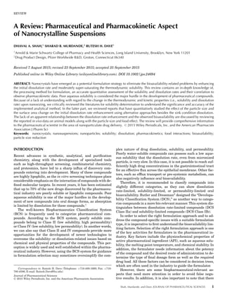
  • 5. REVIEW 5 attrition during particle size reduction. According to the litera- ture, solubility may range from one-fold to several fold, based on particle size.17–19 Bioavailability enhancement associated with nanocrystalline API is attributed to an increase in the dissolu- tion rate because of the enlargement of SA and some increase in solubility based on particle size. This solubility enhance- ment should be in fair agreement with what would be expected based on the Ostwald–Freundlich equation. A change in solu- bility is more significant when particle size is reduced to below 100 nm, which can also be described by the Ostwald–Freundlich equation. Rapid dissolution associated with a nanoparticulate system is clear evidence of a generation of transient super sat- uration of a solution compared with the bulk solubility of a stable crystal form. In the case of crystalline nanoparticles, the degree of supersaturation is low compared with high energy amorphous solids, as particle size has limited impact on satu- ration solubility. Determining accurate solubility is vital to characterize the effectiveness of the formulation. There are several challenges associated with the accurate determination of solubility of any formulation, as it varies case by case. Accurate measurement is significantly more complicated in the case of nanoparticulate systems, as they have the tendency to remain in suspended form in the solution after using conventional approaches. It is almost impossible to visualize the presence of nanoparticles in the filtrate with the naked eye. The intrinsic solubility of poorly soluble compounds is extremely low in number; therefore, the presence of a couple of undissolved particles can lead to a signif- icant error in measurement. While reviewing the literature for determining solubility by a separation-based method, we have found the absence of a validated universal method for accurate solubility determination. This makes it even more challenging when dealing with particle size in the nanometer range, as com- pared with the micronized or bulk particles. The following are the general challenges associated with solubility determination of the nanoparticulate system. 1. No standard method is available in the literature for sol- ubility determination, 2. Difficulty in separation of the dissolved and undissolved nanocrystals/particles because of smaller size. 3. Confirmation that equilibrium is attained or not. 4. Reproducibility of results. 5. Validation of method for accuracy. In addition to the above challenges, one also has to consider other process parameters which vary with the physicochemical properties of the active drug substance for solubility determina- tion. For instance, if the API is weakly acidic or basic, then the pH of the solution plays an important role. It is difficult to deter- mine whether or not equilibrium is attained in this particular case. Several researches have published different approaches for the solubility determination of nanocrystals. Although nu- merous methods for the separation of dissolved and undissolved nanoparticles have been reported in the literature, these are the most common approaches used described in Figure 1. An aque- ous solubility determination by separation-based approach is widely accepted, has been used in industry and academia for many decades, and is the most convenient way to determine solubility. Typically, it is a two-step process: initially, an excess amount of drug is dispersed in an aqueous or buffer solution. The equilibrium is established by shaking or stirring the solu- tion at a specific rpm for a specific time and temperature, at which we want to determine the solubility. Usually the sam- ples are withdrawn after 24 and 48 h. Samples were either cen- trifuged or filtered from syringe filters. A sample analysis was performed using the HPLC and UV. In the case of crystalline nanoparticles, the research articles listed in the Table 2 have reported the solubility determination data for nanosuspensions and nanocrystals by utilizing a separation-based approach as their primary method for solubility determination. The most commonly reported approaches for solubility determination of nanoparticles are by shake-flask method at a specific temper- ature. Most of them have overestimated the thermodynamic solubility associated with nanocrystals, which is why it is im- portant to consider some additional factors during solubility determination when particle size is reduced to nano from mi- cron range. The selection of appropriate pore size filters with respect to the particle size of crystals, and the selection of an appropriate spectroscopic analytical method, is the key factor that needs to be taken in to consideration for accurate solubility determination of crystalline nanoparticles. Bernard Van Eerdenbrugh reported that the UV spectra is a reliable tool to determine the concentration of micronized par- ticles, however, it is not a reliable tool for the determination of the concentration of nanosuspensions, as it is overestimat- ing the actual solubility data. With particles in the nanome- ter range, they itself absorbs the UV light. Therefore, the ab- sorbance data are the mixture of the dissolved and undissolved nanosized particles.32 Today, the measurement of the dissolved drug concentration using an in situ UV probe is the preferred noninvasive method because of its sophistication in terms of contingency and its ability to record data from the start of dis- solution. However, absorption of light from particles is size de- pendent and it is having a great influence on smaller particles. With felodipine as a model drug, an observation has been made that both nanoparticulates of felodipine and free felodipine in the solution absorb light in a similar way, which results in an overestimation of dissolved concentration than what was actu- ally dissolved.33 The results were also dramatic, even for the second derivative of UV spectra. Moreover, the generation of nanoparticles occurs when working with nanosuspensions or supersaturated systems, so caution should be taken. The solubility associated with crystalline nanoparticles is moderately higher (10%–15%)16 compared with what is re- ported in Table 2.34–44 In the case of indomethacin, the reported solubility enhancement was nearly twofold higher. The same is true in the case of oridonin,36 reccardin D,38 and simvastatin39 where reported solubility was substantially higher as compared with bulk crystal solubility. One possible reason behind this misleading data may be the use of an inappropriate separating method and/or analytical method. Nanoparticles with an aver- age size of 200–400 nm can easily pass through 0.22 or 0.45 :m pore sized filters. Determining the concentration of such a fil- tered solution using UV leads to a further over-estimation of actual data because of the mixture of absorbance of both the undissolved and dissolved particles. Moreover, centrifugation at moderate speed of about 10,000–30,000 rpm is not suffi- cient to suspend particles in the nanometer range, therefore, the resultant supernatant contains a mixture of dissolved and undissolved particles. It is noticeable that care should be taken in choosing syringe filters for appropriate pore size. Juene- mann et al.45 were able to show in their study a differentiation DOI 10.1002/jps.24694 Shah, Murdande, and Dave, JOURNAL OF PHARMACEUTICAL SCIENCES
  • 6. 6 REVIEW Table2.ExperimentalSolubilityDeterminationwithSeparation-BasedApproach DrugMethodFiltration ParticleSizeand AnalyticalInstrumentReportedSolubilityReference Indomethacin (IND) Shake-flaskat25°C0.2:mUVNSa:5.86±1.2mg/100mL34 Physicalmix:2.30±0.51mg/100mL Poly1:0.91±0.26mg/100mL Poly2:1.44±0.34mg/100mL Indomethacin (IND) Shake-flaskfor12hinacetate bufferpH5 NMbUVIND–physicalmix:4.89±0.18:g/mL35 P.size:80:mNano-IND/F68:6.43±0.06:g/mL 580±30nmNano-IND/F127:4.80±0.0:g/mL 580±20nmNano-IND/polysorbate:80:10.9±1.54:g/mL Notdetermined Oridonin(ORI)Constant-temperatureshaker at37°Cand100rpmin phosphatebuffersolution (pH7.4) 0.22:mmicropore film UVORI(commercial):99±2:g/mL36 P.size:200–400nmORInanocrystal:170±10:g/mL However,authorreportedslowdecreasein solubilityafterachievingsupersaturationfor NC.c Candesartan cilexetil 24hstirring,followedby centrifugationat25,000rpm inphosphatebuffer containing0.7%Tween20 (pH6.5) 0.2:mUV P.size:223.5±5.4 Bulk:125±6.9:g/mL NC:2805±29.5:g/mL 22.44-foldimprovement 37 RiccardinDStirringinPBS(pH7.4)buffer at25°Cand100rpm 0.22:m microporous membranefilter HPLC P.size:184.1±3.15nm P.size:815.37±9.65nm Bulk:0.6192±0.0245:g/mL38 NS(evaporativeprecipitationintoaqueous solution):242.1±12.1:g/mL NS(microfluidization):31.5±1.9:g/mL SimvastatinPowderform:shake-flask methodat37°C, 0.22:mWhatman filter Supernatant UV P.size:300.3nm Solubilityenhancement:36.14-fold39 NS:centrifugationat 10,000rpm FenofibrateShakeflaskmethod,suspension equilibratedat37°Cfor72h 0.22:mmembrane filter HPLCSolubility40 NS:P.size:606nmConc.SDS%(W/V)Bulk-solubility (:g/mL) 0.00.34±0.03 0.11.42±0.10 0.1511.95±0.16 0.226.38±0.39 0.376.11±0.38 0.4131.95±1.68 0.5183.09±2.17 0.7290.43±5.70 Continued Shah, Murdande, and Dave, JOURNAL OF PHARMACEUTICAL SCIENCES DOI 10.1002/jps.24694
  • 7. REVIEW 7 Table2.Continued DrugMethodFiltration ParticleSizeand AnalyticalInstrumentReportedSolubilityReference Atorvastatin calcium Shakeflaskmethod,for24h and37°CinDIwater 0.1:mmembrane filter UV41 P.size:commercial:38.3± 0.6:m 142.2±0.5:g/mL TurraxR =21.5±0.03:m185.1±1.2:g/mL HPHd:3.12±0.05:m299.8±0.6:g/mL HPH(20cycles@1500bar: =0.446±0.02:m 386.5±0.7:g/mL MeloxicamShakeflaskmethod,for24h and37°CinDIwater,stirred samplewerefurther centrifugedat10,000rpmfor 15min 0.22:mnylon- membrane filter UV42 P.size:Raw:4.4±0.50:g/mL Raw:46.39±7.37:mPhysicalmixture:5.83±0.62:g/mL Sonicated:0.259±0.03 :m Spray-driedNC:21.84±0.78:g/mL Sonicated+HPH:0.212± 0.04:m Spray-dried:0.178±0.02:g/mL LuteinSampleswerekeptonshaker 100rpmand25°Cdistilled water(pH=5.5)anddistilled watercontainingsurfactant solution(0.05%,w/w PlantacareR 2000,pH= 10.5).Sampleswere centrifugedat23,800gfor2h 0.2:mfilterUVDIwater:<0.054:g/mL DIwater+surfactant:0.54:g/mL nanocrystal:upto14.3(>264-fold thanwaterand>26.3-foldwater withsurfactant) 43 P.size:429nm QuercetinSampleswerekeptonashake at37°Cand100rpm.Sample werewithdrawnandtransfer toultrafreetubewithcutoff of10kDaandcentrifugedat 20,000gfor30minat4°C NMHPLCBulk:10.28g/mL44 P.size:PM:16.42g/mL Dried-EPAS:282.6±50.3 nm EPAS(evaporativeprecipitationinto aqueoussolution):422.4g/mL HPH:213.6±29.3nmHPH:278.6g/mL a NS,nanosuspensions. b NM,notmentioned. c NC,nanocrystal d HPH,high-pressurehomogenization. DOI 10.1002/jps.24694 Shah, Murdande, and Dave, JOURNAL OF PHARMACEUTICAL SCIENCES
  • 8. 8 REVIEW Figure 1. Reported approaches for the solubility determination. between suspended submicron colloidal particles and molecu- larly dissolved particles. It has been reported that the results of solubility and dissolution of nanocrystals are comparable with each other if the analysis is performed with filters having pore size ࣘ 0.1 :m. Using larger 0.2 and 0.45 :m filters, the nanocrystalline system shows apparent supersaturated behav- ior because of the combinatory effect of dissolved and undis- solved nanocrystalline particles. In other words, it has been demonstrated that filters with a bigger pore size may not be sufficient enough to hold back the colloidal particles. An ex- cellent demonstration has been reported by carrying out the study of a selection of appropriate filters and their impact on observed in vitro results. It was also compared with an in silico model for more precise justification of the experimental data. Overall, the conclusion and recommendation has been made for using filters with smaller pore sizes (i.e., 0.1 and 0.02 :m) when dealing with nanoparticles. Kinetic solubility determination by nephelometric or turbidi- metric methods during early screening of the drug molecule was introduced earlier by Bevan and Lloyd.46 Bernard Van Eerden- brugh critically evaluated different methods for the solubility determination of nanocrystals using four model compounds: itraconazole, loviride, phenytoin, and naproxen. The data ob- tained show that separation-based methodologies were not suf- ficient to determine solubility, as the data were not in fair agree- ment with the Ostwald–Freundlich equation. Noninvasive analytical techniques, such as light scattering and turbidime- try, were found to be more reliable for appropriate understand- ing of the solubility behavior of crystalline nanoparticles. In the case of amorphous nanosuspensions, Lindfors et al.33 have determined the solubility by plotting scattering intensity with the drug concentrations. Presence of excessive stabilizers in nanosystems also tends to generate the scattering of intensity. Hence, the method that Lindfors demonstrated is not accurate enough because of the summation of scattering intensities that are generated by dissolved nanoparticles and micelles. In or- der to eliminate this, Van Eerdenbrugh et al.16 have chosen the point of intersection at which scattering is exclusively due to the dissolved drug concentration and determine the solu- bility. Measures solubility data by scattering, for loviride was comparable to bulk solubility data. It was not feasible to determine solubility for itraconazole from the turbidimetry method, as the method cannot be distinguished because of very low solubility. Solubility determination for phenytoin can be determined precisely with both light scattering and turbidime- try. In the case of naproxen, determined solubility values were slightly less than the unmilled compound but the results were within the standard margin of error. Hence, this shows that both turbidimetry and light scattering methods can be applied to obtain more realistic solubility data for crystalline nanopar- ticles. Scattering can be used more precisely for compounds that have low scattering intensities and turbidimetry can be applied on compounds having higher scattering intensities. Moreover, the solubility enhancement was in fair agreement with the theoretical prediction from the Ostwald–Freundlich equation. For the theoretical calculation, the interfacial ten- sion was estimated (interfacial tension of typical pharmaceu- tical API ranges between 5 and 50 mN/m) as described in the reference literature.47 Experimental data and theoretical pre- dictions were also in agreement with the Ostwald–Freundlich equation, suggesting that solubility enhancement should be marginal in the case of crystalline nanoparticles and both the light scattering and turbidimetry are good noninvasive meth- ods for the solubility determination of nanocrystals. However, there are certain assumptions and limitations associated with these analytical methods. In most cases, the dynamic light scat- tering is used to measure the light scattering intensity. The instrument assumes each particle as a sphere and carries out the determination. When the particles are not spherical ini- tially or if the particles tend to change shape during disso- lution or solubilization, they may have different results than those measured by the instrument. Therefore, the impact of particle shape is a factor that also needs to be considered, as it plays major role during dissolution and solubilization. Anhalt et al.48 has reported a method for the solubility determination by real time measuring the light scattering. The main focus was to determine the equilibrium solubility and dissolution rate of a crystalline nanosuspension with different particle size using FBT as a model compound. The solubility results of the light scattering method were fair enough to justify it as a reliable analytical tool for the solubility measurement. Reported sol- ubility enhancement for nanocrystals was around 10%–15%, Shah, Murdande, and Dave, JOURNAL OF PHARMACEUTICAL SCIENCES DOI 10.1002/jps.24694
  • 9. REVIEW 9 Table 3. Alternative Approaches for Accurate Solubility Determination Analytical Method, Compound and Particle Size Solubility Determination Solubility Enhancement Ratio Reference Light scattering 48 Fenofibrate 8.69 ± 0.78 :g/ml 140 nm 10.38 ± 0.01 :g/ml 1.19 270 nm 8.70 ± 0.24 :g/ml 1.00 1070 nm 9.62 ± 0.50 :g/ml 1.11 Light scattering and turbidimetry 16 Loviride (162 nm) 0.0108 mg/mL 1.10 Scattering – 0.0119 mg/mL Itraconazole (220 nm) 0.0047 mg/mL 1.15 Scattering – 0.0054 mg/mL Phenytoin (406 nm) 0.0667 mg/mL 1.07 Scattering – 0.0711 ± 0.007 mg/mL Turbidity – 0.07 ± 0.0034 mg/mL 1.05 Naproxen (288 nm) 0.2116 mg/mL 0.97 Scattering – 0.2053 ± 0.003 mg/mL Turbidity – 0.2083 ± 0.0024 mg/mL 0.97 Separation-based methodology (ultracentrifugation, filtration—0.1 :m, 0.02 :m, and dissolution) 51 Griseofulvin-micro 7.63 ± 0.89 :g/mL 1.10 362 nm 8.40 ± 0.25 :g/mL 122 nm 9.99 ± 0.15 :g/mL 1.30 Compound X-micro 65.17 ± 1.58 :g/mL 0.97 238 nm 63.41 ± 1.26 :g/mL 93 nm 89.06 ± 6.36 :g/mL 1.36 Fenofibrate-micro 0.74 ± 0.27 :g/mL 1.11 290 nm 0.82 ± 0.26 :g/mL Celecoxib-micro 1.00 ± 0.03 :g/mL 1.11 341 nm 1.11 ± 0.03 :g/mL which justifies the previously reported solubility data by Van Eerdenbrugh.16 However, one should consider certain limita- tions before utilizing this light scattering method. Most impor- tantly, the sample has to be ultra clean; the presence of any dust and debris may lead to the generation of scattering intensity that affects to the observed data. The use of a plastic cuvette is also a potential source of error. Moreover, as light scattering is less sensitive to small particles, the results are slanted towards the larger sized particles as they have a tendency to generate more scattering of light. Besides these methods, other analyti- cal techniques like potentiometry and pulse polarography have also been reported for real time solubility measurement.49,50 However, these methods are not universal and can be used for a fewer number APIs with certain properties (i.e., electroac- tive). In the case of potentiometric measurement, each time a specific electrode has to use for the specific API. In general, separation-based methods are universally ac- cepted as they do not require a high level of experimental skill. Murdande et al.51 has reported three different approaches (ul- tracentrifugation, ultracentrifugation with filter, dissolution) to determine the accurate solubility. Ultracentrifugation was used after optimizing different parameters such as speed, time, and temperature for the satisfactory separation of dissolved and undissolved nanocrystals. Syringe filters were also used (i.e., 0.1 and 0.02 :m) based on the particle size to determine sol- ubility from the supernatant of the ultra-centrifuged sample. In addition, dissolution data at the end of the experiment was also evaluated for the total dissolved concentration based on the theoretical knowledge that nanocrystals should reach the satu- ration level at equilibrium. Results from all three methods were similar and in fair agreement with the Ostwald–Freundlich equation. The results from the solubility determination using the separation-based approach described above are in Table 3. Interfacial solubility (the concentration in a boundary layer of a spherical particle) plays an important role. As the particle size decreases, the SA increases proportionally, along with the in- terfacial tension. Sun et al.52 have determined the equilibrium solubility of coenzyme Q10 nanocrystals and bulk drug in three different dissolution media, which were mixtures of different concentrations of tween 20 and isopropanol. Solubility usually leads via diffusion and the driving force would be the difference between the solubility at the boundary layer and the bulk drug concentration. Based on observations, they had also proposed a solubility model for nanocrystals and bulk drugs.52 Another separation-based approach is the determination of solubility and/or drug release with the application of equi- librium dialysis. The published research has applied this concept, mainly for the nanoparticle-based formulation of large molecules and a few for small molecules, and de- termined the equilibrium solubility and drug release from nanoparticulate formulation. The most common approaches in- clude: sac dialysis,53,54 side by side dialysis,55–57 and reverse dialysis.58,59 In dialysis, separation is achieved by means of a semi-permeable membrane using molecular weight cut-off membrane. Assuming that the dialysis membrane is perme- able to free API only, Frank et al.60 reported the quantification DOI 10.1002/jps.24694 Shah, Murdande, and Dave, JOURNAL OF PHARMACEUTICAL SCIENCES
  • 10. 10 REVIEW of molecularly dissolved, poorly soluble drug by using the equi- librium dialysis method and determined the apparent solubility (molecularly dissolved API + miscellany solubilized drug). The solubility of the drug in both the Hanks Balance salt solutions and the supplementary salts (HBSS++) and fasted-state sim- ulated intestinal fluid (FaSSIF) buffer were nearly the same within the margin of error.60 It is well known that nanoparti- cles have a rapid release dissolution kinetics. Efforts have been made to determine the interplay of the diffusion rate, size of the molecular weight cutoff (MWCO) membrane, and concen- tration difference of the donor and receiver compartment by Moreno-Bautista and Tam.61 Decreasing the dialysis cassettes to smaller MWCO showed a reduction in the diffusion rate. The compounds having a molecular weight of around 200–400 Da showed a diffusion rate profile several times higher with 10 kDa membrane compared to 2 kDa membrane MWCO. The cassettes having a pore size smaller than the drug molecule should be suitable for the evaluation of the dissolution rate of nanopar- ticles. Furthermore, the release profile from the dialysis cas- settes failed to catch some necessary events, such as the burst effect and lag time, which leads to a question the suitability of the method for true discrimination of the rapid release kinetics of submicron size colloidal particles. This situation prompted Zambito et al.62 to ask question whether the dialysis is a re- liable method for studying drug release from nanoparticulate systems. Their study concluded that for nanoparticles, the dial- ysis method was insufficient in expressing the discrimination and founded results were deceptive. Because of a reversible in- teraction between the drug and dispersed nanoparticles, the rate of permeation through the dialysis membrane was neg- ligible as compared with the plain drug solution. Hence, the kinetic release was found to be far less and unrealistic. With such a study, the release kinetic is governed by the dialysis membrane (not dependent on the drug release from colloidal particles), which makes it unreliable for the in vitro predictions and means the data may not be very discriminative. DISSOLUTION Dissolution is known as a significant tool for the analysis of pharmaceutical formulations. Indeed, it has somewhat poten- tial to predict the in vivo performance. Prediction of the extent of absorption can be made from the rate of drug release in the gastrointestinal tract.63 A drug candidate has to pass through some pre-requisites steps before being absorbed into the sys- temic circulation. Dissolution is one of the essential steps for effective absorption of a drug candidate and is a key parame- ter for assessing the onset of action of an oral dosage form. As per the well-known BCS, classification compounds belonging to Class II and IV are poorly soluble in nature, especially ones belonging to Class II, which have a dissolution rate of limited absorption because of their low aqueous solubility.64 In another words, the rate of absorption is proportional to the rate of disso- lution in the gastrointestinal tract. For a given drug moiety that dissolves in the gastrointestinal tract, its dissolution can be tai- lored by either generating amorphous systems or reducing the particle size to the micron or submicron level (i.e., nanometer). The motivation for the development of nanosystems has been generated from the increased number of new chemical entities with poor aqueous solubility. For such low aqueous-soluble com- pounds, even micronization is not sufficient enough to eliminate the problem. The reduction of particle size leads to an increase in the SA of the drug particle. The SA enhancement factor from micron to nanoparticle is about 10-fold. Thus, significant en- hancement in SA has a positive impact on the dissolution rate of a drug particle. This positive effect on the dissolution rate can create a higher concentration difference between the gut lu- men and the blood, which increases the absorption via passive diffusion and leads to an improved therapeutic response. Direct proportionality of the rate of dissolution with respect to specific SA has been well documented by the Noyes–Whitney equation. As a follow up to this, Nernst–Brunner and Danckwerts also proposed modified equations to describe the dissolution behav- ior a solid powder. Noyes − Whitney Equation : dM dt = k(Cs − C) (1) Nernst − Brunner Equation : dM dt = S D h (Cs − C) (2) Danckwerts surface − renewal model : dM dt = S √ DP (Cs − C) (3) This section will provide an overview of the different estab- lished methods to assess the dissolution for nanocrystals. As of now, it is well understood that particle size is inversely pro- portional to SA and the dissolution rate has a direct relation- ship with the change in SA. The major challenge is to develop a discriminative dissolution method for poorly water-soluble drugs. Earlier reports have indicated that there is an alteration of wetting behavior with particle size reduction. In the case of nanoparticles, the wetting phenomenon is more prominent, which leads the dissolution rate to increase more quickly. More- over, the reduction in the diffusional distance upon size reduc- tion generates a moderate spring effect that quickly achieves supersaturation level with respect to particle size during dis- solution (Fig. 2). Therefore, the traditional methods are inad- equate to determine the actual dissolution rate enhancement for nanoparticles which intensify the need for an appropriate method to perform the dissolution study of nanoparticles. Sink Condition (Conventional) Dissolution Sink condition dissolution condition is defined as the volume of the dissolution medium which can dissolve more than three times the amount of the dose used in the dissolution study.65 In most cases, the dissolution study is performed using a USP type – I or II apparatus. Usually for a study of a drug release profile, the dissolution is carried out in 900 mL of dissolution media (i.e., in a different pH or in deionized water) at 37 ± 0.5°C temperature and by rotating at a specific rpm (i.e., 100 rpm). Samples are withdrawn at specific time intervals throughout the dissolution study. After each sample withdrawal, the same amount of the fresh dissolution media, which is equilibrated at the same temperature, is introduced to maintain perfect sink condition. In most cases, samples will be filtered prior to anal- ysis. However, for the nanoparticles, sink condition dissolution will provide good qualitative and less quantitative information regarding the dissolution behavior of the formulation. So, the comparison of dissolution profiles between nanosuspension and microsuspension will show the impact of particle size on disso- lution velocity. The literature has reported a higher dissolution Shah, Murdande, and Dave, JOURNAL OF PHARMACEUTICAL SCIENCES DOI 10.1002/jps.24694
  • 11. REVIEW 11 Figure 2. In vitro surface dissolution, (A) surface image dissolution in correspondence with diffusional layer thickness (h), and (B) intrinsic dissolution profile in correspondence with the surface image dissolution. rate for the nanosuspensions,66,67 which dissolve instanta- neously as compared with micronized suspension. Hence, sink condition dissolution is an appropriate tool for QC analysis. However, because of the instantaneous dissolution behavior of nanocrystals during sink condition, it is challenging to quan- titatively discriminate dissolution rate enhancement with re- spect to particle size. In addition, the long sampling interval during conventional dissolution also adds to the difficulty. In response to these problems, few scientists have tried alterna- tive approaches. Flow-Through Cell The flow-through cell dissolution apparatus demonstrated the capability of studying the dissolution of all kinds of formula- tions, such as tablets, capsules, and powders.68,69 Heng et al.70 have carried out dissolution studies and compared the dissolu- tion rate by asking the question, “What is the suitable disso- lution method for drug nanoparticles?” Initial dissolution rates were determined for the paddle, basket, and flow-through ap- paratus. The initial dissolution rate was calculated from the following equation: dM dt Nanoparticles dM dt Unprocessed = slope %dissolved time nanoparticles slope %dissolved Time unprocessed The paddle and basket dissolution apparatus were described as inappropriate for nanoparticles because of their tendency to form aggregates. The ratios obtained for the initial dissolution rate enhancement in both cases did not agree with the model predicted values. Dissolution studies via the dialysis process (membrane with MWCO 12–14 kDa) were found to be very slow and having a limited ability for size discrimination. As a result, there was no significant difference noted in the dissolution pro- files between nanoparticles and the unprocessed powder. Based on the analysis, the flow-through cell set-up was described as appropriate for dissolution of nanoparticulate powder by mini- mizing the wetting problems.71 The experimental data for the dissolution profiles were able to discriminate well with the un- processed powder. The initial reported dissolution rate ratio was the average number of the triplicate study and was in agreement (6.95 vs. 7.97) with model predicted value. The au- thors concluded it as the most suitable analytical process for the dissolution of nanoparticulate powders. However, selection of an appropriate flow rate is required for proper discrimination because a change in the flow rate can generate change in the release behavior. Note that the presence of air can also affect the flow rate during the study, which may alter the dissolu- tion behavior. Therefore, the study should be carried out with caution. Alternative Approaches Nanosuspensions/nanocrystals exhibit instantaneous dissolu- tion behavior, making it difficult to monitor them with conven- tional methods, which requires sample withdrawals, as well as dilution and filtration steps before quantitative analysis. In situ fiber optic probes are not able to provide an accurate quantita- tive determination because of the generation of absorbance by both the dissolved and undissolved nanocrystalline particles.32 Recently, Kayaert et al.72 reported an alternative approach by studying the dissolution behavior of nanosuspensions using a solution calorimeter. The use of a solution calorimeter for dis- solution study has been previously reported for polymer and polymeric blends.73 This dissolution study was examined based on appraising the change in the heat throughout the process. In other words, heat required for the dissolution process is mea- sured. The methodology consists of sealing the breakable glass ampoule along with nanosuspension using bees wax. The glass vial should be kept in the sample cell, surrounded by the me- dia in which the dissolution study is intended to perform. After reaching the required temperature, the glass vial should be broken, and the dissolution process is started. The rate of dis- solution can be then assessed by converting the raw data (i.e., temperature vs. time and cumulative heat vs. time) to percent- age dissolved versus time. The dissolution process for nanosus- pensions was observed to be extremely fast. It takes just 20 s to complete the dissolution process for nanosuspension, whereas in the case of crude suspension, it was found to be between 8 and 15 min For the traditional dissolution apparatus (i.e., basket and paddle), the earliest sample one can withdraw is at 2 min. Hence, the solution calorimeter unquestionably pro- vides the advantage of real time measurement compared with traditional methods. Consumption and/or production of heat can be monitored from the beginning of the dissolution process without disturbing (i.e., sample withdrawal) the system. There are drawbacks with this method, which need to be considered for an accurate evaluation of the data generated. The method DOI 10.1002/jps.24694 Shah, Murdande, and Dave, JOURNAL OF PHARMACEUTICAL SCIENCES
  • 12. 12 REVIEW measures the summation of the total heat change, which in- cludes contributions from the breakage of the glass ampoule, heat change from other excipients in the formulations, and heat change due to the dissolution process. Therefore, a careful eval- uation of the data is warranted by considering all these con- tributing factors. In addition, the reported dissolution time is far shorter (i.e., 20 s to 15 min) using this method, but a single experiment requires additional time for the equilibration before the test and also requires post-test time after the dissolution, making it more time consuming than the conventional dissolu- tion methods. Furthermore, the instrument is very subtle and requires a skilled operator. Recently, another separation-based approach was reported by Shah et al.74 using a modified dial- ysis method known as pulsatile microdialysis (PMD)75 for the characterization of supersaturation and precipitation behavior of poorly soluble drugs. Here, the pore size for the dialysis probe was approximately 18 kDA (ß2 nm). Hence, the final sample should contain only the dissolved drug. This analytical method can be useful for characterization of high energy solid forms (amorphous) or nanoamorphous33 forms because of the advan- tage of quick sampling (i.e., 10 s). As a more convenient direct sampling method, PMD may not be much useful for the study of nanocrystals with the size range of 150–400 nm because of the availability of the syringe filters with pore size 0.1 :m. The use of 0.02 :m (ß20 nm) anotop syringe filters for the dissolution study has also been reported in the literature.45 As a result, it may be of great interest if the comparison of the sample anal- ysis from PMD and the sample analysis from 0.02 :m syringe filters has been carried out to describe the usefulness of the method more precisely. Another separation-based approach has been reported by Murdande et al.51 by modifying the traditional sink dissolu- tion to various non-sink conditions. Non-sink conditions can be generated by pre-dissolving API in the dissolution me- dia prior to perform the dissolution experiment. Recently, an- other literature has also reported the dissolution of nanocrys- talline suspension for poorly soluble molecules using non-sink conditions.76 Because of the lack of discrimination during the initial phase of dissolution by sink dissolution, the goal was to slow down the dissolution velocity of nanocrystals and mi- cronized crystals in such way that the initial dissolution phase becomes more discriminative. Here, the samples were filtered with both 0.02 :m (for nano) and 0.1 :m (for micro) after es- tablishing the filter binding capacity of the each model com- pounds. As the saturation level increases, the rate of disso- lution decreases, creating a more discriminating portrayal of the initial dissolution rate enhancement (i.e., at 75% satura- tion level the initial rate of dissolution, 10 :m:362 nm:122 nm were 1.0:1.8:3.6). The observed data look more promising com- pared with the data obtained under sink conditions. However, filtration from 20 nm pore size of filters should be performed carefully because of the chance of blockage or clogging of the pores by larger size particles. From our knowledge, all these approaches are reported specifically for nanocrystals. One can select any analytical method listed above based on their re- quirements and the availability of resources. PK BEHAVIOR OF CRYSTALLINE NANOPARTICLES Different formulations have different effects on the PK profile of the same drug. Besides chemical properties, a change in the physical property (i.e., particle size reduction) may also have a considerable impact on altering the PK behavior of a molecule. The reduction of the particle size increases the dissolution ve- locity and may have some effect on in vitro solubility. Similar behavior was observed in the case of in vivo parameters, such as AUC (+ve), Cmax (+ve), Tmax (−ve), and fed/fast variability (−ve). A reduction of particle size improves the dissolution rate, which increases the absorption of the drug in the body, and it leads to an increase in PK performance. Compounds belong- ing to the BCS Class II have issues related to poor solubility, dissolution-rate-limited absorption, and bioavailability. Parti- cle size reduction can improve the bioavailability of such com- pounds by improving the dissolution rate. For the nanocrystals, an increase in the oral bioavailability of the compounds can be attributed to an increase in the SA.77 Improvement in Oral Bioavailability An improvement in the dissolution rate and adhesion to the gut wall can be considered the main contributing factors for enhancing bioavailability and overall PK performance. After oral administration, the formulation disintegrates and begins the dissolution process. The dissolution rate resembles the concentration gradient in the physiological environment. As a result, an improvement in the dissolution rate leads to an enhancement in the absorption, and finally, in the bioavail- ability of the compound. Because of the smaller size of the nanoparticles, they tend to adhere to the gut wall.78 Bioavail- ability enhancement can be achieved both actively and pas- sively. The classic example for bioavailability enhancement has been represented in the case of danazol, which is poorly sol- uble (<1 :g/mL) and one of the most challenging compound to work within the pharmaceutical industry.79 In vivo studies of nanosuspensions of danazol (169 nm) in beagle dogs have shown enormous enhancement in Cmax and a 16-fold enhance- ment in relative bioavailability, compared with the micronized suspension.12 Therefore, it is understood that by reducing the particle size, the solubility issue associated with danazol can be solved. Moreover, it can also be delivered at a lower dose by enhancing the therapeutic outcome of the compound. FBT is another classic example of a poorly soluble drug with a high log P (i.e., 5.3) value. FBT is used in hypercholesterolemia and has a solubility and dissolution-rate-limited absorption property. Bioavailability of FBT is attributed to the dissolution behavior of particles in the gastric environment. A comparison of the PK profile of nanocrystals and coarse powder demonstrated signif- icant enhancement in the rate of absorption after oral adminis- tration. The Cmax of nanocrystals was observed to be five times higher than that of the coarse powder, along with a higher AUC and Tmax.80 Zuo et al.40 made an effort to compare formulated nanocrystalline particles with the commercial nanocrystalline formulation LipidilTM -ez with respect to in vitro and in vivo behavior. No significant difference was observed between the two nanocrystalline formulations, suggesting that variability will be less when particle size is reduced to nanometer range. Certainly, the re-dispersion behavior of dried nanocrystals also plays a contributing role by demonstrating reversible or irre- versible aggregation behavior. Bioavailability can be altered by several factors in the gastric environment, such as pH change, food effect, the presence of salts, and so on. In all the reported cases reported in the Table 4,12,40,80–88 a significant enhance- ment in dissolution rate was observed for the nanocrystals, Shah, Murdande, and Dave, JOURNAL OF PHARMACEUTICAL SCIENCES DOI 10.1002/jps.24694
  • 13. REVIEW 13 Table 4. Effect on In Vivo Parameters Compound Animal Model PK Parameters Comment Reference Aripiprazole Beagle dogs NSa (350 nm) Coarse suspension 71% enhancement in relative bioavailability 81 Tmax (h) 1.04 ± 0.24 3.33 ± 1.50 Cmax (ng/mL) 137.37 ± 17.38 43.10 ± 11.68 AUC (ng h/mL) 618.15 ± 81.28 386.06 ± 78.54 Nitrendipine Rat NS (209 nm) Tablet Fivefold enhancement in relative bioavailability 82 Tmax (h) 1 ± 0 2.2 ± 1.17 Cmax (:g/mL) 3.65 ± 0.43 0.60 ± 0.11 AUC (:g h/mL) 17.12 ± 2.59 3.44 ± 0.47 Danazol Beagle dogs NS (169 nm) Coarse suspension 16-fold enhancement in relative bioavailability 12 Tmax (h) 1.5 ± 0.3 1.7 ± 0.4 Cmax (:g/mL) 3.01 ± 0.80 0.20 ± 0.06 AUC (:g h/mL) 16.5 ± 3.2 1.0 ± 0.4 Naproxen Rat NS (270 nm) Unmilled suspension ß1.25-fold enhancement in relative bioavailability 83 Tmax (min) 23.7 ± 5.1 33.5 ± 2.9 Cmax (:g/mL) 187 ± 18 126 ± 4 AUC (:g min/mL) 19,062 ± 573 15228 ± 994 Apigenin Rat NCb (400—800 nm) Coarse powder ß3.5-fold enhancement in relative bioavailability 84 Tmax (min) 90 ± 14 120 ± 16 Cmax (:g/mL) 5.4 ± 0.6 1.5 ± 0.2 AUC (:g min/mL) 1509 ± 196 445 ± 45 Cefpodoxime proxetil Rabbit SDNSc (<300 nm) Microsuspension ß1.6-fold enhancement in relative bioavailability 85 Tmax (h) 0.75 ± 0.11 1.75 ± 0.68 Cmax (:g/mL) 18.36 ± 2.03 10.88 ± 1.01 AUC (mg h/mL) 47.55 ± 4.33 29.78 ± 3.47 Baicalein Rat Baicalin NC (335 nm) Coarse powder ß1.6-fold enhancement in relative bioavailability 86 Tmax (h) 0.92 ± 0.38 1.67 ± 0.29 Cmax (:g/mL) 11.12 ± 1.25 7.18 ± 1.25 AUC (:g h/mL) 119.25 ± 20.26 71.41 ± 4.38 Simvastatin Rat NC (387 nm) Coarse powder ß1.5-fold enhancement in relative bioavailability 87 Tmax (h) 1.99 ± 0.05 2.88 ± 0.08 Cmax (ng/mL) 450.3 ± 140.5 300.2 ± 67.01 AUC (ng h/mL) 1110.3 ± 280.62 770.9 ± 110.3 Fenofibrate Rabbit NC (460 nm) Coarse powder ß4.7-fold enhancement in relative bioavailability 80 Tmax (h) ß0.4 ß1 Cmax (:g/mL) 536.66 ± 35.09 113.46 ± 29.05 AUC (:g h/mL) 134.38 ± 6.47 28.41 ± 5.52 BMS-347070 Beagle dog NC Micronized ß2.7-fold enhancement in relative bioavailability 88 Tmax (h) 2 3 Cmax (ng/mL) 1475 ± 375 483 ± 79 AUC (ng h/mL) 28,613 ± 3850 10,870 ± 1651 Fenofibrate Beagle dog SDNCd LipidilTM-ez No significant difference in relative bioavailability 40 Tmax (h) 2.8 ± 1.8 5.2 ± 9.2 Cmax (ng/mL) 2075.2 ± 1101.1 2349.5 ± 1050.5 AUC (ng h/mL) 30,496 ± 6541 34,035.9 ± 19,286.4 a NS, nanosuspension. b NC, nanocrystal. c SDNS, spray-dried nanosuspension. d SDNC, spray-dried nanocrystals. which caused a significant reduction in the time to reach max- imum concentration, an improvement in the rate of absorp- tion, peak plasma concentration, and enhancement in relative bioavailability. Food Effect In most cases, food increases the bioavailability of the drug by increasing bile secretion and increasing the duration of gas- tric emptying time. For a drug molecule’s clinical efficacy and DOI 10.1002/jps.24694 Shah, Murdande, and Dave, JOURNAL OF PHARMACEUTICAL SCIENCES
  • 14. 14 REVIEW Table 5. Food Effect PK Parameter Fed Fast Compound NSa Refb NS Ref Reference ELND-006 Tmax (h) 1.4 3 1.4 1.8 90 Cmax (ng/mL) 365 159 294 49.4 AUC (ng h/mL) 3063 1767 2430 315 Fc 110 63.4 87.2 11.3 Fed Fast NCd Jetmilled NC Jetmilled Cilostazole Tmax (h) 1 1 1.3 ± 0.5 1 91 Cmax (ng/mL) 4872 ± 112 2901 ± 314 5371 ± 1173 1029 ± 218 AUC (ng h/mL) 13,589 ± 3895 10,669 ± 3417 17,832 ± 4994 2875 ± 587 F 0.67 ± 0.22 0.53 ± 0.21 0.86 ± 0.29 0.15 ± 0.04 Fed Fast SNCDe SDDf SNCD SDD Ziprasidone Cmax (ng/mL) 260 285 416 140 92 AUC (ng h/mL) 1911 1949 2044 879 a NS, nanosuspensions. b Ref, reference sample. c Relative bioavailability. d NC, nanocrystal. e SNCD, solid nanocrystalline dispersion. f SDD, solid amorphous spray-dried dispersion. future success, persistence in oral bioavailability can only be achieved by eliminating the food effect.89 The presence of bile salts in the gastric environment also has an impact on the dis- solution behavior of the molecule. It has been reported that the presence of food reduces the dissolution behavior of micronized particles in certain cases as reported in Table 5.90–92 The ratio of bioavailability of the fed to fast state was observed to be close to 1 in the case of nanocrystals, while micronized formulation of the same drug showed a significantly higher ratio (approx. sixfold) of the fed to fast state by demonstrating the poten- tial food effect after oral administration to an animal model with the same dosing. Bile salt secretion usually increases in the fed state.93 A change in concentration of the bile salt in the stomach leads to a change in dissolution behavior and slows down or enhances absorption of the drug as depicted in Figure 3. This phenomenon causes a large variability from pa- tient to patient and also from fed state to fast state. Micronized or larger particles that have shown improved absorption in the fed state might be because of the micelle formation. For the nanocrystals though, they are not helping in improving the in- trinsic solubility, but they provide an advantage by enhancing the initial dissolution rate because of the larger SA. The higher rate of dissolution leads to an increased rate of absorption, and eventually, enhancement in the overall bioavailability, irrespec- tive of the fed or fast state. Hence, it can be stated that the bile salts concentrations have significantly less effect on the absorp- tion behavior of nanoparticles. The same observation has been reported for cilostazole91 [fed/fast ratio—0.78 (nano vs. 3.53 jet-milled)]. Thombre et al.92 have reported the PK behavior of an anti-psychotic drug ziprasidone in beagle dogs by conduct- ing the fed- and fasted-state bioavailability experiment for the both solid nanocrystalline dispersion and the commercial blend (amorphous spray dried dispersion). The commercial blend has shown a twofold improvement in bioavailability in the fed state as compared with the fasted state, indicating the failure of the commercial blend to prevent variability in absorption behavior irrespective of the food state. A similar food effect was observed with human data. On the other hand, nanocrystals have proven to be efficient enough to prevent the variability from fed to fast state by generating a similar bioavailability profile in beagle dogs. From the reported literature, it can be concluded that vul- nerability in the variation of oral bioavailability for potential drugs can be avoided with reduction in particle size to nanome- ter scale. Particle Size Effect: Micro Versus Nano The effect of particle size on in vitro and in vivo performance is well documented elsewhere.91,94–96 Different particle sizes have a different effect on the initial dissolution rate, the rate of ab- sorption, and on oral bioavailability. Moreover, particles with nanometer size range may have the tendency to pass through both active and passive diffusion pathway, which increases the chances for sub-micron particles to reach to peak plasma con- centration with a reduction in time compared to macro/large particles. Sun et al.97 have shown the effects of particle size, ranging from micron to nanometer size, on Cmax, Tmax, and AUC.98 A large increase in bioavailability was observed for the both nanoparticles (i.e., 300 and 750 nm) as compared with the micron size particles and coarse powder, which is reported in Table 6. The rise in bioavailability can be attributed to in- creased SA and adhesion behavior of nanosized particles. There Shah, Murdande, and Dave, JOURNAL OF PHARMACEUTICAL SCIENCES DOI 10.1002/jps.24694
  • 15. REVIEW 15 Figure 3. Effect of food on dissolution of nanocrystals and microcrystals. Table 6. Particle Size Effect: Micro versus Nano PK Parameter Compound Size (nm) Tmax (h) Cmax (ng/mL) AUC (:g h/mL) Reference Itraconazole 300 3 712 ± 121 9967 ± 2527 97 750 4 501 ± 73 8649 ± 1580 5500 3 218 ± 86 1271 ± 398 Powder – 40 ± 9 197 ± 61 Nitrendipine 200 1.0 ± 0.0 3.65 ± 0.43 17.12 ± 2.59 98 620 1.0 ± 0.0 2.75 ± 0.50 14.36 ± 2.75 2700 0.8 ± 0.1 1.74 ± 0.26 8.19 ± 0.85 4100 0.7 ± 0.1 1.19 ± 0.08 7.41 ± 0.46 20,200 3.4 ± 1.2 1.04 ± 0.21 6.89 ± 1.15 Powder 4.1 ± 2.4 0.42 ± 0.08 2.76 ± 0.46 was significant difference observed in the dissolution profiles between both of the nanoparticles (i.e., 300 and 750 nm), but no significant difference has been reported in the PK profiles. The same observation was reported by Xia et al.98 by studying effect of particle size on the oral bioavailability of nitrendipine in the rat model. The overall bioavailability was reported to be a ninefold increase compared with the coarse powder and a threefold increase as compared with the micronized particles. Comparing the PK parameters for both nanoparticles (200 vs. 620 nm), a slight improvement was noted in Cmax, Tmax, and AUC. Based on the literature data, an interesting area for a further research is possible, by lowering the particle size down to the 50 nm range with a narrow size distribution and mea- suring the relative bioavailability. This may have more pro- nounced effect on the bioavailability because of the further im- provement in the thermodynamic solubility suggested by the Ostwald–Freundlich equation. A comparison of adhesion rates, along with the various particle size ranges, starting from 50 nm to micron level, will also be an interesting area to explore for further understanding the particle size effect on the bioavail- ability. SUMMARY Drug nanocrystal formulation is a well-established and suc- cessful approach, and it is described in the initial phase of this review article. Nanocrystal formulation approach is more promising because most of the major pharmaceutical compa- nies have adopted this approach because of its effectiveness and convenience. During the early phase development and during formulation development for pre-clinical studies, it is more con- venient to formulate the nanocrystal for the determination of the exposure and the PK parameters to confirm the candidacy DOI 10.1002/jps.24694 Shah, Murdande, and Dave, JOURNAL OF PHARMACEUTICAL SCIENCES
  • 16. 16 REVIEW of the drug molecule. It is also important to know the thermody- namic and the kinetic behavior with respect to size reduction. Several approaches have been tried by researchers for the accu- rate determination of thermodynamic solubility (Tables 2 and 3). Some have reported suspicious results due to generation of artifacts by the analytical method for the solubility determi- nation. Despite the shortcomings of some of the conventional methods for determining solubility (specifically for nanoparti- cles), others have reported excellent results by adopting newer approaches for the solubility determination. The boost in ther- modynamic solubility can be observed only when the particle size is reduced to approximately 50–80 nm, in the remainder of the cases, only kinetic boost (i.e., the dissolution rate) can be observed without greatly impacting the thermodynamic sol- ubility. As the nanocrystals have a high dissolution velocity, the traditional dissolution method is not suitable to discrimi- nate the quantitative difference in the dissolution behavior for the nanoparticle. However, there are several alternative ap- proaches that have been described in this review article for discriminative analysis of the dissolution rate enhancement upon nanosizing. The initial dissolution rate enhancement as- sociated with particle size is a potential parameter for the esti- mation of bioavailability enhancement. More advanced work is warranted in this area to understand the behavior of nanocrys- tals and to avoid excessive animal studies. The effect of food is found to be negligible in the case of nanocrystals as compared with microcrystals. The particle size is shown to be a dominant characteristic in governing dissolution and bioavailability per- formances. Furthermore, some issues such as rate of adhesion along with particle size change, and the mechanism of diffusion (i.e., active or passive) across the cell membrane still require additional study. The field of nanocrystal is growing rapidly and is receiving the attention of researchers. In the coming decade, nano formulations have great potential to go beyond its present level, largely because of having the advantage of delivering large and small drug molecules at specific targets. Conflict of interest: The authors reports no conflict of interest. REFERENCES 1. Lipinski C. 2002. Poor aqueous solubility—An industry wide problem in drug discovery. Am Pharm Rev 5:82–85. 2. Butler JM, Dressman JB. 2010. The developability classification sys- tem: Application of biopharmaceutics concepts to formulation develop- ment. J Pharm Sci 99(12):4940–4954. 3. Stella VJ, Rajewski RA. 1997. Cyclodextrins: Their future in drug formulation and delivery. Pharm Res 14(5):556–567. 4. Jayne M, Lawrence GDR. 2000. Microemulsion-based media as novel drug delivery systems. Adv Drug Deliv Rev 45:89–121. 5. Serajuddin ATM. 1999. Solid dispersion of poorly water-soluble drugs: Early promises, subsequent problems, and recent break- throughs. J Pharm Sci 88(10):1058–1066. 6. Dave RH, Shah DA, Patel PG. 2014. Development and evaluation of high loading oral dissolving film of aspirin and acetaminophen. J Pharm Sci Pharmacol 1(2):11. 7. Rabinow BE. 2004. Nanosuspensions in drug delivery. Nat Rev Drug Discov 3(9):785–796. 8. Merisko-Liversidge E, Liversidge GG, Cooper ER. 2003. Nanosiz- ing: A formulation approach for poorly-water-soluble compounds. Eur J Pharm Sci 18(2):113–120. 9. Verma S, Gokhale R, Burgess DJ. 2009. A comparative study of top-down and bottom-up approaches for the preparation of mi- cro/nanosuspensions. Int J Pharm 380(1–2):216–222. 10. Kesisoglou F, Panmai S, Wu Y. 2007. Nanosizing—Oral formulation development and biopharmaceutical evaluation. Adv Drug Deliv Rev 59(7):631–644. 11. Amidon GL, Lennernas H, Shah VP, Crison JR. 1995 A theoretical basis for a biopharmaceutic drug classification: The correlation of in vitro drug product dissolution and in vivo bioavailibilty. Pharm Res 12(3):413–420. 12. Liversidge GG, Cundy KC. 1995. Particle size reduction for im- provement of oral bioavailability of hydrophobic drugs: I. Absolute oral bioavailability of nanocrystalline danazol in beagle dogs. Int J Pharm 125(1):91–97. 13. Noyes AA, Whitney WR. 1897. The rate of solution of solid sub- stances in their own solutions. J Am Chem Soc 19(12):930–934. 14. Kipp JE. 2004. The role of solid nanoparticle technology in the parenteral delivery of poorly water-soluble drugs. Int J Pharm 284(1– 2):109–122. 15. M¨uller RH, Benita S, B¨ohm B, Eds. 1998. Emulsions and nanosus- pensions for the formulation of poorly soluble drugs. Stuttgart Med- pharm Scientific Publishers, Germany. 16. Van Eerdenbrugh B, Vermant J, Martens JA, Froyen L, Humbeeck JV, Van den Mooter G, Augustijns P. 2010. Solubility increases asso- ciated with crystalline drug nanoparticles: Methodologies and signifi- cance. Mol Pharm 7(5):1858–1870. 17. Dai WG, Dong LC, Song YQ. 2007. Nanosizing of a drug/ carrageenan complex to increase solubility and dissolution rate. Int J Pharm 342(1–2):201–207. 18. Hecq J, Deleers M, Fanara D, Vranckx H, Amighi K. 2005. Prepara- tion and characterization of nanocrystals for solubility and dissolution rate enhancement of nifedipine. Int J Pharm 299(1–2):167–177. 19. M¨uller RH, Peters K. 1998. Nanosuspensions for the formulation of poorly soluble drugs: I. Preparation by a size-reduction technique. Int J Pharm 160(2):229–237. 20. Moschwitzer JP. 2012. Drug nanocrystals in the commercial phar- maceutical development process. Int J Pharm 453(1):142–156. 21. Salazar J, Muller RH, M¨oschwitzer JP. 2014. Combinative particle size reduction technologies for the production of drug nanocrystals. J Pharm 2014:14. 22. Weber U. 2010. The effect of grinding media performance on milling a water-based color pigment. Chem Eng Technol 33(9):1456–1463. 23. Singh SK, Srinivasan KK, Gowthamarajan K, Singare DS, Prakash D, Gaikwad NB. 2011. Investigation of preparation parameters of nanosuspension by top-down media milling to improve the dissolution of poorly water-soluble glyburide. Eur J Pharm Biopharm 78(3):441– 446. 24. Junghanns JU, Muller RH. 2008. Nanocrystal technology, drug de- livery and clinical applications. Int J Nanomed 3(3):295–309. 25. Krause KP, Muller RH. 2001. Production and characterisation of highly concentrated nanosuspensions by high pressure homogenisa- tion. Int J Pharm 214(1–2):21–24. 26. Innings F, Tr¨ag˚ardh C. 2007. Analysis of the flow field in a high- pressure homogenizer. Exp Ther Fluid Sci 32(2):345–354. 27. Keck CM, Muller RH. 2006. Drug nanocrystals of poorly soluble drugs produced by high pressure homogenisation. Eur J Pharm Bio- pharm 62(1):3–16. 28. Bevilacqua A, Costa C, Corbo MR, Sinigaglia M. 2009. Effects of the high pressure of homogenization on some spoiling micro-organisms, representative of fruit juice microflora, inoculated in saline solution. Lett Appl Microbiol 48(2):261–267. 29. Moschwitzer J, Muller RH. 2006. New method for the effective pro- duction of ultrafine drug nanocrystals. J Nanosci Nanotechnol 6(9– 10):3145–3153. 30. Sucker Heinz LM. 1987. Pharmaceutical colloidal hydrosols for in- jection. A61K 09/10 ed., United Kingdom: Sandoz. 31. Auweter H, Andr´e V, Horn D, L¨uddecke E. 1998. The function of gelatin in controlled precopitation processes of nanosize particles. J Dispersion Sci Technol 19(2–3):163–184. 32. Van Eerdenbrugh B, Alonzo DE, Taylor LS. 2011. Influence of parti- cle size on the ultraviolet spectrum of particulate-containing solutions: Shah, Murdande, and Dave, JOURNAL OF PHARMACEUTICAL SCIENCES DOI 10.1002/jps.24694
  • 17. REVIEW 17 Implications for in-situ concentration monitoring using UV/Vis fiber- optic probes. Pharm Res 28(7):1643–1652. 33. Lindfors L, Forssen S, Skantze P, Skantze U, Zackrisson A, Olsson U. 2006. Amorphous drug nanosuspensions. 2. Experimental determi- nation of bulk monomer concentrations. Langmuir 22(3):911–916. 34. Rezaei Mokarram A, Kebriaee Zadeh A, Keshavarz M, Ahmadi A, Mohtat B. 2010. Preparation and in-vitro evaluation of indomethacin nanoparticles. Daru 18(3):185–192. 35. Sarnes A, Ostergaard J, Jensen SS, Aaltonen J, Rantanen J, Hir- vonen J, Peltonen L. 2013. Dissolution study of nanocrystal powders of a poorly soluble drug by UV imaging and channel flow methods. Eur J Pharm Sci 50(3–4):511–519. 36. Gao L, Zhang D, Chen M, Zheng T, Wang S. 2007. Preparation and characterization of an oridonin nanosuspension for solubility and dissolution velocity enhancement. Drug Dev Ind Pharm 33(12):1332– 1339. 37. Detroja C, Chavhan S, Sawant K. 2011. Enhanced antihyperten- sive activity of candesartan cilexetil nanosuspension: Formulation, characterization and pharmacodynamic study. Sci Pharm 79(3):635– 651. 38. Liu G, Zhang D, Jiao Y, Guo H, Zheng D, Jia L, Duan C, Liu Y, Tian X, Shen J, Li C, Zhang Q, Lou H. 2013. In vitro and in vivo evaluation of riccardin D nanosuspensions with different particle size. Colloids Surfs B Biointerfaces 102:620–626. 39. Pandya VM, Patel JK, Patel DJ. 2011. Formulation, optimization and characterization of simvastatin nanosuspension prepared by nano- precipitation technique. Der Pharmacia Lettre 3(2):129–140. 40. Zuo B, Sun Y, Li H, Liu X, Zhai Y, Sun J, He Z. 2013. Preparation and in vitro/in vivo evaluation of fenofibrate nanocrystals. Int J Pharm 455(1–2):267–275. 41. Arunkumar N, Deecaraman M, Rani C, Mohanraj KP, Venkatesku- mar K. 2010. Formulation development and in vitro evaluation of nanosuspensions loaded with Atorvastatin calcium. Asian J Pharm 4(1):28–33. 42. Raval AJ, Patel MM. 2011. Preparation and characterization of nanoparticles for solubility and dissolution rate enhancement of meloxicam. Intl Res J Pharm 1(2):42–49. 43. Mitri K, Shegokar R, Gohla S, Anselmi C, Muller RH. 2011. Lutein nanocrystals as antioxidant formulation for oral and dermal delivery. Int J Pharm 420(1):141–146. 44. Gao L, Liu G, Wang X, Liu F, Xu Y, Ma J. 2011. Preparation of a chemically stable quercetin formulation using nanosuspension tech- nology. Int J Pharm 404(1–2):231–237. 45. Juenemann D, Jantratid E, Wagner C, Reppas C, Vertzoni M, Dress- man JB. 2011. Biorelevant in vitro dissolution testing of products con- taining micronized or nanosized fenofibrate with a view to predicting plasma profiles. Eur J Pharm Biopharm 77(2):257–264. 46. Bevan CD, Lloyd RS. 2000. A high-throughput screening method for the determination of aqueous drug solubility using laser nephelometry in microtiter plates. Anal Chem 72(8):1781–1787. 47. Lindfors L, Forssen S, Westergren J, Olsson U. 2008. Nucleation and crystal growth in supersaturated solutions of a model drug. J Col- loid Interface Sci 325(2):404–413. 48. Anhalt K, Geissler S, Harms M, Weigandt M, Fricker G. 2012. Development of a new method to assess nanocrystal dissolution based on light scattering. Pharm Res 29(10):2887–2901. 49. Rosenblatt KM, Douroumis D, Bunjes H. 2007. Drug release from differently structured monoolein/poloxamer nanodispersions studied with differential pulse polarography and ultrafiltration at low pres- sure. J Pharm Sci 96(6):1564–1575. 50. Mora L, Chumbimuni-Torres KY, Clawson C, Hernandez L, Zhang L, Wang J. 2009. Real-time electrochemical monitoring of drug release from therapeutic nanoparticles. J Control Release 140(1):69–73. 51. Murdande SB, Shah DA, Dave RH. 2015. Impact of nanosizing on solubility and dissolution rate of poorly soluble pharmaceuticals. J Pharm Sci 104(6):2094–2102. 52. Sun J, Wang F, Sui Y, She Z, Zhai W, Wang C, Deng Y. 2012. Effect of particle size on solubility, dissolution rate, and oral bioavailability: Evaluation using coenzyme Q10 as naked nanocrystals. Int J Nanomed 7:5733–5744. 53. Avgoustakis K, Beletsi A, Panagi Z, Klepetsanis P, Karydas AG, Ithakissios DS. 2002. PLGA-mPEG nanoparticles of cisplatin: In vitro nanoparticle degradation, in vitro drug release and in vivo drug resi- dence in blood properties. J Control Release 79(1–3):123–135. 54. Saarinen-Savolainen P, J¨arvinen T, Taipale H, Urtti A. 1997. Method for evaluating drug release from liposomes in sink conditions. Int J Pharm 159(1):27–33. 55. Johnston MJ, Edwards K, Karlsson G, Cullis PR. 2008. Influence of drug-to-lipid ratio on drug release properties and liposome integrity in liposomal doxorubicin formulations. J Liposome Res 18(2):145–157. 56. Henriksen I, Sande SA, Smistad G, ˚Agren T, Karlsen J. 1995. In vitro evaluation of drug release kinetics from liposomes by fractional dialysis. Int J Pharm 119(2):231–238. 57. Ammoury N, Fessi H, Devissaguet JP, Puisieux F, Benita S. 1990. In vitro release kinetic pattern of indomethacin from Poly(D, L-Lactide) nanocapsules. J Pharm Sci 79(9):763–767. 58. Chidambaram N, Burgess DJ. 1999. A novel in vitro release method for submicron-sized dispersed systems. AAPS PharmSci 1(3):32–40. 59. Muthu MS, Singh S. 2009. Poly (D, L-lactide) nanosuspensions of risperidone for parenteral delivery: Formulation and in-vitro evalua- tion. Curr Drug Deliv 6(1):62–68. 60. Frank KJ, Westedt U, Rosenblatt KM, H¨olig P, Rosenberg J, M¨agerlein M, Brandl M, Fricker G. 2012. Impact of FaSSIF on the solubility and dissolution-/permeation rate of a poorly water-soluble compound. Eur J Pharm Sci 47(1):16–20. 61. Moreno-Bautista G, Tam KC. 2011. Evaluation of dialysis mem- brane process for quantifying the in vitro drug-release from colloidal drug carriers. Colloids Surf A 389(1–3):299–303. 62. Zambito Y, Pedreschi E, Di Colo G. 2012. Is dialysis a reliable method for studying drug release from nanoparticulate systems?—A case study. Int J Pharm 434(1–2):28–34. 63. Kataoka M, Itsubata S, Masaoka Y, Sakuma S, Yamashita S. 2011. In vitro dissolution/permeation system to predict the oral absorption of poorly water-soluble drugs: Effect of food and dose strength on it. Biol Pharm Bull 34(3):401–407. 64. Shahbaziniaz M, Foroutan SM, Bolourchian N. 2013. Dissolution rate enhancement of clarithromycin using ternary ground mixtures: Nanocrystal formation. Iran J Pharm Res 12(4):587–598. 65. Brown CK, Friedel HD, Barker AR, Buhse LF, Keitel S, Cecil TL, Kraemer J, Morris JM, Reppas C, Stickelmeyer MP, Yomota C, Shah VP. 2011. FIP/AAPS Joint Workshop Report: Dissolution/in vitro release testing of novel/special dosage forms. Indian J Pharm Sci 73(3):338–353. 66. Badawi AA, El-Nabarawi MA, El-Setouhy DA, Alsammit SA. 2011. Formulation and stability testing of itraconazole crystalline nanopar- ticles. AAPS PharmSciTech 12(3):811–820. 67. Song J, Wang Y, Song Y, Chan H, Bi C, Yang X, Yan R, Zheng Y. 2014. Development and characterisation of ursolic acid nanocrys- tals without stabilizer having improved dissolution rate and in vitro anticancer activity. AAPS PharmSciTech 15(1):11–19. 68. Neisingh SE, Sam AP, de Nijs H. 1986. A dissolution method for hard and soft gelatin capsules containing testosterone undecanoate in oleic acid. Drug Dev Ind Pharm 12(5):651–663. 69. Nicklasson M, Orbe A, Lindberg J, Borg˚a B, Magnusson AB, Nilsson G, Ahlgren R, Jacobsen L. 1991. A collaborative study of the in vitro dissolution of phenacetin crystals comparing the flow through method with the USP Paddle method. Int J Pharm 69(3):255–264. 70. Heng D, Cutler DJ, Chan HK, Yun J, Raper JA. 2008. What is a suit- able dissolution method for drug nanoparticles? Pharm Res 25(7):1696– 1701. 71. Langenbucher F, Benz D, Kurth W, M¨oller H, Otz M. 1989. Stan- dardized flow-cell method as an alternative to existing pharmacopoeial dissolution testing. Pharm Ind 51:1276–1281. 72. Kayaert P, Li B, Jimidar I, Rombaut P, Ahssini F, Van den Mooter G. 2010. Solution calorimetry as an alternative approach for dissolution testing of nanosuspensions. Eur J Pharm Biopharm 76(3):507–513. DOI 10.1002/jps.24694 Shah, Murdande, and Dave, JOURNAL OF PHARMACEUTICAL SCIENCES
  • 18. 18 REVIEW 73. Conti S, Gaisford S, Buckton G, Conte U. 2006. Solution calorimetry to monitor swelling and dissolution of polymers and polymer blends. Thermochim Acta 450(1–2):56–60. 74. Shah KB, Patel PG, Khairuzzaman A, Bellantone RA. 2014. An improved method for the characterization of supersaturation and pre- cipitation of poorly soluble drugs using pulsatile microdialysis (PMD). Int J Pharm 468(1–2):64–74. 75. Bellantone RA. 2012. Method for use of microdialysis. Patent US8333107. G01N 15/08. 76. Liu P, De Wulf O, Laru J, Heikkila T, van Veen B, Kiesvaara J, Hir- vonen J, Peltonen L, Laaksonen T. 2013. Dissolution studies of poorly soluble drug nanosuspensions in non-sink conditions. AAPS Pharm- SciTech 14(2):748–756. 77. Hu J, Johnston KP, Williams RO, 3rd. 2004. Nanoparticle engi- neering processes for enhancing the dissolution rates of poorly water soluble drugs. Drug Dev Ind Pharm 30(3):233–245. 78. Ponchel G, Montisci M-J, Dembri A, Durrer C, Duchˆene D. 1997. Mucoadhesion of colloidal particulate systems in the gastro-intestinal tract. Eur J Pharm Biopharm 44(1):25–31. 79. Pedersen BL, Mullertz A, Brondsted H, Kristensen HG. 2000. A comparison of the solubility of danazol in human and simulated gas- trointestinal fluids. Pharm Res 17(7):891–894. 80. Ige PP, Baria RK, Gattani SG. 2013. Fabrication of fenofibrate nanocrystals by probe sonication method for enhancement of dissolu- tion rate and oral bioavailability. Colloids Surf B Biointerfaces 108:366– 373. 81. Xu Y, Liu X, Lian R, Zheng S, Yin Z, Lu Y, Wu W. 2012. Enhanced dissolution and oral bioavailability of aripiprazole nanosuspensions prepared by nanoprecipitation/homogenization based on acid-base neu- tralization. Int J Pharm 438(1–2):287–295. 82. Xia D, Quan P, Piao H, Sun S, Yin Y, Cui F. 2010. Prepara- tion of stable nitrendipine nanosuspensions using the precipitation- ultrasonication method for enhancement of dissolution and oral bioavailability. Eur J Pharm Sci 40(4):325–334. 83. Liversidge GG, Conzentino P. 1995. Drug particle size reduction for decreasing gastric irritancy and enhancing absorption of naproxen in rats. Int J Pharm 125(2):309–313. 84. Zhang J, Huang Y, Liu D, Gao Y, Qian S. 2013. Preparation of apigenin nanocrystals using supercritical antisolvent process for disso- lution and bioavailability enhancement. Eur J Pharm Sci 48(4–5):740– 747. 85. Gao Y, Qian S, Zhang J. 2010. Physicochemical and pharmacoki- netic characterization of a spray-dried cefpodoxime proxetil nanosus- pension. Chem Pharm Bull 58(7):912–917. 86. Zhang J, Lv H, Jiang K, Gao Y. 2011. Enhanced bioavailability after oral and pulmonary administration of baicalein nanocrystal. Int J Pharm 420(1):180–188. 87. Jiang T, Han N, Zhao B, Xie Y, Wang S. 2012. Enhanced disso- lution rate and oral bioavailability of simvastatin nanocrystal pre- pared by sonoprecipitation. Drug Dev Ind Pharmacy 38(10):1230– 1239. 88. Yin SX, Franchini M, Chen J, Hsieh A, Jen S, Lee T, Hussain M, Smith R. 2005. Bioavailability enhancement of a COX-2 inhibitor, BMS- 347070, from a nanocrystalline dispersion prepared by spray-drying. J Pharm Sci 94(7):1598–1607. 89. Prajapati HN, Dalrymple DM, Serajuddin AT. 2012. A comparative evaluation of mono-, di- and triglyceride of medium chain fatty acids by lipid/surfactant/water phase diagram, solubility determination and dispersion testing for application in pharmaceutical dosage form devel- opment. Pharm Res 29(1):285–305. 90. Quinn K, Gullapalli RP, Merisko-Liversidge E, Goldbach E, Wong A, Liversidge GG, Hoffman W, Sauer JM, Bullock J, Tonn G. 2012. A formulation strategy for gamma secretase inhibitor ELND006, a BCS class II compound: Development of a nanosuspension formulation with improved oral bioavailability and reduced food effects in dogs. J Pharm Sci 101(4):1462–1474. 91. Jinno J, Kamada N, Miyake M, Yamada K, Mukai T, Odomi M, Toguchi H, Liversidge GG, Higaki K, Kimura T. 2006. Effect of particle size reduction on dissolution and oral absorption of a poorly water- soluble drug, cilostazol, in beagle dogs. J Control Release 111(1–2):56– 64. 92. Thombre AG, Caldwell WB, Friesen DT, McCray SB, Sutton SC. 2012. Solid nanocrystalline dispersions of ziprasidone with en- hanced bioavailability in the fasted state. Mol Pharm 9(12):3526– 3534. 93. Lentz KA. 2008. Current methods for predicting human food effect. AAPS journal 10(2):282–288. 94. Rao GC, Kumar MS, Mathivanan N, Rao ME. 2004. Nanosuspen- sions as the most promising approach in nanoparticulate drug delivery systems. Pharmazie 59(1):5–9. 95. Ghosh I, Bose S, Vippagunta R, Harmon F. 2011. Nanosuspension for improving the bioavailability of a poorly soluble drug and screening of stabilizing agents to inhibit crystal growth. Int J Pharm 409(1– 2):260–268. 96. Dong Y, Ng WK, Shen S, Kim S, Tan RB. 2009. Preparation and characterization of spironolactone nanoparticles by antisolvent precip- itation. Int J Pharm 375(1–2):84–88. 97. Sun W, Mao S, Shi Y, Li LC, Fang L. 2011. Nanonization of itra- conazole by high pressure homogenization: Stabilizer optimization and effect of particle size on oral absorption. J Pharm Sci 100(8):3365– 3373. 98. Xia D, Cui F, Piao H, Cun D, Jiang Y, Ouyang M, Quan P. 2010. Effect of crystal size on the in vitro dissolution and oral absorption of nitrendipine in rats. Pharm Res 27(9):1965–1976. Shah, Murdande, and Dave, JOURNAL OF PHARMACEUTICAL SCIENCES DOI 10.1002/jps.24694