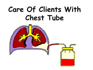
4. PURPOSE
• 1. To permit drainage of air and fluid
from the pleural cavity
• 2. To establish normal negative
pressure in the pleural cavity for lung
expansion
• 3. To equalize pressure on both sides
of the thoracic cavity
• 4. To provide continuous suction to
prevent tension pneumothorax
5. SITES FOR CHEST TUBE INSERTION
1. Thoracic surgery.
• 2 chest tube inserted – Anterior chest
tube & Posterior chest tube
2. Anterior chest :
• Upper/anterior chest wall
• Inserted in the 2nd intercostal space to
remove the air arising from the pleural
cavity
6. CON’T
3.Posterior chest tube :
• Placed at the posterior chest in the 8th
or 9th intercostal space at the mid-
axilllary line.
• Indication to remove serogeneous fluid
at the lower area of pleural cavity
• Diameter of tube in the lower section
is wider or longer compare to the
upper tube.
7. CON’T
4.Pneumothorax :
• Tube placed at
the 2nd or 3rd
intercostal space
along midclavicle
or anterior
axillary line.
8. TYPES OF CHEST DRAINAGE SYSTEM
Chest Drainage
System
1 bottle 2 bottles 3 bottles Pleurovac
13. FUNCTION OF PLEURAL
DRAINAGE SYSTEM
Inspiration
Intrapleural pressure
Air and fluid
move into bottle
Pleural space
becomes negative
Lungs reexpand
14. PRINCIPLES OF THE CHEST TUBE
1. Gravity
2. Under Water Seal
3. Suction
15. 1. Gravity
• Enhances flow
from high to low
• Chest drain is
placed below
client’s bed
16. 2. Water Seal
• Is a barrier to prevents backflow into
pleural space.
• Rod – depth determines the negative
pressure
• Air bubbles is released through the rod
• Air vent – to allow drained air to
escape to prevent pressure build up
17. 3. Suction
• Is a pull force
• MUST be in another bottle
• Purpose for the suction when :
- Gravity drainage is not enough.
- Patient’s respiration and cough are too
weak
- Air leak is fast into the pleural space
- Need to speed up removal from pleural
space
19. Pre-procedure care
1. Confirm :
• Open thoracotomy – during surgery
• Closed thoracotomy – at patient’s
bedside.
2. Inform patient
3. Check for consent
4. X-ray – with report to determine the
affected lung
24. DURING – INSERTION OF CHEST
TUBE
Procedure :
1. Chest tubes can be inserted in the ER,
client’s bedside,or in OT
2. In OT the chest tube is inserted via the
thoracotomy insertion.
3. In ER, client’s bedside the client is
placed in the sitting position or is
lying down with the affected side
elevated
25. CON’T
4. The area is prepared with antiseptic
solution, and the site is infiltrated with
a local anesthetic agent.
5. After a small incision is made, one or
two chest tubes are inserted into the
pleural space.
26. CON’T
6. One catheter is placed anteriorly
through the 2nd intercostal space; the
other is placed posteriorly through the
8th or 10th space to drain fluid and
blood.
7. The tubes are sutured to the chest
wall, and the puncture wound is
covered with an airtight dressing.
27. CON’T
8. During insertion, the tubes are kept
clamped
9. After the tubes are in place in the
pleural space, they are connected to
drainage tubing and pleural drainage
and clamp is removed.
10. Each tube may be connected to a
separate drainage system and suction.
28. CON’T
11. More commonly, a Y connecter is
used to attach both chest tubes to the
same drainage system.
29. DURING THE PROCEDURE –
NURSING RESPONSIBILITIES
1. Observe respiration.
2. Reduce anxiety
3. Monitor saturation
4. Prepare the under water seal
5. Connect the closed system fast
30. POST PROCEDURE CARE
1. Respiratory status :
- Vital signs (15 min x 1 hour,30 mins x 1
hour,1 hr x 4 hours)
- Respiration rate,pattern and rhythm
- Color, chest pain, rapid pulse.
- Check saturation
- Administer oxygen when necessary.
31. POST PROCEDURE CARE –
RESPIRATORY STATUS
2. Auscultate :
- Every 2 hours
- Listen for breath sound
- Listen for increased area of absent
breath sound
- Place patient in flowler’s or high
fowler’s.
32. POST PROCEDURE CARE –
ANXIETY
• Due to fear of pain and complication.
• Increase the need for oxygen
• Explain to the patient – care of tube,
the fluid drained and frequent checks.
• Pay attention to their needs.
• Allow relatives to stay.
33. POST PROCEDURE CARE –
WOUND STATUS
• Change the gauze when necessary
• Strict aseptic technique.
• Skin integrity – redness,swelling and
loose suture
34. POST PROCEDURE CARE –
TUBING
1. Intact and taped
2. Maintain patency.
- Check for obstruction
35. CON’T
1. Teach the patient on how to care for
the tubing.
- Place a pillow between patient and
tubing.
- Instruct the patient to cough if tube is
blocked
- Milking and stripping of the tube when
blocked
36. POST PROCEDURE CARE –
CLAMPS
• Use rubber tips.
• Clamped at the bedside.
• Clamping :
- During transfer
- Not > 1 min
- Upon doctor’s orders.
37. POST PROCEDURE CARE –
WATER SEAL
• Place below patient’s chest wall
• Fill with sterile water
• Rod must be immersed 2 cm in water
• Observe for the fluctuation of water
level.
38. CON’T
1. Fluctuation (tidaling)
• To ensure patency of system.
• Stops when :
- Lung is fully expanded (36-72 hours)
- When there is an obstruction
• Check for obstruction.
- Tubing – kinked
- Patients position
- Ask patient to take a deep breath and
cough
39. CON’T
2. Observe for bubbling :
- Intermittent bubbling is normal
- Continuous bubbling is abnormal.
- Check for
• Wound
• Tube
• Connection
- If rapid bubbling without air leak –
inform the doctor immediately
40. CON’T
3. Drainage output :
- 70 – 100 mls/hour
- Observe for change in drainage colour.
- Mark the amount.
• Mark the time of measurement and the fluid
level on the drainage chamber according to
the prescribed orders
• Marking intervals may range from once per
hour to every 8 hours.
• Any change in the quantity or characteristics
of drainage ( eg. Clear yellow to bloody )
should be reported to Dr.
41. CON’T
3. Drainage output (con’t)
- Document in the I/O chart
- Change bottle every 24 hours or when
full
42. POST PROCEDURE CARE –
SUCTION APPARATUS
1. Low suction pump :
- Must be controlled
- Suction valve / meter is inserted for wall
suction.
- Check for bubbling.
- If no bubbles :
• Clamp the chest tube to check for air
leaks
• Check the tubing and connection.
• Observe patient’s condition while chest
tube is clamped
43. POST PROCEDURE CARE –
SAFETY
1. Tube :
- Prevent kinking
- Place a pillow as a barrier
- Never clamp unnecessarily.
- Assist patient during ambulation the
first time
44. CON’T
2. Bottle :
- Bottles must be below chest level
- Keep bottle in a basin
- Inform relatives and housekeeping
regarding bottles
- Bed must be locked
- Activity should be limited to avoid injury
45. POST PROCEDURE CARE –
AMBULATION
- Explain to client
- Encourage change of position to
promote drainage.
- Can sit up, get in and out of bed.
- Stop the suction
- No need to clamp the tube.
- Maintain chest drain below chest wall.
46. POST PROCEDURE CARE –
DEEP BREATHING AND ARM
EXERCISE
1. On the 1st post op day.
2. When patient is not in severe pain
3. Enhances lung expansion – expels air
and fluid
47. CON’T
4. Prevents stiffness of the arm
5. Assist patient .
- Deep breathing exercise
- Support when patient is coughing
- Abdominal breathing
48. POST PROCEDURE CARE -
COMFORT
• Administer analgesic in the first 24 hours
• Allow position that is comfortable for the
patient.
• Assist patient in providing self-care
49. REMOVAL OF CHEST TUBE
• The chest tubes are removed when the
lungs are reexpanded and fluid
drainage.
• Assessment :
• - X-ray is done to check the progress
• - Clamp for 2 hours
• Chest tube is removed.
50. EMERGENCY CARE
1. Bleeding Post Chest tube insertion :
- Observe wound dressing
- Observe drainage
- Inform the surgeon immediately
51. CON’T
2. Dislodgement :
- From insertion site – place a gauze
immediately over the wound
- From connection – clamp the chest
tube immediately.
52. CON’T
3. Bottle breaks :
- Identify patient’s problem –
pneumothorax or hemothorax.
- Observe patient fortension
pneumothorax
- Place the tube in saline immediately.
- Unclamp immediately – prevent
respiratory distress