SlideShare a Scribd company logo
1 of 38
FRACTURAS DISTALES DE
       FÉMUR


 Dr. Carlos Alejandro Brambila Botello R2TYO
OBJETIVOS
•   INTRODUCCION

•   GENERALIDADES

•   REPASO ANATOMICO

•   MECANISMO DE LESION y LESIONES ASOCIADAS.

•   CLASIFICACION AO Y TRATAMIENTOS

•   TRATAMIENTOS ESPECIFICOS

•   COMPLICACIONES TARDIAS

•   CONCLUSION

•   PREGUNTAS.
INTRODUCCION

• Lafractura metafisaria distal del fémur es una fractura compleja
 que se puede presentar de forma intra articular o extra
 articular o en combinación y que se delimita hasta los 15cm
 arriba de la articulación de la rodilla, la mayoría de estas
 fracturas son de difícil manejo y siempre es un reto importante
 para cualquier cirujano ortopédico.
GENERALIDADES
• Constituyen   el 7% de todas las fracturas de
 fémur.

• Antesde la Osteosintesis se trataban de
 forma conservadora mediante la tracción
 esquelética y tenían buen resultado.

• Las
    desventajas: prolongada inmovilización y
 una rigidez de la rodilla.

• Actualmente la osteosintesis tiene el objetivo
 de una rapida MOVILIZACION y UNA
 BUENA RECONSTRUCCION ARTICULAR.
REPASO ANATOMICO

Aductor Mayor


                Gastrocnemios
ARTERIA FEMORAL
                  ANATOMIA              N. CIATICO


                      ARTERIA Y VENAS
                         POPITLEAS


                                                     N. PERONEO
                                                       COMÚN
MECANISMO DE LESION
• La mayoría son de alta energía a
 excepción de la que presentan los
 ancianos.

• Sepuede producir en varo, valgo o
 en caso de una rotación forzada
 con carga axial acompañada.

• La  mayoría de las fracturas se
 producen en accidentes de trafico y
 también se asocian a caídas de
 altura.
LESIONES ASOCIADAS
• Buscar:Fracturas del Acetabulo, cuello
 femoral y diafisis femoral.

• El
   varo o valgo forzado produce lesiones
 de los ligamentos de las rodillas.

• Se puede encontrar lesiones en meseta
 tibial.

• La lesión a tejidos superficiales
 secundario a una exposición.

• La   lesión vascular es poco frecuente.
CLASIFICACIÓN

                 ARTICULAR
            SIMPLE ó UNICONDILAR
 EXTRA-
ARTICULAR
                        ARTICULAR COMPLEJA
                           ó BICONDILEA
EXTRA-ARTICULARES
EXTRA-ARTICULARES
EXTRA-ARTICULARES
ARTICULAR
                  SIMPLE ó UNICONDILAR


CONDILO LATERAL
ARTICULAR
                 SIMPLE ó UNICONDILAR


CONDILO MEDIAL
ARTICULAR
SIMPLE ó UNICONDILAR
ARTICULAR COMPLEJA ó BICONDILEA
TRATAMIENTOS ESPECIFICOS
• En   las técnicas de fijación de
 implantes es indispensable el uso de
 clavillos Kirshner para la reducción
 articular.

• El uso de este implemento nos
 ayuda a utilizar tornillos canulados
 6.5 para reducir los fragmentos de
 fractura.
TORNILLO DE COMPRESIÓN DINÁMICA Y
PLACA CONDILEA


• INDICACIONESPARA COLOCAR UN DCS O UNA PLACA
 CONDILEA DE SOSTÉN

   • FRACTURAS   EXTRA-ARTICULARES COMPLEJAS

   •   FRACTURAS INTRA-ARTICULAR SIMPLE


       DCS

                    PLACA CONDILEA DE SOSTÉN
• A distal portion of the medial condyle should be intact for
• Intercondylar fractures*               9the DHS/DCStheScrew to gain goodby turning the handle clock-
                                               Insert Lag lag screw purchase.
• Supracondylar fractures*                                                         3 95° Condylar
                                         wise until thenot exist, a SYNTHESUsing the DCS Drill Guide, insert the central guide pin
                                          If these conditions do 0 mark on the assembly aligns with the


TECNICA PARA COLOCAR UN DCS
• Unicondylar fractures*
                                         lateral cortex. The threadedparallel to lag screw now lies in the A-P view, and
                                                                                   (C) tip of the the distal K-wire (A)
                                          Plate or Condylar Buttress Plate should be considered.

                                         10 mm from the medial parallelThe laganteriormay be (B) in the axial view. Do not
                                                                                   cortex. to the screw K-wire
                                                                                DCS Technique (Continued)
                                         inserted an additional 5 insert the guide pin too far medially; consider the inclination
                                                                                    mm in porotic bone, for increased
                                          Note: This procedure requires image intensification.
                                         holding power.                            of the medial wall of the distal femur. In the sagittal plane, guide pin, and
                                                                                                                                  4 Slide the Direct Measuring Device over the
                                          Surgical Technique                                                                      determine guide pin insertion depth. Calibration on the
                                                                                   the central guide pin enters the distal femur providespointreading.
                                          1 Reduce the fracture. The fracture can be temporarily                                  measuring device at a a direct
                is parallel to A
    A-P view: C s parallel to A          Note: withaDHS/DCS Guide Pins or anterior to thanmidline between the condyles, and in line
                                          stabilized If     lag screw 5 mm shorter the reaming and
                                                                                   Steinmann pins.

                                       DCS Technique
                                          Place these pins so they do not interfere with subsequent
                                         tappingofdepth implant assembly. (See illustrations 65 axis, approximately 2 cm from the knee joint.
                                          positioning the DCS is used (in thisthe shaft mm), insert it an
                                                                                   with case,
                                                                              (Continued)
                                         additional 5 3mm, pinsimplantConfirm In on the assembly aligns guide pin under image
                                          accompanying step for proper
                                          intercondylar fractures, the
                                                                        until thereplaced with
                                                                                        5 mark
                                                                                    positioning.)
                                                                            should be             placement of the central
                                         with the6.5 mm Cancellous Bone Screws with washers. If it is not parallel to the knee joint axis,
                                          independent lateral cortex.
                                                                                   intensification.


                                                                                                                                                                              DCS TECHNIQUE
                                                                                                                   DCS TECHNIQUE
  Axial view: C is parallel to B
                                                                                   insert a new DHS/DCS Guide Pin. joint fragments were not previously reduce
                                                                                                                        13 If the
                                         10 Before removing the assembly, align the handle so                           independent 6.5 mm Cancellous Bone Screws, the D
                                          2 is parallel with the femoral shaft
                                                                                   Notes: the axis when viewed
                                         it To determine direction of the central guide pin, flex
                                                                                                                                  8 Select the correct length DHS/DCS Lag Screw and
                                                                                                                        Compressing calculate reaming be inserted into the screw scre
                                                                                                                                         Screw may Insertion Assembly.                             lag
                                                                                                                                                            2
                                                                                                                                  assemble the Lag Screw depth, tapping depth (See “Assem-
                                          knee to 90°, and mark the axis of the knee joint by placing a it is designed for use with the DHS/DCS 20.) Slide theand lag
                                                                                        Because                                   5 To
                                         laterally. over the condyles (A).proper placement of the DCS porotic bone,thesubtractthe screw.reamed .hole..Seat .the80to avoid
                                          K-wire distally This allows Place a second K-wire                              Plate bling theguide pin and into the.thevery carefully Long
                                                                                                                                         insert 10 mm from
                                                                                                                                  length, Instrumentation,” page reading. For example:    assembly

                                         1
                                                                                                                                  over
                                         onto the lagcondyles (B).
                                          anteriorly over the
                                                               screw.              instruments and implants,ping the lag screwGuidehole .to. .center..and.. stabilize..the mm
                                                                                                                         the DHS/DCS Direct reading .. ..and.. .. . .. .. .. . 70 mm
                                                                                                                                  Centeringb. Reamerthe Pin, . . .
                                                                                                                                            a. thread.
                                                                                                                                             Sleeve in setting
                                                                                                                                                                       ..
                                                                                                                                  assembly.c. Tapping depth (optional) . . . . . . . . . . 70 mm
                                          Note: Placement of the DHS/DCS Guidenotdetermines
                                                                                    Pin an alternate pin, must be used.                         Lag screw length . . . . . . . . . . . . . . . . 70 mm*
                                                                                        This guide pin remains in place throughout Screw will be used, allow for additional
                                          placement of the DCS implant assembly. Misplacement of
                                          the guide pin can result in varus/valgus or rotational                                  Wrench while advancing theprocedure.
                                                                                                                                                        the lag screw.
                                                                                                                                  Note: Keep continuous forward pressure on the DHS/DCS
                                                                                                                                  *If the Compression
                  B                       malalignment of the fracture fragments.If it is inadvertently withdrawn, reinsert itofimmediately. a lag screw 5 mm
                                                                                                                                  compression the fracture by selecting
                                                                                                                                  shorter (in this case, 65 mm) and inserting it an additional
                                                                                   (See “Reinserting the DHS/DCS Guide Pin,” screw by turning the handle clock-
                                                                                                                                  5 mm.
                                                                                                                                  9 Insert the lag
                                                                                                                                                     page 21.)
                 A
                                                                                                                                             wise until the 0 mark on the assembly aligns with the9
                                                                                                                                             lateral cortex. The threaded tip of the lag screw now lies
                                                                                                                                             10 mm from the medial cortex. The lag screw may be
                                         3 Using the DCS Drill Guide, insert the central guide pin                                           inserted an additionalTriple Reamer. (See “Assembling the
                                                                                                                                             6 Assemble the DCS 5 mm in porotic bone, for increased
                                         (C) parallel to the distal K-wire (A) in the A-P view, and                                          holding power. page 18.) Set the reamer to the correct
                                                                                                                                             Instrumentation,”
                                         parallel to the anterior K-wire (B) in the axial view. Do not                                       depth. Insert the DCS Triple Reamer into the Power Drive
                                                                                                                                             Note: theaLarge Quick Coupling attachment. Slide and reamer
                                                                                                                                             using If lag screw 5 mm shorter than reaming the
                                         insert the guide pin too far medially; consider the inclination
                                         11 Remove the DHS/DCS Wrench and Long Centering                                           14 Further interfragmentary compression aligns
                                                                                                                                        10   tapping depth is used (in this case, 65 mm), the lag itcan be ac
                                                                                                                                             over the guide pin to simultaneously drill for insert screw,
                                                                                                                                                                                                     an


                                                                                                                                                                                                      3
                                         of the medial wall of the distal femur. In the sagittal plane,                                      additional 5 mm, until the 5 mark on the assembly
                                                                                                                                             ream for the plate barrel, and countersink for the plate/barrel
                                         Sleeve. Slide the appropriate DCS Plate onto the guide
                                         the central guide pin enters the distal femur at a point                                  by using twothe to the preset depth. When reaming in dense bone, throu
                                                                                                                                             with 6.5 mm Cancellous Bone Screws
                                                                                                                                             junction lateral cortex.
                   is parallel to A
       A-P view: C s parallel to A       anterior to the midline between the condyles, and in line
                                         shaft/lag screw assembly. Loosen and remove the Coupling
                                         with the shaft axis, approximately 2 cm from the knee joint.                              distal roundBefore removing the DCS Plate.theto prevent
                                                                                                                                             10
                                                                                                                                                    holes of the DCS Triplealign handle so
                                                                                                                                             continuously irrigate
                                                                                                                                                                    the assembly,
                                                                                                                                                                                   Reamer
                                                                                                                                             thermal necrosis.
                                         Confirm placement of the central guide pin under image                                              it is parallel with the femoral shaft axis when viewed
                                         Screw and Guide Shaft. Use the Power Drive in reverse,
                                         intensification. If it is not parallel to the knee joint axis,                                      laterally. This allows proper placement of the DCS Plate
                                         insert a new DHS/DCS Guide Pin.
      Axial view: C is parallel to B     with the Jacobs Chuck attachment, to withdraw the guide
                                         Notes:
                                                                                                                                             onto the lag screw.

                                         pin.Because it is designed for use with the DHS/DCS
                                         instruments and implants, the DHS/DCS Guide Pin, and
                                                                                                                                                7 If necessary, use the Tap Assembly to tap to the
                                         not an alternate pin, must be used.
                                                                                                                                                predetermined depth, which can be seen through the window
                                             This guide pin remains in place throughout the procedure.
                                                                                                                                                in the Short Centering Sleeve. (See “Assembling the
                                         If it is inadvertently withdrawn, reinsert it immediately.                                             Instrumentation,” page 19.)
                                         (See “Reinserting the DHS/DCS Guide Pin,” page 21.)
                                                                                                           9
                                                                                                                                                11 Remove the DHS/DCS Wrench and Long Centering
                                                                                                                                                Sleeve. Slide the appropriate DCS Plate onto the guide
                                                                                                                                                shaft/lag screw assembly. Loosen and remove the Coupling
TECNICA PARA COLOCAR UNA
PLACA CONDILEA DE SOSTÉN
ENCLAVADO ENDOMEDULAR
      RETROGRADO
•   ESTA INDICADO EN FRACTURAS EXTRA-ARTICULARES (33-
    A) ó EN OCASIONES FRACTURAS ARTICULARES SIMPLES
    (33-C1-33-C2)

•   DESVENTAJAS: UNA REDUCCION ARTICULAR PB DEFICIENTE
FIJACION EXTERNA CON PONTEO TEMPORAL.


tornillo y el clavo de Steinmann. A continuación, reduzca la
fractura tirando longitudinalmente con una ligamentotaxis ba-
lanceada. Seguidamente, introduzca dos tornillos en la
tibia, comenzando desde la barra medial. Para la profilaxis del
pie equino, introduzca, en ángulo desde encima, otro tornillo de
                                                                                             cerradas con daño grave a los tejidos blandos


Schanz en el primer y quinto huesos metatarsianos.
Puente de la articulación de la rodilla
Introduzca dos tornillos de Schanz en el fémur distal, desde una
dirección lateral o ventral, y en la porción proximal de la tibia,
desde una dirección anteromedial. Conéctelos mediante la téc-
nica barra-barra.
               CUANDO?


                                                          Politraumatizados.

                                                                               expuestas.
                                        • INDICACIONES:

                                                          • Pacientes

                                                                               • Fracturas

                                                                                             • Fracturas
COMPLICACIONES TARDIAS

• PSEUDOARTROSIS

• ARTROSIS TEMPRANA   DE LA RODILLA

• RIGIDEZ

• DEFORMIDADES
             LINEARES COMO GENOVARO O
 GENORECURVATUM
PREGUNTAS

•   1.- A QUE SEGMENTO DE LA CLASIFICACION AO
    CORRESPONDEN LAS FRACTURAS DE FEMUR
    DISTAL ?

     a)   32.A
     b)   33.A
     c)   34.B
     d)   35.C
PREGUNTAS

•   1.- A QUE SEGMENTO DE LA CLASIFICACION AO
    CORRESPONDEN LAS FRACTURAS DE FEMUR
    DISTAL ?

     a)   32.A
     b)   33.A
     c)   34.B
     d)   35.C
PREGUNTAS

•   2.- CUALES SON LOS 2 GRANDES MUSCULOS QUE
    SE INSERTAN EN LA PARTE POSTERIOR DEL FEMUR
    DISTAL?

     a)   ISQUIOTIBIAL Y SEMIMEMBRANOSO
     b)   SEMIMEMBRANOS Y SEMITENDINOSO
     c)   GRACIL Y SARTORIO
     d)   GASTROCNEMIOS
PREGUNTAS

•   2.- CUALES SON LOS 2 GRANDES MUSCULOS QUE
    SE INSERTAN EN LA PARTE POSTERIOR DEL FEMUR
    DISTAL?

     a)   ISQUIOTIBIAL Y SEMIMEMBRANOSO
     b)   SEMIMEMBRANOS Y SEMITENDINOSO
     c)   GRACIL Y SARTORIO
     d)   GASTROCNEMIOS
PREGUNTAS

•   3.- CUAL ES EL TRATAMIENTO DE ELECCION PARA
    LA FRACTURA 33B1.1?

     a)   COLOCACION DE PLACA CONDILEA DE SOSTÉN
     b)   COLOCACION DE CLAVO DINAMICO DE CONDILO
     c)   COLOCACION DE TORNILLO DE COMPRESION
     d)   ENCLAVADO ENDOMEDULAR
PREGUNTAS

•   3.- CUAL ES EL TRATAMIENTO DE ELECCION PARA
    LA FRACTURA 33B1.1?

     a)   COLOCACION DE PLACA CONDILEA DE SOSTÉN
     b)   COLOCACION DE CLAVO DINAMICO DE CONDILO
     c)   COLOCACION DE TORNILLO DE COMPRESION
     d)   ENCLAVADO ENDOMEDULAR
PREGUNTAS


4.- CUAL ES EL IMPLANTE DE ELECCION EL LAS
FRACTURAS EXTRA-ARTICULES MULTIFRAGMETARIAS
EN FEMUR DISTAL?
  a)COLOCACION DE PLACA CONDILEA DE SOSTEN
  b)COLOCACION DE CLAVO DINAMICO DE CONDILO
  c)COLOCACION DE TORNILLO DE COMPRESION
  d)ENCLAVADO ENDOMEDULAR
PREGUNTAS


4.- CUAL ES EL IMPLANTE DE ELECCION EL LAS
FRACTURAS EXTRA-ARTICULES MULTIFRAGMETARIAS
EN FEMUR DISTAL?
  a)COLOCACION DE PLACA CONDILEA DE SOSTEN
  b)COLOCACION DE CLAVO DINAMICO DE CONDILO
  c)COLOCACION DE TORNILLO DE COMPRESION
  d)ENCLAVADO ENDOMEDULAR
PREGUNTAS

•   5.- CUANDO ESTA INDICADO EL FIJADOR EXTERNO
    DE PONTEO EN LAS FRACTURAS DISTALES DE
    FEMUR ?

     a)   AO: IC1,MT1,NV0
     b)   AO: IC3,MT3,NV3
     c)   AO: IO1,MT1,NV0
     d)   SOLO B Y C SON CORRECTAS.
PREGUNTAS

•   5.- CUANDO ESTA INDICADO EL FIJADOR EXTERNO
    DE PONTEO EN LAS FRACTURAS DISTALES DE
    FEMUR ?

     a)   AO: IC1,MT1,NV0
     b)   AO: IC3,MT3,NV3
     c)   AO: IO1,MT1,NV0
     d)   SOLO B Y C SON CORRECTAS.

More Related Content

What's hot

Fractura de astragalo
Fractura de astragaloFractura de astragalo
Fractura de astragaloJuanjo Targa
 
Fractura de meseta tibial
Fractura de meseta tibialFractura de meseta tibial
Fractura de meseta tibialEfrén Quintero
 
Fx cadera
Fx caderaFx cadera
Fx caderatopaken
 
Fracturas de meseta tibial
Fracturas de meseta tibialFracturas de meseta tibial
Fracturas de meseta tibialyaneira varela
 
Fracturas de muñecayenny
Fracturas de muñecayennyFracturas de muñecayenny
Fracturas de muñecayennygfmb100
 
FRACTURA DE ROTULA
FRACTURA DE ROTULAFRACTURA DE ROTULA
FRACTURA DE ROTULAoytkinesio
 
Fractura de Muñeca
Fractura de MuñecaFractura de Muñeca
Fractura de MuñecaJuanjo Targa
 
Fractura de femur distal
Fractura de femur distalFractura de femur distal
Fractura de femur distalCAMILA AZOCAR
 
Fracturas de cadera
Fracturas de caderaFracturas de cadera
Fracturas de caderajdelvallea
 
Fracturas de muñeca y mano um 2011
Fracturas de muñeca y mano um 2011Fracturas de muñeca y mano um 2011
Fracturas de muñeca y mano um 2011jdelvallea
 
principios de osteosintesis
principios de osteosintesisprincipios de osteosintesis
principios de osteosintesiselmer narvaez
 

What's hot (20)

Fracturas del tercio proximal del humero
Fracturas del tercio proximal del humeroFracturas del tercio proximal del humero
Fracturas del tercio proximal del humero
 
Fx femur distal
Fx femur distalFx femur distal
Fx femur distal
 
Fractura de astragalo
Fractura de astragaloFractura de astragalo
Fractura de astragalo
 
Fractura de meseta tibial
Fractura de meseta tibialFractura de meseta tibial
Fractura de meseta tibial
 
Fx cadera
Fx caderaFx cadera
Fx cadera
 
Fracturas de meseta tibial
Fracturas de meseta tibialFracturas de meseta tibial
Fracturas de meseta tibial
 
Fracturas de muñecayenny
Fracturas de muñecayennyFracturas de muñecayenny
Fracturas de muñecayenny
 
Luxación traumática de la rodilla y de Rótula
Luxación traumática de la rodilla y de RótulaLuxación traumática de la rodilla y de Rótula
Luxación traumática de la rodilla y de Rótula
 
FRACTURA DE ROTULA
FRACTURA DE ROTULAFRACTURA DE ROTULA
FRACTURA DE ROTULA
 
Fractura De Cadera
Fractura De CaderaFractura De Cadera
Fractura De Cadera
 
Pseudoartrosis
PseudoartrosisPseudoartrosis
Pseudoartrosis
 
Fractura de Muñeca
Fractura de MuñecaFractura de Muñeca
Fractura de Muñeca
 
Fracturas de la mano
Fracturas de la manoFracturas de la mano
Fracturas de la mano
 
Fracturas del radio distal
Fracturas del radio distalFracturas del radio distal
Fracturas del radio distal
 
Fractura de femur distal
Fractura de femur distalFractura de femur distal
Fractura de femur distal
 
Fracturas de cadera
Fracturas de caderaFracturas de cadera
Fracturas de cadera
 
Fracturas de muñeca y mano um 2011
Fracturas de muñeca y mano um 2011Fracturas de muñeca y mano um 2011
Fracturas de muñeca y mano um 2011
 
principios de osteosintesis
principios de osteosintesisprincipios de osteosintesis
principios de osteosintesis
 
Fractura de muñeca
Fractura de muñecaFractura de muñeca
Fractura de muñeca
 
Amputaciones del pie
Amputaciones del pieAmputaciones del pie
Amputaciones del pie
 

Viewers also liked

Viewers also liked (19)

Fracturas femur distal
Fracturas femur distal Fracturas femur distal
Fracturas femur distal
 
Fractura de diafisis femoral
Fractura de diafisis femoralFractura de diafisis femoral
Fractura de diafisis femoral
 
Fracturas supracondileas
Fracturas supracondileasFracturas supracondileas
Fracturas supracondileas
 
Fractura del femur distal
Fractura del femur distalFractura del femur distal
Fractura del femur distal
 
Fracturas de Diáfisis femoral
Fracturas de Diáfisis femoralFracturas de Diáfisis femoral
Fracturas de Diáfisis femoral
 
Fractura de diáfisis femoral
Fractura de diáfisis femoralFractura de diáfisis femoral
Fractura de diáfisis femoral
 
Fractura Del Extremo Distal Y Diafisis Del Femur Walter
Fractura Del Extremo Distal Y Diafisis Del Femur WalterFractura Del Extremo Distal Y Diafisis Del Femur Walter
Fractura Del Extremo Distal Y Diafisis Del Femur Walter
 
Lesiones de pie en el deportista pptx
Lesiones de pie en el deportista pptxLesiones de pie en el deportista pptx
Lesiones de pie en el deportista pptx
 
Esguince
EsguinceEsguince
Esguince
 
Esguinces y luxaciones 2010 [modo de compatibilidad]
Esguinces y luxaciones 2010 [modo de compatibilidad]Esguinces y luxaciones 2010 [modo de compatibilidad]
Esguinces y luxaciones 2010 [modo de compatibilidad]
 
Traumatismo del miembro inferior y superior
Traumatismo del miembro inferior y superiorTraumatismo del miembro inferior y superior
Traumatismo del miembro inferior y superior
 
Esguince de tobillo. 2 modulo loterzo
Esguince de tobillo. 2 modulo loterzoEsguince de tobillo. 2 modulo loterzo
Esguince de tobillo. 2 modulo loterzo
 
Esguinces en traumatologia
Esguinces en traumatologiaEsguinces en traumatologia
Esguinces en traumatologia
 
Fractura diáfisis femoral
Fractura diáfisis femoralFractura diáfisis femoral
Fractura diáfisis femoral
 
esguince
esguinceesguince
esguince
 
Fracturas, esguinces y luxaciones
Fracturas, esguinces y luxacionesFracturas, esguinces y luxaciones
Fracturas, esguinces y luxaciones
 
Líquido sinovial
Líquido sinovialLíquido sinovial
Líquido sinovial
 
Articulaciones del miembro inferior
Articulaciones del miembro inferiorArticulaciones del miembro inferior
Articulaciones del miembro inferior
 
esguinces
esguincesesguinces
esguinces
 

Similar to Fracturas de Femur Distal

8 use of pythagorean theorem # 9 single implant case
8 use of pythagorean theorem # 9 single implant case8 use of pythagorean theorem # 9 single implant case
8 use of pythagorean theorem # 9 single implant caseDePlaque
 
Surgical technique for gama nail
Surgical technique for gama nailSurgical technique for gama nail
Surgical technique for gama nailorthopedic Implant
 
Trochanteric and subtrochanteric non union dr mahmoud hadhoud
Trochanteric and subtrochanteric non union   dr mahmoud hadhoudTrochanteric and subtrochanteric non union   dr mahmoud hadhoud
Trochanteric and subtrochanteric non union dr mahmoud hadhoudMahmoud Hadhoud
 
Dvr digital radius
Dvr digital radiusDvr digital radius
Dvr digital radiusmaruchan-doc
 
Lt0520 d peek bio-pushlock knotless anchor for bankart repair
Lt0520 d   peek bio-pushlock knotless anchor for bankart repairLt0520 d   peek bio-pushlock knotless anchor for bankart repair
Lt0520 d peek bio-pushlock knotless anchor for bankart repairdrnaula
 
Freedom pck revision knee system by meril life
Freedom pck revision knee system by meril lifeFreedom pck revision knee system by meril life
Freedom pck revision knee system by meril lifeRajRathore36
 
200330 Controversies in the intramedullary nailing of tibia
200330 Controversies in the intramedullary nailing of tibia200330 Controversies in the intramedullary nailing of tibia
200330 Controversies in the intramedullary nailing of tibiaDr MADAN MOHAN
 
Intra-articular Distal Radius Fractures
Intra-articular Distal Radius FracturesIntra-articular Distal Radius Fractures
Intra-articular Distal Radius FracturesArun Shanbhag
 
Lt0460 b achilles suturebridge
Lt0460 b   achilles suturebridgeLt0460 b   achilles suturebridge
Lt0460 b achilles suturebridgedrnaula
 
MENISCUS SURGERY.pptx
MENISCUS SURGERY.pptxMENISCUS SURGERY.pptx
MENISCUS SURGERY.pptxRitesh Gupta
 
Pearls and pitfalls with im nailing of proximal tibia fractures
Pearls and pitfalls with im nailing of proximal tibia fracturesPearls and pitfalls with im nailing of proximal tibia fractures
Pearls and pitfalls with im nailing of proximal tibia fracturesBipulBorthakur
 
Instability in TKR
Instability in TKRInstability in TKR
Instability in TKRRishi Poudel
 
Fracture of the distal radius
Fracture of the distal radiusFracture of the distal radius
Fracture of the distal radiusMd Ashiqur Rahman
 
200331 Evolving concepts in treatment of supracondylar fractures in children
200331 Evolving concepts in treatment of supracondylar fractures in children200331 Evolving concepts in treatment of supracondylar fractures in children
200331 Evolving concepts in treatment of supracondylar fractures in childrenDr MADAN MOHAN
 
The Indirect Ophthalmoscope
The Indirect OphthalmoscopeThe Indirect Ophthalmoscope
The Indirect OphthalmoscopeDrAbdelLatifsiam
 

Similar to Fracturas de Femur Distal (20)

8 use of pythagorean theorem # 9 single implant case
8 use of pythagorean theorem # 9 single implant case8 use of pythagorean theorem # 9 single implant case
8 use of pythagorean theorem # 9 single implant case
 
Surgical technique for gama nail
Surgical technique for gama nailSurgical technique for gama nail
Surgical technique for gama nail
 
Trochanteric and subtrochanteric non union dr mahmoud hadhoud
Trochanteric and subtrochanteric non union   dr mahmoud hadhoudTrochanteric and subtrochanteric non union   dr mahmoud hadhoud
Trochanteric and subtrochanteric non union dr mahmoud hadhoud
 
Dvr digital radius
Dvr digital radiusDvr digital radius
Dvr digital radius
 
PPT.pptx
PPT.pptxPPT.pptx
PPT.pptx
 
Lt0520 d peek bio-pushlock knotless anchor for bankart repair
Lt0520 d   peek bio-pushlock knotless anchor for bankart repairLt0520 d   peek bio-pushlock knotless anchor for bankart repair
Lt0520 d peek bio-pushlock knotless anchor for bankart repair
 
Fractures of the olecranon
Fractures of the olecranonFractures of the olecranon
Fractures of the olecranon
 
Freedom pck revision knee system by meril life
Freedom pck revision knee system by meril lifeFreedom pck revision knee system by meril life
Freedom pck revision knee system by meril life
 
200330 Controversies in the intramedullary nailing of tibia
200330 Controversies in the intramedullary nailing of tibia200330 Controversies in the intramedullary nailing of tibia
200330 Controversies in the intramedullary nailing of tibia
 
Lima 2013 final
Lima 2013 finalLima 2013 final
Lima 2013 final
 
Intra-articular Distal Radius Fractures
Intra-articular Distal Radius FracturesIntra-articular Distal Radius Fractures
Intra-articular Distal Radius Fractures
 
103330.pdf
103330.pdf103330.pdf
103330.pdf
 
Lt0460 b achilles suturebridge
Lt0460 b   achilles suturebridgeLt0460 b   achilles suturebridge
Lt0460 b achilles suturebridge
 
MENISCUS SURGERY.pptx
MENISCUS SURGERY.pptxMENISCUS SURGERY.pptx
MENISCUS SURGERY.pptx
 
Pearls and pitfalls with im nailing of proximal tibia fractures
Pearls and pitfalls with im nailing of proximal tibia fracturesPearls and pitfalls with im nailing of proximal tibia fractures
Pearls and pitfalls with im nailing of proximal tibia fractures
 
PFNA NAW.pptx
PFNA NAW.pptxPFNA NAW.pptx
PFNA NAW.pptx
 
Instability in TKR
Instability in TKRInstability in TKR
Instability in TKR
 
Fracture of the distal radius
Fracture of the distal radiusFracture of the distal radius
Fracture of the distal radius
 
200331 Evolving concepts in treatment of supracondylar fractures in children
200331 Evolving concepts in treatment of supracondylar fractures in children200331 Evolving concepts in treatment of supracondylar fractures in children
200331 Evolving concepts in treatment of supracondylar fractures in children
 
The Indirect Ophthalmoscope
The Indirect OphthalmoscopeThe Indirect Ophthalmoscope
The Indirect Ophthalmoscope
 

Fracturas de Femur Distal

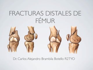
  • 1. FRACTURAS DISTALES DE FÉMUR Dr. Carlos Alejandro Brambila Botello R2TYO
  • 2. OBJETIVOS • INTRODUCCION • GENERALIDADES • REPASO ANATOMICO • MECANISMO DE LESION y LESIONES ASOCIADAS. • CLASIFICACION AO Y TRATAMIENTOS • TRATAMIENTOS ESPECIFICOS • COMPLICACIONES TARDIAS • CONCLUSION • PREGUNTAS.
  • 3. INTRODUCCION • Lafractura metafisaria distal del fémur es una fractura compleja que se puede presentar de forma intra articular o extra articular o en combinación y que se delimita hasta los 15cm arriba de la articulación de la rodilla, la mayoría de estas fracturas son de difícil manejo y siempre es un reto importante para cualquier cirujano ortopédico.
  • 4. GENERALIDADES • Constituyen el 7% de todas las fracturas de fémur. • Antesde la Osteosintesis se trataban de forma conservadora mediante la tracción esquelética y tenían buen resultado. • Las desventajas: prolongada inmovilización y una rigidez de la rodilla. • Actualmente la osteosintesis tiene el objetivo de una rapida MOVILIZACION y UNA BUENA RECONSTRUCCION ARTICULAR.
  • 6. ARTERIA FEMORAL ANATOMIA N. CIATICO ARTERIA Y VENAS POPITLEAS N. PERONEO COMÚN
  • 7. MECANISMO DE LESION • La mayoría son de alta energía a excepción de la que presentan los ancianos. • Sepuede producir en varo, valgo o en caso de una rotación forzada con carga axial acompañada. • La mayoría de las fracturas se producen en accidentes de trafico y también se asocian a caídas de altura.
  • 8. LESIONES ASOCIADAS • Buscar:Fracturas del Acetabulo, cuello femoral y diafisis femoral. • El varo o valgo forzado produce lesiones de los ligamentos de las rodillas. • Se puede encontrar lesiones en meseta tibial. • La lesión a tejidos superficiales secundario a una exposición. • La lesión vascular es poco frecuente.
  • 9. CLASIFICACIÓN ARTICULAR SIMPLE ó UNICONDILAR EXTRA- ARTICULAR ARTICULAR COMPLEJA ó BICONDILEA
  • 13. ARTICULAR SIMPLE ó UNICONDILAR CONDILO LATERAL
  • 14. ARTICULAR SIMPLE ó UNICONDILAR CONDILO MEDIAL
  • 16. ARTICULAR COMPLEJA ó BICONDILEA
  • 17. ARTICULAR COMPLEJA ó BICONDILEA
  • 18. ARTICULAR COMPLEJA ó BICONDILEA
  • 19.
  • 20.
  • 21.
  • 22. TRATAMIENTOS ESPECIFICOS • En las técnicas de fijación de implantes es indispensable el uso de clavillos Kirshner para la reducción articular. • El uso de este implemento nos ayuda a utilizar tornillos canulados 6.5 para reducir los fragmentos de fractura.
  • 23. TORNILLO DE COMPRESIÓN DINÁMICA Y PLACA CONDILEA • INDICACIONESPARA COLOCAR UN DCS O UNA PLACA CONDILEA DE SOSTÉN • FRACTURAS EXTRA-ARTICULARES COMPLEJAS • FRACTURAS INTRA-ARTICULAR SIMPLE DCS PLACA CONDILEA DE SOSTÉN
  • 24. • A distal portion of the medial condyle should be intact for • Intercondylar fractures* 9the DHS/DCStheScrew to gain goodby turning the handle clock- Insert Lag lag screw purchase. • Supracondylar fractures* 3 95° Condylar wise until thenot exist, a SYNTHESUsing the DCS Drill Guide, insert the central guide pin If these conditions do 0 mark on the assembly aligns with the TECNICA PARA COLOCAR UN DCS • Unicondylar fractures* lateral cortex. The threadedparallel to lag screw now lies in the A-P view, and (C) tip of the the distal K-wire (A) Plate or Condylar Buttress Plate should be considered. 10 mm from the medial parallelThe laganteriormay be (B) in the axial view. Do not cortex. to the screw K-wire DCS Technique (Continued) inserted an additional 5 insert the guide pin too far medially; consider the inclination mm in porotic bone, for increased Note: This procedure requires image intensification. holding power. of the medial wall of the distal femur. In the sagittal plane, guide pin, and 4 Slide the Direct Measuring Device over the Surgical Technique determine guide pin insertion depth. Calibration on the the central guide pin enters the distal femur providespointreading. 1 Reduce the fracture. The fracture can be temporarily measuring device at a a direct is parallel to A A-P view: C s parallel to A Note: withaDHS/DCS Guide Pins or anterior to thanmidline between the condyles, and in line stabilized If lag screw 5 mm shorter the reaming and Steinmann pins. DCS Technique Place these pins so they do not interfere with subsequent tappingofdepth implant assembly. (See illustrations 65 axis, approximately 2 cm from the knee joint. positioning the DCS is used (in thisthe shaft mm), insert it an with case, (Continued) additional 5 3mm, pinsimplantConfirm In on the assembly aligns guide pin under image accompanying step for proper intercondylar fractures, the until thereplaced with 5 mark positioning.) should be placement of the central with the6.5 mm Cancellous Bone Screws with washers. If it is not parallel to the knee joint axis, independent lateral cortex. intensification. DCS TECHNIQUE DCS TECHNIQUE Axial view: C is parallel to B insert a new DHS/DCS Guide Pin. joint fragments were not previously reduce 13 If the 10 Before removing the assembly, align the handle so independent 6.5 mm Cancellous Bone Screws, the D 2 is parallel with the femoral shaft Notes: the axis when viewed it To determine direction of the central guide pin, flex 8 Select the correct length DHS/DCS Lag Screw and Compressing calculate reaming be inserted into the screw scre Screw may Insertion Assembly. lag 2 assemble the Lag Screw depth, tapping depth (See “Assem- knee to 90°, and mark the axis of the knee joint by placing a it is designed for use with the DHS/DCS 20.) Slide theand lag Because 5 To laterally. over the condyles (A).proper placement of the DCS porotic bone,thesubtractthe screw.reamed .hole..Seat .the80to avoid K-wire distally This allows Place a second K-wire Plate bling theguide pin and into the.thevery carefully Long insert 10 mm from length, Instrumentation,” page reading. For example: assembly 1 over onto the lagcondyles (B). anteriorly over the screw. instruments and implants,ping the lag screwGuidehole .to. .center..and.. stabilize..the mm the DHS/DCS Direct reading .. ..and.. .. . .. .. .. . 70 mm Centeringb. Reamerthe Pin, . . . a. thread. Sleeve in setting .. assembly.c. Tapping depth (optional) . . . . . . . . . . 70 mm Note: Placement of the DHS/DCS Guidenotdetermines Pin an alternate pin, must be used. Lag screw length . . . . . . . . . . . . . . . . 70 mm* This guide pin remains in place throughout Screw will be used, allow for additional placement of the DCS implant assembly. Misplacement of the guide pin can result in varus/valgus or rotational Wrench while advancing theprocedure. the lag screw. Note: Keep continuous forward pressure on the DHS/DCS *If the Compression B malalignment of the fracture fragments.If it is inadvertently withdrawn, reinsert itofimmediately. a lag screw 5 mm compression the fracture by selecting shorter (in this case, 65 mm) and inserting it an additional (See “Reinserting the DHS/DCS Guide Pin,” screw by turning the handle clock- 5 mm. 9 Insert the lag page 21.) A wise until the 0 mark on the assembly aligns with the9 lateral cortex. The threaded tip of the lag screw now lies 10 mm from the medial cortex. The lag screw may be 3 Using the DCS Drill Guide, insert the central guide pin inserted an additionalTriple Reamer. (See “Assembling the 6 Assemble the DCS 5 mm in porotic bone, for increased (C) parallel to the distal K-wire (A) in the A-P view, and holding power. page 18.) Set the reamer to the correct Instrumentation,” parallel to the anterior K-wire (B) in the axial view. Do not depth. Insert the DCS Triple Reamer into the Power Drive Note: theaLarge Quick Coupling attachment. Slide and reamer using If lag screw 5 mm shorter than reaming the insert the guide pin too far medially; consider the inclination 11 Remove the DHS/DCS Wrench and Long Centering 14 Further interfragmentary compression aligns 10 tapping depth is used (in this case, 65 mm), the lag itcan be ac over the guide pin to simultaneously drill for insert screw, an 3 of the medial wall of the distal femur. In the sagittal plane, additional 5 mm, until the 5 mark on the assembly ream for the plate barrel, and countersink for the plate/barrel Sleeve. Slide the appropriate DCS Plate onto the guide the central guide pin enters the distal femur at a point by using twothe to the preset depth. When reaming in dense bone, throu with 6.5 mm Cancellous Bone Screws junction lateral cortex. is parallel to A A-P view: C s parallel to A anterior to the midline between the condyles, and in line shaft/lag screw assembly. Loosen and remove the Coupling with the shaft axis, approximately 2 cm from the knee joint. distal roundBefore removing the DCS Plate.theto prevent 10 holes of the DCS Triplealign handle so continuously irrigate the assembly, Reamer thermal necrosis. Confirm placement of the central guide pin under image it is parallel with the femoral shaft axis when viewed Screw and Guide Shaft. Use the Power Drive in reverse, intensification. If it is not parallel to the knee joint axis, laterally. This allows proper placement of the DCS Plate insert a new DHS/DCS Guide Pin. Axial view: C is parallel to B with the Jacobs Chuck attachment, to withdraw the guide Notes: onto the lag screw. pin.Because it is designed for use with the DHS/DCS instruments and implants, the DHS/DCS Guide Pin, and 7 If necessary, use the Tap Assembly to tap to the not an alternate pin, must be used. predetermined depth, which can be seen through the window This guide pin remains in place throughout the procedure. in the Short Centering Sleeve. (See “Assembling the If it is inadvertently withdrawn, reinsert it immediately. Instrumentation,” page 19.) (See “Reinserting the DHS/DCS Guide Pin,” page 21.) 9 11 Remove the DHS/DCS Wrench and Long Centering Sleeve. Slide the appropriate DCS Plate onto the guide shaft/lag screw assembly. Loosen and remove the Coupling
  • 25. TECNICA PARA COLOCAR UNA PLACA CONDILEA DE SOSTÉN
  • 26. ENCLAVADO ENDOMEDULAR RETROGRADO • ESTA INDICADO EN FRACTURAS EXTRA-ARTICULARES (33- A) ó EN OCASIONES FRACTURAS ARTICULARES SIMPLES (33-C1-33-C2) • DESVENTAJAS: UNA REDUCCION ARTICULAR PB DEFICIENTE
  • 27. FIJACION EXTERNA CON PONTEO TEMPORAL. tornillo y el clavo de Steinmann. A continuación, reduzca la fractura tirando longitudinalmente con una ligamentotaxis ba- lanceada. Seguidamente, introduzca dos tornillos en la tibia, comenzando desde la barra medial. Para la profilaxis del pie equino, introduzca, en ángulo desde encima, otro tornillo de cerradas con daño grave a los tejidos blandos Schanz en el primer y quinto huesos metatarsianos. Puente de la articulación de la rodilla Introduzca dos tornillos de Schanz en el fémur distal, desde una dirección lateral o ventral, y en la porción proximal de la tibia, desde una dirección anteromedial. Conéctelos mediante la téc- nica barra-barra. CUANDO? Politraumatizados. expuestas. • INDICACIONES: • Pacientes • Fracturas • Fracturas
  • 28. COMPLICACIONES TARDIAS • PSEUDOARTROSIS • ARTROSIS TEMPRANA DE LA RODILLA • RIGIDEZ • DEFORMIDADES LINEARES COMO GENOVARO O GENORECURVATUM
  • 29. PREGUNTAS • 1.- A QUE SEGMENTO DE LA CLASIFICACION AO CORRESPONDEN LAS FRACTURAS DE FEMUR DISTAL ? a) 32.A b) 33.A c) 34.B d) 35.C
  • 30. PREGUNTAS • 1.- A QUE SEGMENTO DE LA CLASIFICACION AO CORRESPONDEN LAS FRACTURAS DE FEMUR DISTAL ? a) 32.A b) 33.A c) 34.B d) 35.C
  • 31. PREGUNTAS • 2.- CUALES SON LOS 2 GRANDES MUSCULOS QUE SE INSERTAN EN LA PARTE POSTERIOR DEL FEMUR DISTAL? a) ISQUIOTIBIAL Y SEMIMEMBRANOSO b) SEMIMEMBRANOS Y SEMITENDINOSO c) GRACIL Y SARTORIO d) GASTROCNEMIOS
  • 32. PREGUNTAS • 2.- CUALES SON LOS 2 GRANDES MUSCULOS QUE SE INSERTAN EN LA PARTE POSTERIOR DEL FEMUR DISTAL? a) ISQUIOTIBIAL Y SEMIMEMBRANOSO b) SEMIMEMBRANOS Y SEMITENDINOSO c) GRACIL Y SARTORIO d) GASTROCNEMIOS
  • 33. PREGUNTAS • 3.- CUAL ES EL TRATAMIENTO DE ELECCION PARA LA FRACTURA 33B1.1? a) COLOCACION DE PLACA CONDILEA DE SOSTÉN b) COLOCACION DE CLAVO DINAMICO DE CONDILO c) COLOCACION DE TORNILLO DE COMPRESION d) ENCLAVADO ENDOMEDULAR
  • 34. PREGUNTAS • 3.- CUAL ES EL TRATAMIENTO DE ELECCION PARA LA FRACTURA 33B1.1? a) COLOCACION DE PLACA CONDILEA DE SOSTÉN b) COLOCACION DE CLAVO DINAMICO DE CONDILO c) COLOCACION DE TORNILLO DE COMPRESION d) ENCLAVADO ENDOMEDULAR
  • 35. PREGUNTAS 4.- CUAL ES EL IMPLANTE DE ELECCION EL LAS FRACTURAS EXTRA-ARTICULES MULTIFRAGMETARIAS EN FEMUR DISTAL? a)COLOCACION DE PLACA CONDILEA DE SOSTEN b)COLOCACION DE CLAVO DINAMICO DE CONDILO c)COLOCACION DE TORNILLO DE COMPRESION d)ENCLAVADO ENDOMEDULAR
  • 36. PREGUNTAS 4.- CUAL ES EL IMPLANTE DE ELECCION EL LAS FRACTURAS EXTRA-ARTICULES MULTIFRAGMETARIAS EN FEMUR DISTAL? a)COLOCACION DE PLACA CONDILEA DE SOSTEN b)COLOCACION DE CLAVO DINAMICO DE CONDILO c)COLOCACION DE TORNILLO DE COMPRESION d)ENCLAVADO ENDOMEDULAR
  • 37. PREGUNTAS • 5.- CUANDO ESTA INDICADO EL FIJADOR EXTERNO DE PONTEO EN LAS FRACTURAS DISTALES DE FEMUR ? a) AO: IC1,MT1,NV0 b) AO: IC3,MT3,NV3 c) AO: IO1,MT1,NV0 d) SOLO B Y C SON CORRECTAS.
  • 38. PREGUNTAS • 5.- CUANDO ESTA INDICADO EL FIJADOR EXTERNO DE PONTEO EN LAS FRACTURAS DISTALES DE FEMUR ? a) AO: IC1,MT1,NV0 b) AO: IC3,MT3,NV3 c) AO: IO1,MT1,NV0 d) SOLO B Y C SON CORRECTAS.

Editor's Notes

  1. \n
  2. \n
  3. \n
  4. \n
  5. \n
  6. \n
  7. \n
  8. \n
  9. \n
  10. \n
  11. \n
  12. \n
  13. \n
  14. \n
  15. \n
  16. \n
  17. \n
  18. \n
  19. \n
  20. \n
  21. \n
  22. \n
  23. \n
  24. \n
  25. \n
  26. \n
  27. \n
  28. \n
  29. \n
  30. \n
  31. \n
  32. \n
  33. \n