SlideShare a Scribd company logo
1 of 79
- Dr. Dona Bhattacharya
1. Introduction
2. Surgical anatomy
3. Classification
4. Etiology
5. Clinical features
6. Management
7. Conclusion
8. References
∏ Area between a superior plane drawn through the FZ sutures
tangential to the skull base and inferior plane at the level of
maxillary occlusal surface
∏ Triangular region with widest dimension facing anterior
∏ Middle 3rd of face is composed of
Paired Bones Unpaired Bones
Maxilla Vomer
Zygomatic bone Ethmoid
Zygomatic process of
temporal bone
Sphenoid (Pterygoid plates)
Palatine bone
Nasal bone
Lacrimal bone
Inferior conchae
∏ Maxilla –central bone; prominent
position where trauma hits face
∏ This structure is analogous to a
matchbox sitting below and anterior
to hard shell containing brain
∏ Act as cushion for trauma directed
towards cranium from anterior or
antero-lateral direction
∆ Areas of weakness act as “crumple zone”.
∆ Sutures
∆Areas of strength: pillars of face
∏ This arrangement with stands force of mastication
from below and protects the vital structure
∏ Bones easily fractured from forces applied from
other directions.
∏ Clinical implications
Soft tissue attachments
1. Alphonso Guerin(1886)
2. Rene Le Fort Fracture classification (1901)
3. Rowe and william classification (1985)
4. Modified Le fort classification (Marciani,1993)
5. Donag,Endress,Mathog classification(1998)
Pitfalls:
a) # caused by loc penetrating missile injuries & gun
shot wounds not
included.
b) Only meant for bilateral # occuring at same level
c) mid palatine split along palatal suture not described
d) Inaccurate prediction of reduction techniques.
Fracture not involving the occlusion
Central region
Nasal bone/ septum (lateral, anterior injuries)
Frontal process of the maxilla
Nasoethmoid
Fronto-orbito-nasal dislocation
Lateral region (zygomatic complex ,arch, dento-alveolar fracture
Fracture involving the occlusion
Dento alveolar
Subzygomatic:
Le Fort (I, II)
Supra zygomatic:
Le Fort III
From: Donat TL et al. Facial Fracture Classification According to Skeletal Support
Mechanisms. Arch Otolaryngol Head Neck Surg 1998;124:1306-1314.
∏ Assault
∏ RTA
∏ Gunshot wounds
∏ Sports
∏ Falls
∏ Industrial accidents
Prevalence of mid-face
fractures
Fracture Type Prevalence
Zygomaticomaxillary complex (tripod fracture) 40 %
LeFort
I 15 %
II 10 %
III 10 %
Zygomatic arch 10 %
Alveolar process of maxilla 5 %
Smash fractures 5 %
Other 5 %
A). Le fort I/ Floating fracture/ Guerin fracture/ Low level
fracture/ Subzygomatic fracture
1. Mobility of maxillary alveolar segment (floating fracture)
2. Pain and tenderness while speaking or clenching
3. Ecchymosis or laceration in labial or buccal vestibule
4. Ecchymosis at GP foramen (Guerin sign)
5. Swelling and oedema of upper lip
6. Mal occlusion
7. Bilateral epistaxis
8. Brusing of palatal tissues (15-20% of cases)
9. On palpation tenderness over buttress area
10. Percussion of teeth – cracked pot sound
Clinical Features
B). Le fort II/ Pyramidal fracture/ Mid level fracture/ Subzygomatic
fracture
1. Oedema mid third of face (Moon face)
2. Paresthesia of cheek
3. Bilateral circumorbital ecchymosis
4. Bilateral subconjunctival haemorrhage
5. Dish face deformity
6. Depressed nose
7. Epistaxis
8. CSF rhinorrhea
9. Limited ocular movement (Diplopia)
10. Mal occlusion
11. Inability to open mouth
12. Step deformity at IO margins
13. Mobility of fractured fragment at nasal bridge and IO margins
14. Percussion of teeth – cracked pot sound
C). Le fort III/ Craniofacial dysfunction/ High level fracture/
Suprazygomatic fracture
1. Oedema of face (Panda facies)
2. Bilateral periorbital edema
3. Bilateral circumorbital ecchymosis (Racoon eyes)
4. Bilateral subconjunctival haemorrhage
5. Dish face deformity
6. Depressed nose, flattening of nose
7. Epistaxis
8. CSF rhinorrhea
9. Limited ocular movement (Diplopia, Enophthalmos)
10. Dystopia, hooding of eyes with antimongloid slant
11. Haemotympanum
12. CSF otorrhoea
13. Mal occlusion – posterior gagging of occlusion
14. Inability to open mouth
15. Mobility of fractured fragment at NF, FZ sutures
16. Tenderness over zygomatic bone, arch and FZ suture
17. Ecchymosis at mastoid process (Battle’s sign)
1. Emergency care and stabilization
2. Initial assessment
3. Definitive treatment
4. Continuing care
∆ Airway immediately evaluated for obstruction
∆Control of oral or nasal bleeding
Possibility of C – spine fracture – endotracheal incubation
should not be attempted
Cervical collar in case of suspected spine fractures
∆Circulation
LeFort I fracture
LeFort I fracture with Mandible fracture
LeFort I fracture with Nasal injury
LeFort II fracture
Lefort III fracture
Panfacial fractures
Nasal Airway
Edentulous Partially Dentate
with space
Fully Dentate
Oral Airway
through portal
cut in Gunning
splints or
dentures
Oral Airway
with tube
displaced
through space
Surgical
Airway
Guided Nasal
Intubation
• fixate maxilla
and mandible
• switch to Oral
Airway for
nasal/NOE
reduction
Premlatha Shetty et al;submental intubation in patients with panfacial fractures;Indian journal of anesthesia,vol
55,issue 3,may 2011
1. History
2. Palpation of entire facial skeleton
3. I/O Examination
4. Ophthalmologic exam / consultation
5. Radiographic examination
After stabilization of patients condition, complete facial
examination is performed.
1. Laceration, bruising , etc.
2. Obvious depressions on nose, check, etc.
3. Facial asymmetry, swelling
4. Nasal discharge (Blood/ CSF)
Features CSF fluid Nasal secretion
History Nasal or sinus surgery, head injury or
intracranial tumour
Sneezing, nasal stuffiness,
itching in the nose or
lacrimation
Flow of discharge A few drops or a stream of fluid gushes
down when bending forward or
straining; can’t be sniffed back
Continuous. No effect of
bending forward or
straining. Can be sniffed
back
Character of
discharge
Thin, watery and clear Slimy (mucus) or clear
(tears)
Taste Sweet Salty
Sugar content More than 30 mg/dl (Compare with
sugar in CSF after lumbar puncture as
sugar is less in CSF in meningitis)
Less than 10 mg/dl
Presence of β2
transferrin
Always present. It is specific for CSF Always absent
Palpation of facial skeleton
Bowstring
test
1. Periorbital edema
2. Periorbital ecchymosis
3. Proptosis
4. Diplopia
5. Pupillary size and shape
6. Sub-conjunctival haemorrhage
7. Lid laceration
8. Visual acuity
9. Dystopia
Inspection Palpation Percussion
Laceration
Ecchymosis
Restricted mouth
opening
Occlusion
Tenderness
Mobility of teeth
Crepitus
Mobility of fractured
fragment
Cracked pot sound
1. OPG
2. OM
3. Lateral skull view
4. Occlusal view for split palate
5. CT Scan
6. 3D CT Scan
7. MRI
∆ Aims of treatment
1. Relieve pain
2. Precise anatomical reduction of the # fragment
3. Stable fixation of the reduced fragment
4. Restore function
5. Restore the dental occlusion
Preoperative planning:
∆ Need for surgical airway
∆ Open/closed method of reduction
∆ Necessity for and type if IMF to be employed in case for
closed reduction
∆ Type of osteosynthesis in case of open method
∆ Need for internal suspension in case of communited #
∆ Timing of surgery
∏ Optimum time for reduction of mid face fracture is 5th to 8th
post injury day
∏ After this with every succeeding day disimpaction become
difficult and open reduction more essential
Open reduction Closed reduction
Displaced # Non displaced #
Multiple # of facial bones Grossly communited #
Edentulous maxillary # - with severe
displacement
Fractures associated with significant
loss of soft tissues
Edentulous maxillary # - opposite to
Edentulous mandibular #
Edentulous maxillary #
Delay of treatment In children with developing dentition
Inter position of soft tissues between
non contacting displaced # segment
Systemic condition contra indicating
IMF
1. Accurate diagnosis
2. Determination of priority of treatment
3. Early reconstruction
4. Wide exposure of vertical and horizontal pillar of face
5. Use of bone graft to restore skeletal form
6. Use of rigid fixation to stabilize # segment
7. Restoration of bony support to over lying soft tissue envelop
1. Intra oral
a) Vestibular
2. Extra oral
a) Lower eye lid incision
i. Sub cilliary
ii. Infra orbital
iii. Trans conjunctival
b) Coronal approach
c) Midface degloving approach
Technique
Advantages
Disadvantage
Indication
Technique
Advantages
Indication
1. Manual reduction
2. Reduction with wires
3. Reduction using disimpaction
forceps
4. Reduction with bone hook
5. Reduction with elastics
1. Simple manipulation by hand
2. Use of dental compound loaded in impression tray
(Dingman and Harding, 1951)
3. Use of rubber dam sheets, long ribbon/strip gauze or
rubber catheter (Propescu and Burlibasa, 1966)
1. Rowe’s maxillary disimpaction forceps
2. Hayton William’s disimpaction forceps
Movements:
1. Downwards – to affect disimpaction
of pterygoid plates down
2. Anterior
3. Combination of forward traction
with rotational movement in both
horizontal and vertical axis
Universal rule
Oculocardiac reflex
Used in delayed cases:
1. Intra oral elastic traction
2. Extra oral elastic traction
Maxillary # fixation
Internal fixation
Direct osterosynthesis
1. Miniplates
2. Intraosseous Wires
- high(FZ,FN)
- Mid(buttress,orbital rim)
- Low(alveolar/midpalatal)
Suspension wires
1. Frontal
2. Circumzygomatic
3. Zygomatic
4. Circumpalatal
5. Infraorbital
6. Piriform aperture
7. Peralveolar
External fixation
Craniomandibular
Craniomaxillary
1. Supraorbital pins
2. Zygomatic pins
3. POP head frame
4. Halo frame
5 . Levant frame
6. Box frame
Intraosseous wires
By Merville & Derome(1976)
Miniplates and screws
These are monocortical, semi-rigid fixation device which
provide 3D stability.
Designs: X, H, L, T, Y
Thickness:0.6-1 mm
Plating system depends on:
1. Rigidity of plate
2. Width and shape
3. Diameter and number of screws
Increase in width provides more stability towards rotational forces.
Type of metal:
a. Stainless steel
b. Titanium
c. Vitallium
Advantages:
1) Easily adaptable
2) Monocortical
3) Functional stability
4) Reduced surgical access
1. Minimum 2 screws required in each bone segment to prevent
rotation in X and Y axis
2. Farther the point of stabilization the more effective the device
is in preventing rotation
3. Large diameter screws are not used because of constraint
imposed by particular anatomic location
4. All screw require adequate intervening bone between adjacent
holes to preserve integrity of screw bone interface
Le fort I: L plates at zygomatic buttress
Curved plate at pyriform aperture
3D plate sometimes to fix buttress #
Le fort II: Linear/Y plate/curved plate along intra orbital rim
L plate at buttress
Le fort III: Linear/Y plate at FN and ZF junction
Harle & duker(1975;Luhr(1979)
0.3-0.6 mm
Used for :
a. FN region
b. Frontal bone
c. Frontal process of maxilla
Sites of application:
a. Linear/T/Y plate at FN region
b. Long curve plate for frontal process of maxilla or frontal bone
Used for retention and alignment
of small fragments or bone
grafts.
Sites of application:
1. Anterior and lateral wall of
maxilla
2. Anterior table of frontal bone
Introduced by Kuffner, 1970
Two types
1. Central
2. Lateral
Usually used for high midface
fracture.
Incision in lateral 3rd/nasal process of
frontal bone
Exposure of zygomatic proces/outer
cortex of frontal bone
Drilling of bur hole and placement of
screw
Passage of SS wire attached to awl;
through incision into maxillary
vestibule
Release of wire and attachment to the
arch bar
Indication: le fort II and III fracture
Incision in maxillary vestibule above
canine
Subperiosteal dissection and
exposure of infra orbital rim
Drill hole and passage of wire above
IO rim and back to oral cavity
Release of wire and attachment to
the arch bar
Also known as buttress wire
Incision in maxillary vestibule below buttress
Exposure of ZM junction
Drill hole and passage of wire
Release of wire and attachment to the arch bar
Cubero Technique
Introduced by Bowerman and
Conroy, 1981
Simple technique for fixing
gunning splint to maxilla
Superior retention, stability and
decreased discomfort
Incision in maxillary vestibule over nasal
spine
Exposure of ANS
Drill hole and passage of wire
Release of wire and attachment to the arch
bar
Incision in maxillary vestibule in canine fossa
Subperiosteal dissection and exposure of
pyriform aperture
Elevation of nasal mucosa and drill hole from
lateral to medial
Passage of wire and attachment to the arch
bar
Drill hole in palatal aspect of splint
Direct wire through alveolus over canine region and
emerge in Buccal Sulcus
Passage of 0.5 mm SS wire and secure to splint
Trend towards ORIF has changed
External fixation is used in cases where there is depressed posterior
displaced #
Principle:
External appliances relies on sandwiching the midface between base of
skull and mandible to provide cantilever support to midface in 3D
following disimpaction and closed reduction.
Disadvantages:
Disadvantage:
1. Heavy
2. Uncomfortable
3. Unstable
Method of
application
Described by Crawford;modified by
Mackenzie & Ray,1970
Secure the frame work to the skull
directly by screw pins
Advantage:
1. Light weight
2. Adjustable
3. Titanium Screw pin
∏ More stable and rigid
∏ Other unstable fracture fragment
can also be attached to vertical rod
∏ Developed at Royal
Melbourne Hospital
∏ Provided simple rigid
craniomaxillary
fixation between
supraorbital rims and
maxilla connected by
central rod attached
at lower end by means
of cast metal splint or
acrylic splint
1. Provide dimensional stability
2. Indications:
1. Grossly communited #
2. Extensive soft tissue loss
3. Bone gap>5mm
3. Sites:
1. Calvarium
2. Illium
3. Rib
1. Resorbable plates
2. Endoscopic management(Harold Hopkins)
3. Distraction osteogenesis(Ilizarov)
Immediate
1. Airway
2. Nasal hemorrhage
3. Ophthalmic complications
4. Inaccurate reduction
5. Insecure fixation
Late complications
1. Non union
2. mal occlusion
3. Cranial nerve dysfunction
4. Secondary nasal deformity
5. Dacrocystitis
6. Facial asymmetry
Due to the complex 3D arrangement of the structures of middle
third of face,management is complicated.Proper reduction of
the # fragments remains the key component.
A proper understanding of the anatomy,fracture patterns, its
clinical presentation and the available treatment modalities is
necessary to successfully treat Le Fort Fractures.
1. Oral & maxillofacial trauma-Fonseca & walker vol 2
2. Oral & maxillofacial surgery-Fonseca vol 3
3. Oral & maxillofacial trauma-Rowe & Williams vol 2
4. Principles of Oral & maxillofacial surgery-Peterson
5. Fractures of middle third of face-Killey & Kay
6. Oral & maxillofacial surgery-Fragiskos
7. Maxillofacial trauma & facial reconstruction-Peter Ward Booth
8. Oral & maxillofacial surgery-Peter Ward Booth: vol 2
9. Chen Lee et al ;Applications of the Endoscope in Facial fracture
Management, seminars in plastics surgery/volume 22, number 1
2008
9. Manual of internal fixation-J Prein
10. Donat TL et al. Facial Fracture Classification According to Skeletal
Support Mechanisms. Arch Otolaryngol Head Neck Surg
1998;124:1306-1314.
11. Mirko S. Gilardino et al;Choice of Internal Rigid Fixation
materials in the treatment of facial fractures; craniomaxillofacial
trauma & reconstruction/volume 2, number 1 2009
12. Khaled M Emara et al ;Methods to shorten the duration of an
external fixator in the management of fractures; World J Orthop
2011 September 18; 2(9): 85-92
13. Chan hum park et al;resorbable skeletal fixation systems for
treating maxillofacial bone fractures; arch otolaryngol head neck
surg/vol 137 (no. 2), feb 2011
14. Premlatha Shetty et al;submental intubation in patients with
panfacial fractures;Indian journal of anesthesia,vol 55,issue 3,may
2011.
Le fort fracture(2)

More Related Content

What's hot

What's hot (20)

Temporomandibular joint ankylosis
Temporomandibular   joint ankylosisTemporomandibular   joint ankylosis
Temporomandibular joint ankylosis
 
Fracture maxilla
Fracture maxillaFracture maxilla
Fracture maxilla
 
Midface fractures
Midface fracturesMidface fractures
Midface fractures
 
Maxillofacial trauma
Maxillofacial traumaMaxillofacial trauma
Maxillofacial trauma
 
Lefort 1 fracture
Lefort 1 fracture Lefort 1 fracture
Lefort 1 fracture
 
Various intermaxillary fixation techniques
Various intermaxillary fixation techniquesVarious intermaxillary fixation techniques
Various intermaxillary fixation techniques
 
Zygomatic Complex Fracture- ZMC
Zygomatic Complex Fracture- ZMCZygomatic Complex Fracture- ZMC
Zygomatic Complex Fracture- ZMC
 
Management of oroantral fistula
Management of oroantral fistulaManagement of oroantral fistula
Management of oroantral fistula
 
Ludwig's angina
Ludwig's anginaLudwig's angina
Ludwig's angina
 
Maxillofacial Trauma and Its Emergency Management
Maxillofacial Trauma and Its Emergency ManagementMaxillofacial Trauma and Its Emergency Management
Maxillofacial Trauma and Its Emergency Management
 
Dentigerous cyst
Dentigerous cystDentigerous cyst
Dentigerous cyst
 
Midface fractures
Midface fracturesMidface fractures
Midface fractures
 
Oral Submucous Fibrosis
Oral Submucous FibrosisOral Submucous Fibrosis
Oral Submucous Fibrosis
 
space infection
space infectionspace infection
space infection
 
Genioplasty
GenioplastyGenioplasty
Genioplasty
 
ORAL SUBMUCOUS FIBROSIS- PPT
ORAL SUBMUCOUS FIBROSIS- PPTORAL SUBMUCOUS FIBROSIS- PPT
ORAL SUBMUCOUS FIBROSIS- PPT
 
Mandibular fractures
Mandibular fracturesMandibular fractures
Mandibular fractures
 
Mandibular Angle Fractures
Mandibular Angle FracturesMandibular Angle Fractures
Mandibular Angle Fractures
 
Temporomandibular joint dislocation
Temporomandibular joint dislocationTemporomandibular joint dislocation
Temporomandibular joint dislocation
 
Case of space infection
Case of space infectionCase of space infection
Case of space infection
 

Similar to Le fort fracture(2)

Treatment of le fort fractures.pptx
Treatment of le fort fractures.pptxTreatment of le fort fractures.pptx
Treatment of le fort fractures.pptxAshishpanda32
 
Management of Facial Fractures in ED
Management of Facial Fractures in EDManagement of Facial Fractures in ED
Management of Facial Fractures in EDtmit2
 
Management of condylar fractures
Management of condylar fracturesManagement of condylar fractures
Management of condylar fracturesdralimohammedhasan
 
Management of maxillofacial injuries
Management of maxillofacial injuriesManagement of maxillofacial injuries
Management of maxillofacial injuriesmanahrsinh rajput
 
Treatment and complications of impactions
Treatment and complications of impactionsTreatment and complications of impactions
Treatment and complications of impactionsSuparn Kelkar
 
Periodontal Flap Surgery
Periodontal Flap SurgeryPeriodontal Flap Surgery
Periodontal Flap SurgeryWendy Jeng
 
Condylar fracture
Condylar fracture Condylar fracture
Condylar fracture sasa425020
 
Controversies in maxillofacial trauma
Controversies in maxillofacial traumaControversies in maxillofacial trauma
Controversies in maxillofacial traumaDr. SHEETAL KAPSE
 
MANAGEMENT OF MANDIBULAR ANGLE FRACTURE.pptx
MANAGEMENT OF MANDIBULAR ANGLE FRACTURE.pptxMANAGEMENT OF MANDIBULAR ANGLE FRACTURE.pptx
MANAGEMENT OF MANDIBULAR ANGLE FRACTURE.pptxGhanshyam Prajapati
 
Naso-orbital-ethmoid (NOE) fractures: Management principles, options and rec...
Naso-orbital-ethmoid (NOE) fractures: Management principles, options  and rec...Naso-orbital-ethmoid (NOE) fractures: Management principles, options  and rec...
Naso-orbital-ethmoid (NOE) fractures: Management principles, options and rec...Dibya Falgoon Sarkar
 
TMJ ANKYLOSIS of the Jaw and its clinical significancies
TMJ ANKYLOSIS of the Jaw and its clinical significanciesTMJ ANKYLOSIS of the Jaw and its clinical significancies
TMJ ANKYLOSIS of the Jaw and its clinical significanciesVamshi392572
 
Dentist in pune.(BDS) MDS- OMFS - Dr. Amit T. Suryawanshi.. Mandibular fractu...
Dentist in pune.(BDS) MDS- OMFS - Dr. Amit T. Suryawanshi.. Mandibular fractu...Dentist in pune.(BDS) MDS- OMFS - Dr. Amit T. Suryawanshi.. Mandibular fractu...
Dentist in pune.(BDS) MDS- OMFS - Dr. Amit T. Suryawanshi.. Mandibular fractu...All Good Things
 
Surgical orthodontics ii /certified fixed orthodontic courses by Indian den...
Surgical orthodontics ii   /certified fixed orthodontic courses by Indian den...Surgical orthodontics ii   /certified fixed orthodontic courses by Indian den...
Surgical orthodontics ii /certified fixed orthodontic courses by Indian den...Indian dental academy
 

Similar to Le fort fracture(2) (20)

Treatment of le fort fractures.pptx
Treatment of le fort fractures.pptxTreatment of le fort fractures.pptx
Treatment of le fort fractures.pptx
 
Management of Facial Fractures in ED
Management of Facial Fractures in EDManagement of Facial Fractures in ED
Management of Facial Fractures in ED
 
Management of Mandibular Fractures
Management of Mandibular FracturesManagement of Mandibular Fractures
Management of Mandibular Fractures
 
Facial fracture
Facial fractureFacial fracture
Facial fracture
 
Case prsentation tmj ankylosis
Case prsentation tmj ankylosisCase prsentation tmj ankylosis
Case prsentation tmj ankylosis
 
Management of condylar fractures
Management of condylar fracturesManagement of condylar fractures
Management of condylar fractures
 
Management of maxillofacial injuries
Management of maxillofacial injuriesManagement of maxillofacial injuries
Management of maxillofacial injuries
 
Treatment and complications of impactions
Treatment and complications of impactionsTreatment and complications of impactions
Treatment and complications of impactions
 
Periodontal Flap Surgery
Periodontal Flap SurgeryPeriodontal Flap Surgery
Periodontal Flap Surgery
 
Condylar fracture
Condylar fracture Condylar fracture
Condylar fracture
 
Controversies in maxillofacial trauma
Controversies in maxillofacial traumaControversies in maxillofacial trauma
Controversies in maxillofacial trauma
 
MANAGEMENT OF MANDIBULAR ANGLE FRACTURE.pptx
MANAGEMENT OF MANDIBULAR ANGLE FRACTURE.pptxMANAGEMENT OF MANDIBULAR ANGLE FRACTURE.pptx
MANAGEMENT OF MANDIBULAR ANGLE FRACTURE.pptx
 
TMJ Ankylosis.pptx
TMJ Ankylosis.pptxTMJ Ankylosis.pptx
TMJ Ankylosis.pptx
 
Naso-orbital-ethmoid (NOE) fractures: Management principles, options and rec...
Naso-orbital-ethmoid (NOE) fractures: Management principles, options  and rec...Naso-orbital-ethmoid (NOE) fractures: Management principles, options  and rec...
Naso-orbital-ethmoid (NOE) fractures: Management principles, options and rec...
 
Lefort 1 osteotomy
Lefort 1 osteotomyLefort 1 osteotomy
Lefort 1 osteotomy
 
TMJ ANKYLOSIS of the Jaw and its clinical significancies
TMJ ANKYLOSIS of the Jaw and its clinical significanciesTMJ ANKYLOSIS of the Jaw and its clinical significancies
TMJ ANKYLOSIS of the Jaw and its clinical significancies
 
Dentist in pune.(BDS) MDS- OMFS - Dr. Amit T. Suryawanshi.. Mandibular fractu...
Dentist in pune.(BDS) MDS- OMFS - Dr. Amit T. Suryawanshi.. Mandibular fractu...Dentist in pune.(BDS) MDS- OMFS - Dr. Amit T. Suryawanshi.. Mandibular fractu...
Dentist in pune.(BDS) MDS- OMFS - Dr. Amit T. Suryawanshi.. Mandibular fractu...
 
Surgical orthodontics ii /certified fixed orthodontic courses by Indian den...
Surgical orthodontics ii   /certified fixed orthodontic courses by Indian den...Surgical orthodontics ii   /certified fixed orthodontic courses by Indian den...
Surgical orthodontics ii /certified fixed orthodontic courses by Indian den...
 
Use of distraction osteogenesis in orthognathic surgery
Use of distraction osteogenesis in orthognathic surgeryUse of distraction osteogenesis in orthognathic surgery
Use of distraction osteogenesis in orthognathic surgery
 
Use of distraction osteogenesis in orthognathic surgery
Use of distraction osteogenesis in orthognathic surgeryUse of distraction osteogenesis in orthognathic surgery
Use of distraction osteogenesis in orthognathic surgery
 

Recently uploaded

DATA STRUCTURE AND ALGORITHM for beginners
DATA STRUCTURE AND ALGORITHM for beginnersDATA STRUCTURE AND ALGORITHM for beginners
DATA STRUCTURE AND ALGORITHM for beginnersSabitha Banu
 
Incoming and Outgoing Shipments in 3 STEPS Using Odoo 17
Incoming and Outgoing Shipments in 3 STEPS Using Odoo 17Incoming and Outgoing Shipments in 3 STEPS Using Odoo 17
Incoming and Outgoing Shipments in 3 STEPS Using Odoo 17Celine George
 
Roles & Responsibilities in Pharmacovigilance
Roles & Responsibilities in PharmacovigilanceRoles & Responsibilities in Pharmacovigilance
Roles & Responsibilities in PharmacovigilanceSamikshaHamane
 
Choosing the Right CBSE School A Comprehensive Guide for Parents
Choosing the Right CBSE School A Comprehensive Guide for ParentsChoosing the Right CBSE School A Comprehensive Guide for Parents
Choosing the Right CBSE School A Comprehensive Guide for Parentsnavabharathschool99
 
Science 7 Quarter 4 Module 2: Natural Resources.pptx
Science 7 Quarter 4 Module 2: Natural Resources.pptxScience 7 Quarter 4 Module 2: Natural Resources.pptx
Science 7 Quarter 4 Module 2: Natural Resources.pptxMaryGraceBautista27
 
ANG SEKTOR NG agrikultura.pptx QUARTER 4
ANG SEKTOR NG agrikultura.pptx QUARTER 4ANG SEKTOR NG agrikultura.pptx QUARTER 4
ANG SEKTOR NG agrikultura.pptx QUARTER 4MiaBumagat1
 
Procuring digital preservation CAN be quick and painless with our new dynamic...
Procuring digital preservation CAN be quick and painless with our new dynamic...Procuring digital preservation CAN be quick and painless with our new dynamic...
Procuring digital preservation CAN be quick and painless with our new dynamic...Jisc
 
ISYU TUNGKOL SA SEKSWLADIDA (ISSUE ABOUT SEXUALITY
ISYU TUNGKOL SA SEKSWLADIDA (ISSUE ABOUT SEXUALITYISYU TUNGKOL SA SEKSWLADIDA (ISSUE ABOUT SEXUALITY
ISYU TUNGKOL SA SEKSWLADIDA (ISSUE ABOUT SEXUALITYKayeClaireEstoconing
 
Keynote by Prof. Wurzer at Nordex about IP-design
Keynote by Prof. Wurzer at Nordex about IP-designKeynote by Prof. Wurzer at Nordex about IP-design
Keynote by Prof. Wurzer at Nordex about IP-designMIPLM
 
MULTIDISCIPLINRY NATURE OF THE ENVIRONMENTAL STUDIES.pptx
MULTIDISCIPLINRY NATURE OF THE ENVIRONMENTAL STUDIES.pptxMULTIDISCIPLINRY NATURE OF THE ENVIRONMENTAL STUDIES.pptx
MULTIDISCIPLINRY NATURE OF THE ENVIRONMENTAL STUDIES.pptxAnupkumar Sharma
 
ACC 2024 Chronicles. Cardiology. Exam.pdf
ACC 2024 Chronicles. Cardiology. Exam.pdfACC 2024 Chronicles. Cardiology. Exam.pdf
ACC 2024 Chronicles. Cardiology. Exam.pdfSpandanaRallapalli
 
Judging the Relevance and worth of ideas part 2.pptx
Judging the Relevance  and worth of ideas part 2.pptxJudging the Relevance  and worth of ideas part 2.pptx
Judging the Relevance and worth of ideas part 2.pptxSherlyMaeNeri
 
Earth Day Presentation wow hello nice great
Earth Day Presentation wow hello nice greatEarth Day Presentation wow hello nice great
Earth Day Presentation wow hello nice greatYousafMalik24
 
Influencing policy (training slides from Fast Track Impact)
Influencing policy (training slides from Fast Track Impact)Influencing policy (training slides from Fast Track Impact)
Influencing policy (training slides from Fast Track Impact)Mark Reed
 
INTRODUCTION TO CATHOLIC CHRISTOLOGY.pptx
INTRODUCTION TO CATHOLIC CHRISTOLOGY.pptxINTRODUCTION TO CATHOLIC CHRISTOLOGY.pptx
INTRODUCTION TO CATHOLIC CHRISTOLOGY.pptxHumphrey A Beña
 
ECONOMIC CONTEXT - PAPER 1 Q3: NEWSPAPERS.pptx
ECONOMIC CONTEXT - PAPER 1 Q3: NEWSPAPERS.pptxECONOMIC CONTEXT - PAPER 1 Q3: NEWSPAPERS.pptx
ECONOMIC CONTEXT - PAPER 1 Q3: NEWSPAPERS.pptxiammrhaywood
 

Recently uploaded (20)

DATA STRUCTURE AND ALGORITHM for beginners
DATA STRUCTURE AND ALGORITHM for beginnersDATA STRUCTURE AND ALGORITHM for beginners
DATA STRUCTURE AND ALGORITHM for beginners
 
Incoming and Outgoing Shipments in 3 STEPS Using Odoo 17
Incoming and Outgoing Shipments in 3 STEPS Using Odoo 17Incoming and Outgoing Shipments in 3 STEPS Using Odoo 17
Incoming and Outgoing Shipments in 3 STEPS Using Odoo 17
 
Roles & Responsibilities in Pharmacovigilance
Roles & Responsibilities in PharmacovigilanceRoles & Responsibilities in Pharmacovigilance
Roles & Responsibilities in Pharmacovigilance
 
Choosing the Right CBSE School A Comprehensive Guide for Parents
Choosing the Right CBSE School A Comprehensive Guide for ParentsChoosing the Right CBSE School A Comprehensive Guide for Parents
Choosing the Right CBSE School A Comprehensive Guide for Parents
 
Science 7 Quarter 4 Module 2: Natural Resources.pptx
Science 7 Quarter 4 Module 2: Natural Resources.pptxScience 7 Quarter 4 Module 2: Natural Resources.pptx
Science 7 Quarter 4 Module 2: Natural Resources.pptx
 
ANG SEKTOR NG agrikultura.pptx QUARTER 4
ANG SEKTOR NG agrikultura.pptx QUARTER 4ANG SEKTOR NG agrikultura.pptx QUARTER 4
ANG SEKTOR NG agrikultura.pptx QUARTER 4
 
Procuring digital preservation CAN be quick and painless with our new dynamic...
Procuring digital preservation CAN be quick and painless with our new dynamic...Procuring digital preservation CAN be quick and painless with our new dynamic...
Procuring digital preservation CAN be quick and painless with our new dynamic...
 
TataKelola dan KamSiber Kecerdasan Buatan v022.pdf
TataKelola dan KamSiber Kecerdasan Buatan v022.pdfTataKelola dan KamSiber Kecerdasan Buatan v022.pdf
TataKelola dan KamSiber Kecerdasan Buatan v022.pdf
 
ISYU TUNGKOL SA SEKSWLADIDA (ISSUE ABOUT SEXUALITY
ISYU TUNGKOL SA SEKSWLADIDA (ISSUE ABOUT SEXUALITYISYU TUNGKOL SA SEKSWLADIDA (ISSUE ABOUT SEXUALITY
ISYU TUNGKOL SA SEKSWLADIDA (ISSUE ABOUT SEXUALITY
 
OS-operating systems- ch04 (Threads) ...
OS-operating systems- ch04 (Threads) ...OS-operating systems- ch04 (Threads) ...
OS-operating systems- ch04 (Threads) ...
 
Keynote by Prof. Wurzer at Nordex about IP-design
Keynote by Prof. Wurzer at Nordex about IP-designKeynote by Prof. Wurzer at Nordex about IP-design
Keynote by Prof. Wurzer at Nordex about IP-design
 
MULTIDISCIPLINRY NATURE OF THE ENVIRONMENTAL STUDIES.pptx
MULTIDISCIPLINRY NATURE OF THE ENVIRONMENTAL STUDIES.pptxMULTIDISCIPLINRY NATURE OF THE ENVIRONMENTAL STUDIES.pptx
MULTIDISCIPLINRY NATURE OF THE ENVIRONMENTAL STUDIES.pptx
 
LEFT_ON_C'N_ PRELIMS_EL_DORADO_2024.pptx
LEFT_ON_C'N_ PRELIMS_EL_DORADO_2024.pptxLEFT_ON_C'N_ PRELIMS_EL_DORADO_2024.pptx
LEFT_ON_C'N_ PRELIMS_EL_DORADO_2024.pptx
 
ACC 2024 Chronicles. Cardiology. Exam.pdf
ACC 2024 Chronicles. Cardiology. Exam.pdfACC 2024 Chronicles. Cardiology. Exam.pdf
ACC 2024 Chronicles. Cardiology. Exam.pdf
 
Judging the Relevance and worth of ideas part 2.pptx
Judging the Relevance  and worth of ideas part 2.pptxJudging the Relevance  and worth of ideas part 2.pptx
Judging the Relevance and worth of ideas part 2.pptx
 
Earth Day Presentation wow hello nice great
Earth Day Presentation wow hello nice greatEarth Day Presentation wow hello nice great
Earth Day Presentation wow hello nice great
 
Influencing policy (training slides from Fast Track Impact)
Influencing policy (training slides from Fast Track Impact)Influencing policy (training slides from Fast Track Impact)
Influencing policy (training slides from Fast Track Impact)
 
Model Call Girl in Tilak Nagar Delhi reach out to us at 🔝9953056974🔝
Model Call Girl in Tilak Nagar Delhi reach out to us at 🔝9953056974🔝Model Call Girl in Tilak Nagar Delhi reach out to us at 🔝9953056974🔝
Model Call Girl in Tilak Nagar Delhi reach out to us at 🔝9953056974🔝
 
INTRODUCTION TO CATHOLIC CHRISTOLOGY.pptx
INTRODUCTION TO CATHOLIC CHRISTOLOGY.pptxINTRODUCTION TO CATHOLIC CHRISTOLOGY.pptx
INTRODUCTION TO CATHOLIC CHRISTOLOGY.pptx
 
ECONOMIC CONTEXT - PAPER 1 Q3: NEWSPAPERS.pptx
ECONOMIC CONTEXT - PAPER 1 Q3: NEWSPAPERS.pptxECONOMIC CONTEXT - PAPER 1 Q3: NEWSPAPERS.pptx
ECONOMIC CONTEXT - PAPER 1 Q3: NEWSPAPERS.pptx
 

Le fort fracture(2)

  • 1. - Dr. Dona Bhattacharya
  • 2. 1. Introduction 2. Surgical anatomy 3. Classification 4. Etiology 5. Clinical features 6. Management 7. Conclusion 8. References
  • 3. ∏ Area between a superior plane drawn through the FZ sutures tangential to the skull base and inferior plane at the level of maxillary occlusal surface ∏ Triangular region with widest dimension facing anterior
  • 4. ∏ Middle 3rd of face is composed of Paired Bones Unpaired Bones Maxilla Vomer Zygomatic bone Ethmoid Zygomatic process of temporal bone Sphenoid (Pterygoid plates) Palatine bone Nasal bone Lacrimal bone Inferior conchae
  • 5. ∏ Maxilla –central bone; prominent position where trauma hits face ∏ This structure is analogous to a matchbox sitting below and anterior to hard shell containing brain ∏ Act as cushion for trauma directed towards cranium from anterior or antero-lateral direction
  • 6. ∆ Areas of weakness act as “crumple zone”. ∆ Sutures ∆Areas of strength: pillars of face
  • 7. ∏ This arrangement with stands force of mastication from below and protects the vital structure ∏ Bones easily fractured from forces applied from other directions. ∏ Clinical implications
  • 9. 1. Alphonso Guerin(1886) 2. Rene Le Fort Fracture classification (1901) 3. Rowe and william classification (1985) 4. Modified Le fort classification (Marciani,1993) 5. Donag,Endress,Mathog classification(1998)
  • 10.
  • 11. Pitfalls: a) # caused by loc penetrating missile injuries & gun shot wounds not included. b) Only meant for bilateral # occuring at same level c) mid palatine split along palatal suture not described d) Inaccurate prediction of reduction techniques.
  • 12. Fracture not involving the occlusion Central region Nasal bone/ septum (lateral, anterior injuries) Frontal process of the maxilla Nasoethmoid Fronto-orbito-nasal dislocation Lateral region (zygomatic complex ,arch, dento-alveolar fracture Fracture involving the occlusion Dento alveolar Subzygomatic: Le Fort (I, II) Supra zygomatic: Le Fort III
  • 13.
  • 14. From: Donat TL et al. Facial Fracture Classification According to Skeletal Support Mechanisms. Arch Otolaryngol Head Neck Surg 1998;124:1306-1314.
  • 15. ∏ Assault ∏ RTA ∏ Gunshot wounds ∏ Sports ∏ Falls ∏ Industrial accidents
  • 16. Prevalence of mid-face fractures Fracture Type Prevalence Zygomaticomaxillary complex (tripod fracture) 40 % LeFort I 15 % II 10 % III 10 % Zygomatic arch 10 % Alveolar process of maxilla 5 % Smash fractures 5 % Other 5 %
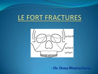
  • 17. A). Le fort I/ Floating fracture/ Guerin fracture/ Low level fracture/ Subzygomatic fracture 1. Mobility of maxillary alveolar segment (floating fracture) 2. Pain and tenderness while speaking or clenching 3. Ecchymosis or laceration in labial or buccal vestibule 4. Ecchymosis at GP foramen (Guerin sign) 5. Swelling and oedema of upper lip 6. Mal occlusion 7. Bilateral epistaxis 8. Brusing of palatal tissues (15-20% of cases) 9. On palpation tenderness over buttress area 10. Percussion of teeth – cracked pot sound Clinical Features
  • 18. B). Le fort II/ Pyramidal fracture/ Mid level fracture/ Subzygomatic fracture 1. Oedema mid third of face (Moon face) 2. Paresthesia of cheek 3. Bilateral circumorbital ecchymosis 4. Bilateral subconjunctival haemorrhage 5. Dish face deformity 6. Depressed nose 7. Epistaxis 8. CSF rhinorrhea 9. Limited ocular movement (Diplopia) 10. Mal occlusion 11. Inability to open mouth 12. Step deformity at IO margins 13. Mobility of fractured fragment at nasal bridge and IO margins 14. Percussion of teeth – cracked pot sound
  • 19. C). Le fort III/ Craniofacial dysfunction/ High level fracture/ Suprazygomatic fracture 1. Oedema of face (Panda facies) 2. Bilateral periorbital edema 3. Bilateral circumorbital ecchymosis (Racoon eyes) 4. Bilateral subconjunctival haemorrhage 5. Dish face deformity 6. Depressed nose, flattening of nose 7. Epistaxis 8. CSF rhinorrhea 9. Limited ocular movement (Diplopia, Enophthalmos) 10. Dystopia, hooding of eyes with antimongloid slant 11. Haemotympanum 12. CSF otorrhoea 13. Mal occlusion – posterior gagging of occlusion 14. Inability to open mouth 15. Mobility of fractured fragment at NF, FZ sutures 16. Tenderness over zygomatic bone, arch and FZ suture 17. Ecchymosis at mastoid process (Battle’s sign)
  • 20. 1. Emergency care and stabilization 2. Initial assessment 3. Definitive treatment 4. Continuing care
  • 21. ∆ Airway immediately evaluated for obstruction ∆Control of oral or nasal bleeding Possibility of C – spine fracture – endotracheal incubation should not be attempted Cervical collar in case of suspected spine fractures ∆Circulation
  • 22. LeFort I fracture LeFort I fracture with Mandible fracture LeFort I fracture with Nasal injury LeFort II fracture Lefort III fracture Panfacial fractures Nasal Airway Edentulous Partially Dentate with space Fully Dentate Oral Airway through portal cut in Gunning splints or dentures Oral Airway with tube displaced through space Surgical Airway Guided Nasal Intubation • fixate maxilla and mandible • switch to Oral Airway for nasal/NOE reduction
  • 23. Premlatha Shetty et al;submental intubation in patients with panfacial fractures;Indian journal of anesthesia,vol 55,issue 3,may 2011
  • 24. 1. History 2. Palpation of entire facial skeleton 3. I/O Examination 4. Ophthalmologic exam / consultation 5. Radiographic examination
  • 25. After stabilization of patients condition, complete facial examination is performed. 1. Laceration, bruising , etc. 2. Obvious depressions on nose, check, etc. 3. Facial asymmetry, swelling 4. Nasal discharge (Blood/ CSF)
  • 26. Features CSF fluid Nasal secretion History Nasal or sinus surgery, head injury or intracranial tumour Sneezing, nasal stuffiness, itching in the nose or lacrimation Flow of discharge A few drops or a stream of fluid gushes down when bending forward or straining; can’t be sniffed back Continuous. No effect of bending forward or straining. Can be sniffed back Character of discharge Thin, watery and clear Slimy (mucus) or clear (tears) Taste Sweet Salty Sugar content More than 30 mg/dl (Compare with sugar in CSF after lumbar puncture as sugar is less in CSF in meningitis) Less than 10 mg/dl Presence of β2 transferrin Always present. It is specific for CSF Always absent
  • 27. Palpation of facial skeleton Bowstring test
  • 28.
  • 29. 1. Periorbital edema 2. Periorbital ecchymosis 3. Proptosis 4. Diplopia 5. Pupillary size and shape 6. Sub-conjunctival haemorrhage 7. Lid laceration 8. Visual acuity 9. Dystopia
  • 30. Inspection Palpation Percussion Laceration Ecchymosis Restricted mouth opening Occlusion Tenderness Mobility of teeth Crepitus Mobility of fractured fragment Cracked pot sound
  • 31. 1. OPG 2. OM 3. Lateral skull view 4. Occlusal view for split palate 5. CT Scan 6. 3D CT Scan 7. MRI
  • 32. ∆ Aims of treatment 1. Relieve pain 2. Precise anatomical reduction of the # fragment 3. Stable fixation of the reduced fragment 4. Restore function 5. Restore the dental occlusion
  • 33. Preoperative planning: ∆ Need for surgical airway ∆ Open/closed method of reduction ∆ Necessity for and type if IMF to be employed in case for closed reduction ∆ Type of osteosynthesis in case of open method ∆ Need for internal suspension in case of communited # ∆ Timing of surgery
  • 34. ∏ Optimum time for reduction of mid face fracture is 5th to 8th post injury day ∏ After this with every succeeding day disimpaction become difficult and open reduction more essential
  • 35. Open reduction Closed reduction Displaced # Non displaced # Multiple # of facial bones Grossly communited # Edentulous maxillary # - with severe displacement Fractures associated with significant loss of soft tissues Edentulous maxillary # - opposite to Edentulous mandibular # Edentulous maxillary # Delay of treatment In children with developing dentition Inter position of soft tissues between non contacting displaced # segment Systemic condition contra indicating IMF
  • 36. 1. Accurate diagnosis 2. Determination of priority of treatment 3. Early reconstruction 4. Wide exposure of vertical and horizontal pillar of face 5. Use of bone graft to restore skeletal form 6. Use of rigid fixation to stabilize # segment 7. Restoration of bony support to over lying soft tissue envelop
  • 37. 1. Intra oral a) Vestibular 2. Extra oral a) Lower eye lid incision i. Sub cilliary ii. Infra orbital iii. Trans conjunctival b) Coronal approach c) Midface degloving approach
  • 38.
  • 39.
  • 40.
  • 41.
  • 44.
  • 45. 1. Manual reduction 2. Reduction with wires 3. Reduction using disimpaction forceps 4. Reduction with bone hook 5. Reduction with elastics
  • 46. 1. Simple manipulation by hand 2. Use of dental compound loaded in impression tray (Dingman and Harding, 1951) 3. Use of rubber dam sheets, long ribbon/strip gauze or rubber catheter (Propescu and Burlibasa, 1966)
  • 47. 1. Rowe’s maxillary disimpaction forceps 2. Hayton William’s disimpaction forceps
  • 48. Movements: 1. Downwards – to affect disimpaction of pterygoid plates down 2. Anterior 3. Combination of forward traction with rotational movement in both horizontal and vertical axis Universal rule Oculocardiac reflex
  • 49. Used in delayed cases: 1. Intra oral elastic traction 2. Extra oral elastic traction
  • 50. Maxillary # fixation Internal fixation Direct osterosynthesis 1. Miniplates 2. Intraosseous Wires - high(FZ,FN) - Mid(buttress,orbital rim) - Low(alveolar/midpalatal) Suspension wires 1. Frontal 2. Circumzygomatic 3. Zygomatic 4. Circumpalatal 5. Infraorbital 6. Piriform aperture 7. Peralveolar External fixation Craniomandibular Craniomaxillary 1. Supraorbital pins 2. Zygomatic pins 3. POP head frame 4. Halo frame 5 . Levant frame 6. Box frame
  • 51.
  • 53. Miniplates and screws These are monocortical, semi-rigid fixation device which provide 3D stability. Designs: X, H, L, T, Y Thickness:0.6-1 mm
  • 54. Plating system depends on: 1. Rigidity of plate 2. Width and shape 3. Diameter and number of screws Increase in width provides more stability towards rotational forces. Type of metal: a. Stainless steel b. Titanium c. Vitallium Advantages: 1) Easily adaptable 2) Monocortical 3) Functional stability 4) Reduced surgical access
  • 55. 1. Minimum 2 screws required in each bone segment to prevent rotation in X and Y axis 2. Farther the point of stabilization the more effective the device is in preventing rotation 3. Large diameter screws are not used because of constraint imposed by particular anatomic location 4. All screw require adequate intervening bone between adjacent holes to preserve integrity of screw bone interface
  • 56. Le fort I: L plates at zygomatic buttress Curved plate at pyriform aperture 3D plate sometimes to fix buttress # Le fort II: Linear/Y plate/curved plate along intra orbital rim L plate at buttress Le fort III: Linear/Y plate at FN and ZF junction
  • 57. Harle & duker(1975;Luhr(1979) 0.3-0.6 mm Used for : a. FN region b. Frontal bone c. Frontal process of maxilla Sites of application: a. Linear/T/Y plate at FN region b. Long curve plate for frontal process of maxilla or frontal bone
  • 58. Used for retention and alignment of small fragments or bone grafts. Sites of application: 1. Anterior and lateral wall of maxilla 2. Anterior table of frontal bone
  • 59.
  • 60. Introduced by Kuffner, 1970 Two types 1. Central 2. Lateral Usually used for high midface fracture.
  • 61. Incision in lateral 3rd/nasal process of frontal bone Exposure of zygomatic proces/outer cortex of frontal bone Drilling of bur hole and placement of screw Passage of SS wire attached to awl; through incision into maxillary vestibule Release of wire and attachment to the arch bar
  • 62. Indication: le fort II and III fracture Incision in maxillary vestibule above canine Subperiosteal dissection and exposure of infra orbital rim Drill hole and passage of wire above IO rim and back to oral cavity Release of wire and attachment to the arch bar
  • 63. Also known as buttress wire Incision in maxillary vestibule below buttress Exposure of ZM junction Drill hole and passage of wire Release of wire and attachment to the arch bar
  • 65. Introduced by Bowerman and Conroy, 1981 Simple technique for fixing gunning splint to maxilla Superior retention, stability and decreased discomfort Incision in maxillary vestibule over nasal spine Exposure of ANS Drill hole and passage of wire Release of wire and attachment to the arch bar
  • 66. Incision in maxillary vestibule in canine fossa Subperiosteal dissection and exposure of pyriform aperture Elevation of nasal mucosa and drill hole from lateral to medial Passage of wire and attachment to the arch bar
  • 67. Drill hole in palatal aspect of splint Direct wire through alveolus over canine region and emerge in Buccal Sulcus Passage of 0.5 mm SS wire and secure to splint
  • 68. Trend towards ORIF has changed External fixation is used in cases where there is depressed posterior displaced # Principle: External appliances relies on sandwiching the midface between base of skull and mandible to provide cantilever support to midface in 3D following disimpaction and closed reduction. Disadvantages:
  • 69. Disadvantage: 1. Heavy 2. Uncomfortable 3. Unstable Method of application
  • 70. Described by Crawford;modified by Mackenzie & Ray,1970 Secure the frame work to the skull directly by screw pins Advantage: 1. Light weight 2. Adjustable 3. Titanium Screw pin
  • 71. ∏ More stable and rigid ∏ Other unstable fracture fragment can also be attached to vertical rod
  • 72. ∏ Developed at Royal Melbourne Hospital ∏ Provided simple rigid craniomaxillary fixation between supraorbital rims and maxilla connected by central rod attached at lower end by means of cast metal splint or acrylic splint
  • 73. 1. Provide dimensional stability 2. Indications: 1. Grossly communited # 2. Extensive soft tissue loss 3. Bone gap>5mm 3. Sites: 1. Calvarium 2. Illium 3. Rib
  • 74. 1. Resorbable plates 2. Endoscopic management(Harold Hopkins) 3. Distraction osteogenesis(Ilizarov)
  • 75. Immediate 1. Airway 2. Nasal hemorrhage 3. Ophthalmic complications 4. Inaccurate reduction 5. Insecure fixation Late complications 1. Non union 2. mal occlusion 3. Cranial nerve dysfunction 4. Secondary nasal deformity 5. Dacrocystitis 6. Facial asymmetry
  • 76. Due to the complex 3D arrangement of the structures of middle third of face,management is complicated.Proper reduction of the # fragments remains the key component. A proper understanding of the anatomy,fracture patterns, its clinical presentation and the available treatment modalities is necessary to successfully treat Le Fort Fractures.
  • 77. 1. Oral & maxillofacial trauma-Fonseca & walker vol 2 2. Oral & maxillofacial surgery-Fonseca vol 3 3. Oral & maxillofacial trauma-Rowe & Williams vol 2 4. Principles of Oral & maxillofacial surgery-Peterson 5. Fractures of middle third of face-Killey & Kay 6. Oral & maxillofacial surgery-Fragiskos 7. Maxillofacial trauma & facial reconstruction-Peter Ward Booth 8. Oral & maxillofacial surgery-Peter Ward Booth: vol 2 9. Chen Lee et al ;Applications of the Endoscope in Facial fracture Management, seminars in plastics surgery/volume 22, number 1 2008
  • 78. 9. Manual of internal fixation-J Prein 10. Donat TL et al. Facial Fracture Classification According to Skeletal Support Mechanisms. Arch Otolaryngol Head Neck Surg 1998;124:1306-1314. 11. Mirko S. Gilardino et al;Choice of Internal Rigid Fixation materials in the treatment of facial fractures; craniomaxillofacial trauma & reconstruction/volume 2, number 1 2009 12. Khaled M Emara et al ;Methods to shorten the duration of an external fixator in the management of fractures; World J Orthop 2011 September 18; 2(9): 85-92 13. Chan hum park et al;resorbable skeletal fixation systems for treating maxillofacial bone fractures; arch otolaryngol head neck surg/vol 137 (no. 2), feb 2011 14. Premlatha Shetty et al;submental intubation in patients with panfacial fractures;Indian journal of anesthesia,vol 55,issue 3,may 2011.

Editor's Notes

  1. Tuberoplasty,sinus lift