ANATOMY OF KNEE JOINT
•Download as PPTX, PDF•
22 likes•840 views

ANATOMY OF KNEE JOINT In this presentation of " Anatomy of Knee Joint" you will know about structures present in Knee Joint. Bones, Joints, Ligaments, Muscles, Mechanism of movements of Knee Joint, Nerve and Blodd supply of Knee Joint.

Recommended
More Related Content
What's hot
What's hot (20)
Similar to ANATOMY OF KNEE JOINT
Similar to ANATOMY OF KNEE JOINT (20)
Recently uploaded
Recently uploaded (20)
ANATOMY OF KNEE JOINT
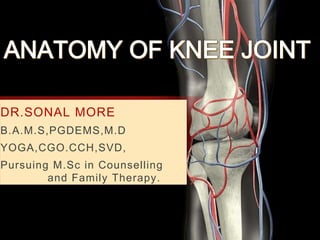
- 1. DR.SONAL MORE B.A.M.S,PGDEMS,M.D YOGA,CGO.CCH,SVD, Pursuing M.Sc in Counselling and Family Therapy.
- 2. - The Knee Joint is the largest. - Most complicated joint in the body. - Most superficial joint. - Hinge type of synovial joint.
- 4. BONES JOINTS LIGAMENTS TENDONS MUSCLES NERVES BLOOD VESSELS KNEE JOINT consist of
- 6. ANATOMIC ORIENTATION TERMS • Closer to the midline MEDIAL • Away from the midline LATERAL • Front side ANTERIOR • Back side of the knee POSTERIOR
- 7. Femur Tibia Patella BONES involve in knee joint are
- 8. FIBULA is not involve directly in the Joint
- 9. Knee joint is Synovial joint enclosed by ligament capsule and contains a fluid called Synovial fluid that lubricates the joint
- 10. It consists of 3 Joints within a single synovial cavity : • Medial Condylar Joint : Between the medial condyle “of the femur” & the medial condyle “of the tibia” . •Lateral Condylar Joint : Between the lateral condyle “of the femur” & the lateral condyle “of the tibia” . •Patellofemoral Joint : Between the patella & the patellar surface of the femur .
- 13. • Condyles of Femur rest on the top surface of Tibia. This surface is called as “TIBIAL PLATEAU”. • Outside half : outside of knee is Lateral tibial plateau Inside half : close to the other knee is Medial tibial plateau. • Patella glides on special groove formed by two femoral condyles called Patello femoral groove.
- 14. ARTICULAR CARTILAGE • Articular Cartilages are material that covers ends of bones of the joints . • These are 6.35mm or 1 quarter inch thick in most large joints. • It is white shiny with rubbery consistency. • Articular cartilages are slippery substance that allows the surfaces to slide against one another without damage to either surface .
- 15. • Function of articular cartilage is to absorb shock and provide extremely smooth surface to facilitate motion. • Articular cartilage are present everywhere where two bony surfaces moves against one another were articulate. • In the knee Articular cartilage covers the ends of femur, top of the tibia and back of the patella.
- 16. LIGAMENTS LIGAMENTS are tough ,fibrous bands of tissues that connects ends of bones together. Two important ligaments are found on either side of the Knee joint. They are : 1. Medial Collateral Ligament [MCL] 2. Lateral Collateral Ligament [LCL]
- 17. • Is a broad band that extends from the medial femoral epicondyle to the medial tibial condyle. • Also called as Tibial collateral ligament MEDIAL COLLATERAL LIGAMENT
- 18. • Extends between the lateral femoral epicondyle and the head of the fibula. • Also called as Fibula collateral ligament. LATERAL COLLATERAL LIGAMENT
- 20. •Inside the Knee joint two other important ligaments are stretched between the femur and tibia. •They are 1. Anterior Cruciate Ligament in the front [ACL] 2. Posterior Cruciate Ligament in the back.[PCL]
- 21. - The weaker of the two cruciate ligaments . - Arise from the anterior intercondylar area of the femur , just posterior to the attachment of medial meniscus intercondylar area of tibia . - The ACL has a relatively poor blood supply. The Anterior Cruciate Ligament
- 22. - The stronger of the two cruciate ligaments. - Arises from the posterior intercondylar area of the tibia THE POSTERIOR CRUCIATE LIGAMENT
- 23. •MCL and LCL prevent the Knee to move too far in a side to side direction. •ACL and PCL controls front and back motion of Knee joint. •ACL keeps tibia sliding too far forward in relation to the Femur. •PCL keeps the tibia sliding too backward in relation to femur.. •Ligaments all taken together are most important structures controlling stability of the Knee.
- 26. MENISCI • Two special types of Ligaments called “MENISCI” sit between the femur and tibia. •They are : 1. Lateral Menisci 2. Medial Menisci • These structures are sometimes referred as the cartilages of the Knee joint. • But Menisci are different from articular cartilages that covers the surface of the joint.
- 30. Imagine ball resting on plate. Here ball is condyles of the femur and flat plate is the tibial plateau. Menisci actually wrap around round the condyles to fill the space between it and flat tibial plateau. • The menisci act like a gasket helping to distribute the weight from the Femur to the tibia. •Without the menisci any weight on the Femur will be concentrated to one point of the tibia. • With a Menisci weight is spread across the tibial surface.
- 31. • Without the Menisci the concentration of force into a small area on the Articular cartilage can damage the surface leading to the degeneration overtime. •In addition to protect the Articular cartilage the menisci help the ligament in stability of Knee joint. • Menisci make the Knee joint more stable by acting like a ‘Wedge Shape’ set against the bottom of the condyle.. • The Menisci are thicker around the outside and its thickness helps to keep the round Femur from rolling on the flat tibia. • The Menisci convert the tibial surface into a shallow socket. And the socket is more stable, more efficient in transmitting weight from the upper body .
- 32. • Menisci enhances the stability of the knee and protect the articular cartilage from excessive concentration of force. • Taken altogether ligaments of the Knee joint are the most Important structures to stabilize the joint. Remember : Ligaments connects bones to bones Without strong and tight ligaments to connect Femur to Tibia Knee joint would be too loose .
- 33. •The Quadriceps femoris muscle, commonly known as the quad muscle, is the strongest muscle of the human body. •It is located in the anterior compartment of the thigh, together with the Sartorius. •The quadriceps femoris muscle translates to “four-headed muscle” from Latin. •It bears this name because it consists of four individual muscles; RECTUS FEMORIS, VASTUS MEDIALIS, VASTUS LATERALIS, AND VASTUS INTERMEDIUS. MUSCLES
- 35. •Out of all four muscles, only the rectus Femoris crosses both the hip and knee joints. •Others cross only the knee joint and these muscles differ in their origin, but share a common Quadriceps femoris tendon which inserts into the patella. •The function of the quadriceps femoris muscle is to extend the leg at the knee joint and to flex the thigh at the hip joint.
- 36. •There are three muscles in the posterior compartment of the thigh: the biceps femoris and two synergistic muscles (the semitendinosus and semimembranosus). These muscles are termed the hamstring group. •The posterior region of the thigh displays similarity with the anterior region of the upper arm in both structure and function. •Biceps Femoris: A similar muscle to the biceps brachii in the upper arm and also double-headed. Two synergistic muscles are associated with the biceps femoris, the semitendinosus and the semimembranosus. Posterior Muscles
- 40. OTHER MUSCLES
- 41. TENDONS • Tendons are similar to ligaments . Except the tendons attached muscles to the bones. • Quadriceps tendon connects the large quadriceps muscles of the thigh to the Patella.
- 42. • This tendon continues across the Patella over Patella and blends into the Patellar Tendon. • Patellar tendon connects the Patella to the Tibia.
- 43. • The Extensor Mechanism is a motor that drives in the Knee and allows us to walk. • It sits in front of the Knee joint and is made up of Patella,Patellor tendon, Quadriceps tendon and Quadriceps muscles. • When these muscles contract they straight the Knee joint such as when you get up from squatting postion. • The way in which Patella fits into the Patellofemoral groove on the front of the Femur and slides as the Knee bends affects overall function of knee. • Patella increases the force exerting by the Quadriceps muscle as the Knee straightens. •Hamstring muscles contract and the Knee bends
- 45. musclemovement Biceps femoris Semitendinosus Semimembranosus Gracilis Sartorius Popliteus Flexion 120 -150 Quadriceps femorisExtension 5 -10 Biceps femorisExternal rotation 30 -40 Sartorius Gracilis Semtendinosus Popliteus Semimembranosus Internal rotation 10
- 46. NERVES
- 47. •The nerves around the knee are motor (move muscles) and sensory (allow you to feel what is h appening). The sensory nerves supply the joint itself as well as the skin over the knee. •Many muscles have both motor and sensory functions.
- 48. SCIATIC NERVE • (L4,5, S1,2,3) is a large nerve which runs down the back of the leg. It is made up of the tibial and common peroneal nerves which branch at different levels of the leg in different people. • The sciatic nerve splits into the tibial and common peroneal nerves above the knee. • The tibial nerve supplies the hamstring muscles (which bend the knee). It also supplies the muscles in the back of the calf • The common peroneal nerve supplies the front compartments of the leg
- 50. TIBIAL NERVE • iIis the larger of the two branches of the sciatic nerve and runs down the back of the knee.
- 51. • The common peroneal nerve is one of two major branches of the sciatic nerves within the buttocks and into the thighs, along with the tibial nerves. • It travels outside of Knee and down the front of the leg and foot. COMMON PERONEAL NERVE
- 53. BLOOD SUPPLY The Femoral artery and the popliteal artery forms artery network surrounding the knee joint , There are 6 main branches : 1.Superior medial genicular artery 2.Superior lateral genicular artery 3.Inferior medial genicular artery 4.Inferior lateral genicular artery 5.Descending genicular artery branch from the femoral artery 6.Recurrent branch of anterior tibial artery The medial genicular arteries penetrate the knee joint Branch from poplitealartery
- 56. KNEE has a unstable and complicated design yet it support the Body’s bone weight in standing and much more than that during walking and running.