
Recommended
More Related Content
What's hot
What's hot (20)
Viewers also liked
Viewers also liked (20)
Similar to Anatomy of the eye
Similar to Anatomy of the eye (20)
Recently uploaded
Recently uploaded (20)
Anatomy of the eye
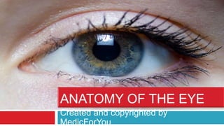
- 1. Created and copyrighted by MedicForYou
- 3. The Eyeball Each eyeball is a cystic structure kept distended by the pressure inside it. Eyeball is not sphere but ablate spheroid. It has anterior and posterior curvature. The maximal convexities of the these curvatures of the eyeball are called the anterior and posterior pole, respectively.
- 4. Dimensions of an adult eyeball Anteroposterior diameter 24 mm Horizontal diameter 23.5 mm Vertical diameter 23 mm Circumference 75 mm Volume 6.5 ml Weight 7 gm
- 5. Coats of the eyeball Three coats: Outer (fibrous coat), Middle (vascular coat) and Inner (nervous coat). Fibrous coat • Dense strong wall • Protects the intraocular contents • Anterior 1/6th transparent part is called cornea • Posterior 5/6th opaque part is called sclera • Junction of the cornea and sclera is called limbus Vascular coat (uveal tissue) • Supplies nutrition • Consists of iris, ciliary body and choroid Nervous coat (retina) • Concerned with visual functions.
- 6. Segments of Eye ball Anterior Segment Anterior Chamber Posterior Chamber Posterior Segment
- 7. Anterior Segment It includes crystalline lens( which is suspended from the ciliary body by zonules), and structures anterior to it, viz., iris, cornea and two aqueous humour-filled spaces anterior and posterior chambers.
- 8. Anterior Segment (…continued) Anterior chamber. Bounded anteriorly by the back of cornea, Bounded posteriorly by the iris and part of ciliary body. About 2.5 mm deep in the centre in normal adults. Shallower in hypermetropes and deeper in myopes. Contains about 0.25 ml of the aqueous humour. Posterior chamber Bounded anteriorly by the posterior surface of iris and part of ciliary body Posteriorly by the crystalline lens and its zonules
- 9. It includes the structures posterior to lens, viz., vitreous humour (a gel like material which fills the space behind the lens), retina, choroid and optic disc. Posterior Segment