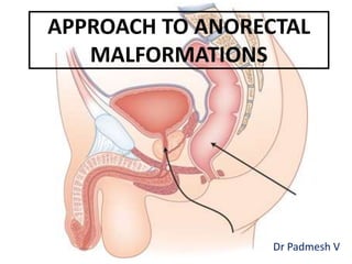
Approach to Ano Rectal Malformations - Dr Padmesh - Neonatology
•Download as PPTX, PDF•
16 likes•3,803 views

This document discusses anorectal malformations (ARMs), which occur in approximately 1 in 4,000 to 5,000 births. ARMs can be low or high lesions depending on whether the rectum has descended through the sphincter complex. Surgical techniques for repair include primary repair for perineal/vestibular fistulas or staged repair for more complex cases involving diverting colostomy, definitive reconstruction, and later colostomy closure. Outcomes depend on the extent and nature of the anomaly as well as surgical expertise, with risks including urinary/fecal incontinence. Thorough evaluation and thoughtful surgical care are important to determine the future of children born with ARMs.

Recommended
More Related Content
What's hot
What's hot (20)
Similar to Approach to Ano Rectal Malformations - Dr Padmesh - Neonatology
Similar to Approach to Ano Rectal Malformations - Dr Padmesh - Neonatology (20)
More from Dr Padmesh Vadakepat
More from Dr Padmesh Vadakepat (20)
Recently uploaded
Recently uploaded (20)
Approach to Ano Rectal Malformations - Dr Padmesh - Neonatology
- 1. APPROACH TO ANORECTAL MALFORMATIONS Dr Padmesh V
- 2. • INTRODUCTION: • Incidence: 1 in 4,000 to 1 in 5,000. • Anorectal malformations (ARMs): common surgically treated anomalies in the newborn period. • Imperforate anus can be divided into: – Low lesions, where the rectum has descended through the sphincter complex, and – High lesions, where it has not descended through sphincter complex. (Sphincter complex: puborectalis, levator ani, external and internal sphincters)
- 3. • EMBRYOLOGY: • Interference in development of anorectal and genitourinary organs at various stages upto 7 to 8 weeks of gestation. • Continued communication between the urogenital tract and rectal portions of the cloacal plate causes rectourethral fistulas or rectovestibular fistulas. • Anomalies in the pelvic floor as well as the external anal sphincter (which are derived from exterior mesoderm). • The higher the rectal pouch, more are the chances of mal- development of the pelvic floor.
- 4. • ETIOLOGY: –Consanguinity, –HLXB9 gene mutations, and –Congenital syndromes • Presence of an ARM Evaluate for VACTERL anomalies (vertebral anomalies, anal atresia, cardiac defects, tracheoesophageal fistula and/or esophageal atresia, renal and radial anomalies, and limb defects). • Most common association: Caudal regression syndrome
- 5. • Associated Malformations • GENITOURINARY • Vesicoureteric reflux • Renal agenesis/dysplasia • Ureteral duplication • Cryptorchidism • Hypospadias • Bicornuate uterus • Vaginal septums • VERTEBRAL • Spinal dysraphism • Tethered chord • Presacral masses • Meningocele • Lipoma • Dermoid • Teratoma • CARDIOVASCULAR • Tetralogy of Fallot • Ventricular septal defect • Transposition of the great vessels • Hypoplastic left-heart syndrome • GASTROINTESTINAL • Tracheoesophageal fistula • Duodenal atresia • Malrotation • Hirschsprung disease • CENTRAL NERVOUS SYSTEM • Spina bifida • Tethered cord
- 6. • CLASSIFICATION • Gross’ Classification: • Earliest classification • Differentiation based on levator muscle, i.e. supralevator – for those above the levator ani or infralevator anomalies, for those below the levator ani.
- 8. • IMPERFORATE ANUS IN MALES: LOW HIGH
- 9. • IMPERFORATE ANUS IN FEMALES: Vestibular fistula Cloaca Fistula between Rectum and Vulval vestibule All three open into a single orifice
- 10. • CLASSIFICATION • Wingspread system • Based on gender • Based on the postulated position of rectum in relation to the pubococcygeal line and ischium. • Position of rectum is determined using plain cross- table radiography in prone position, with the bottom up.
- 11. • CLASSIFICATION • Wingspread system
- 12. • CLASSIFICATION • Krickenbeck system • Divided according to clinical presentation. • Divided into major clinical groups and rare/regional variants
- 13. • CLASSIFICATION • International Classification
- 14. • PRENATAL EVALUATION • Prenatal diagnosis of most ARMs is rare. • Presence of a cystic abdominal/pelvic mass in combination with gastrointestinal and urologic anomaly should heighten the suspicion for cloacal anomalies. • The use of fetal magnetic resonance imaging (MRI) after 20 weeks of gestation.
- 15. • POSTNATAL EVALUATION • Thorough physical examination. • A clear understanding of the potential locations for perineal fistulas is essential. • Boys: • Perineal body • Median raphe should be carefully inspected • Scrotum • Penis
- 16. • Meconium expressed from urethral meatus is indicative of a fistulous connection between rectum and the urinary tract.
- 17. • POSTNATAL EVALUATION • GIRLS: • Determine – Number of orifices – Location of perineal orifices • Presence of 3 orifices does not exclude an ARM. • Presence of 2 orifices is indicative of a urogenital sinus. – Genitourinary tract & reproductive tract share a common channel, – Gastrointestinal tract has separate opening in normal location. • Single orifice: Cloacal anomaly. – Reproductive, urinary, and GIT share a common orifice. – Most complex form of female ARM – Requires extensive evaluation and staged corrective surgery
- 18. • Female Urogenital sinus : 2 orifices
- 19. • Cloacal anomaly : 1 orifice
- 20. • Normal position of anus on perineum is approx halfway (0.5 ratio) between coccyx and scrotum or introitus. • If fistulous connections are difficult to identify, wait for 24 hours meconium descends facilitates visualization. • If Cutaneous fistula can be seen, it is a low lesion. • Majority of ARMs do not require emergent surgical intervention. • Timing of surgery typically after first 24 to 48 hours. • During the first 24 hours, gas and meconium descend through the rectum to either the lowest level of the pouch or into the fistula more accurate assessment of extent of anomaly.
- 21. • RADIOLOGIC EVALUATION • Invertogram at 24 hours of life: Determine distance betwn rectum & perineal skin. • Originally: infants held in inverted position column of gas in rectum rises as high as possible with radio-opaque probe at anal dimple. • Now: Newborns placed in prone position with hips and knees flexed to chest . Infant stays in this position with bottom up for some time, and radio-opaque marker is placed at anal dimple. • Cross-table radiograph (Minimum 3 minutes duration) Distance between rectal gas column and bead measured. • Rectal gas bubble – <1-2 cm from perineal skin Low lesion – >1-2 cm from perineal skin High lesion
- 23. • RADIOLOGIC EVALUATION • Fluoroscopy: contrast study • To detect a recto-urinary, rectovaginal, or rectoperineal fistula • Fistula is termed – Low : if below the levator ani plane – High : if it is below the pubococcygeal line (PCL)
- 24. • SURGICAL TECHNIQUES: • A) Primary Repair • Patients with perineal/vestibular fistulas. • Options: –Cutback Anoplasty –Posterior sagittal anorectoplasty (PSARP). • Two weeks after repair, the neo-anus is calibrated using a dilator, and serial dilations performed.
- 25. • SURGICAL TECHNIQUES: • B) Staged Repair • Rectourethral/rectovesicular fistulas, cloacal anomalies, rare variants, or complex life- threatening associated anomalies. • These stages are divided into: – 1. Performing a colostomy for enteric diversion, – 2. Definite reconstruction, – 3. Subsequent restoration of enteric continuity via colostomy reversal.
- 26. • SURGICAL TECHNIQUES: • Staged Repair • Stage 1: Diverting Colostomy. (Double-barrel colostomy) – Proximal end : Ostomy. – Distal end: Mucous fistula.(Mucous fistula subsequently used to perform distal colostogram before the second stage)
- 27. • SURGICAL TECHNIQUES: • Staged Repair • Stage 2: Definitive reconstruction: (At about 1 year age) • Two main approaches for reconstruction of ARM: – PSARP (posterior sagittal anorectoplasty) – LAARP (Laparoscopy-Assisted Anorectoplasty)
- 28. • SURGICAL TECHNIQUES: • Staged Repair • Stage3: Colostomy Closure. – Once the reconstruction has been performed and healed, the colostomy is reversed. – Typically 4 to 6 weeks after reconstruction.
- 31. • OUTCOMES: • Depends on – (1) Extent and nature of the anomaly, – (2) surgical expertise, – (3) the presence of associated syndromic anomalies, – (4) compliance with an effective and aggressive bowel management program.
- 32. • OUTCOMES: • 1. Urinary Incontinence • Rates of urinary incontinence ranges between 0% for low anomalies to 10% for high anomalies. • 2. Fecal Incontinence • Low ARMs had good bowel function with no soiling, whereas one-third of those with “high” or “intermediate” anomalies had fecal soiling. • 3. Sexual Function and Fertility • Variable outcomes
- 33. Willis J. Potts: “No congenital anomaly requires more thoughtful surgical care than imperforate anus or atresia of the rectum. In practically every case the future of a child so unfortunate as to be born with an imperforate anus is determined by the adequacy and skill of the first operation”
- 34. THANK YOU!!!