Rho Kinase Promotes Alloimmune Responses by Regulating the Proliferation and ...
ACTH and MSH suppress immunity
1. Proc. Nail. Acad. Sci. USA
Vol. 89, pp. 782-786, January 1992
Immunology
Immunosuppressive effects of corticotropin and melanotropin
and their possible significance in human immunodeficiency
virus infection
(neuroimmunomodulation/neuropeptldes/AIDS/neutral endopeptidase 24.11)
ERIC M. SMITH*t, THOMAS K. HUGHES, JR.t, FARHAD HASHEMIt, AND GEORGE B. STEFANOt
Departments of *Psychiatry and tMicrobiology, University of Texas Medical Branch, Galveston, TX 77550; and tMultidisciplinary Center for the Study of
Aging, State University of New York, Old Westbury, NY 11568
Communicated by Berta Scharrer, October 14, 1991
ABSTRACT The activation of human granulocytes and
invertebrate immunocytes was found to be suppressed by
corticotropin (ACTH) and melanotropin (MSH). In spontane-
ously active granulocytes both neuropeptides caus significant
conformational changes indicative ofinactivity plusa reduction
in their locomotion. Significant inactivation of human granu-
locytes by ACTH required 2 hr, that by MSH only 20 min. The
addition to the incubation medium of phosphoramidon, a
specific inhibitor of neutral endopeptidase 24.11, blocked
inactivation ofgranulocytes by ACTH. Radlolmmunosy for
MSH of supernatant fluids from granulocytes incubated with
ACTH demonstrated atime-dependent increase inMSH. These
data strongly indicate that the effect ofACTH is largely due to
its conversion to MSH by granulocyte-associated neutral en-
dopeptidase. Parallel experiments with immune from the
mollusc Mytilus edulis gave similar results, indicating the
universality of this phenomenon. Our finding that the human
immunodeflciency virus, among several viruses, induces
ACTH and MSH production in H9 T-lymphoma cells suggests
an important role of these neuropeptides in the immunosup-
pression characteristic of such infections.
Broadly based comparative studies demonstrate that neu-
ropeptides play important roles in immunoregulatory pro-
cesses (1, 2). Not only do they convey neural directives to the
immune system but also they function as autoregulatory
factors within the immune system (1-3). Many neuropeptides
tested thus far have shown stimulatory effects on granulocyte
(4) and invertebrate immunocyte (5) activity as determined by
conformational and locomQtory responses. Two neuropep-
tides, corticotropin (ACTH) and melanotropin (MSH), how-
ever, demonstrate inhibitory effects (6).
MSH is a proteolytic'cleavage product of proopiomelano-
cortin (POMC), the polyprotein precursor for ACTH, and
corresponds to ACTH-(1-13). Although this precursor-
product relationship of ACTH and MSH is well documented
in the brain and neuroendocrine system, it has only been
suggested in regard to the immune system (4). Some previous
reports are consistent with the concept that some of the
effects of ACTH on the immune system are due to its
conversion to MSH. For example, the inhibitory effect of
MSH on polymorphonuclear cell migration and superoxide
dismutatase induction is much more potent than that of
ACTH (4). ACTH can directly alter immune responses,
including the in vitro production of antibody (7, 8) and
induction of interferon y (IFN-y) (9), inhibition of macro-
phage activation (10), induction oftumor necrosis factor (11),
and modulation of invertebrate hemocyte phagocytotic ac-
tivity (12).
Conversion ofACTH to MSH is suggested by the fact that
pharmacological doses and relatively long periods oftime are
required to obtain the effects reported in the present study.
Furthermore, proteolytic enzymatic activities, such as neu-
tral endopeptidase 24.11 (NEP; EC 3.4.24.11), also known as
the lymphoid surface antigen CD10 (13), are present in high
quantities in granulocytic cells of human and invertebrate
origins (14). Also, proteolytic processing of ACTH has been
reported in leukocytes stimulated by bacterial lipopolysac-
charide (15). Thus, there is a precedent to suggest that ACTH
could be processed to MSH within the lymphoid system.
The present study examines the mechanism of the inhibi-
tory effects ofACTH and MSH on granulocyte conformation
and locomotory responses. We present data that both sub-
stances contribute to the inhibitory effects, but MSH is the
more potent and faster acting of the two. It also provides
evidence that ACTH is processed to MSH in this system by
a granulocyte-associated cell surface enzyme, NEP. The
participation of these hormones in the immunosuppression
seen in certain viral diseases is suggested by our finding that
the human immunodeficiency virus (HIV) induces highly
significant levels ofACTH and MSH in a cultured T-cell line.
MATERIALS AND METHODS
Cells. Human blood was obtained from volunteer donors at
the Dana-Farber Cancer Institute for cellular image analysis
or the University of Texas Medical Branch Blood Bank for
granulocyte preparation. Granulocyte pellets were prepared
by Ficoll-Hypaque density gradient centrifugation and dex-
tran sedimentation (16) and utilized within 6 hr of collection
as noted elsewhere in detail (2, 5).
H9 cells and HIV-1 (strain SK-1) were gifts from Miles
Cloyd (University ofTexas Medical Branch, Galveston) and
cultured by standard methods as previously described (17).
H9 cells (3.5 x 10⁵) were inoculated with HIV at an estimated
multiplicity of infection of 1. The cultures were incubated at
370C for the indicated times. Supernatant fluids were saved
for radioimmunoassay and the cells were saved for indirect
immunofluorescent staining as previously described for
ACTH and p24 antigens (18). For comparative studies with
immunocytes ofthe mollusc Mytilus edulis (mussel) subtidal
animals were collected from the shore ofWading River, Long
Island Sound, New York.
Cellular Inactivation Assay. The examination of the effect
of ACTH and MSH on cellular preparations was carried out
as previously described (5, 14). Briefly, blood or hemolymph
was incubated on albumin-coated slides with the agents and
Abbreviations: ACTH, corticotropin (adrenocorticotropic hor-
mone); MSH, melanotropin (melanocyte-stimulating hormone);
POMC, proopiomelanocortin; IFN-y, interferon 'y; NEP, neutral
endopeptidase 24.11; HIV, human immunodeficiency virus.
782
The publication costs ofthis article were defrayed in part by page charge
payment. This article must therefore be hereby marked "advertisement"
in accordance with 18 U.S.C. §1734 solely to indicate this fact.
2. Proc. Natl. Acad. Sci. USA 89 (1992) 783
the angiotensin-converting enzyme inhibitor captopril (19) or
the NEP inhibitor phosphoramidon (13) at 370C for human
and at room temperature for the invertebrate cells for the
specified incubation times.
Microscopy. The cell preparations were examined by use of
phase-contrast and Nomarski optics, coupled with a Zeiss
Axiophot microscope. Measurements were taken by utilizing
the Zeiss Videoplan/Vidas and American Innovision image
analysis systems as previously described (20). Before the
various cells were recorded, specific images were converted
to binary images after "frame grabbing." Simultaneously,
specific cells were photographed with a time-lapse video
synchronization system (JVC). Changes in cellular confor-
mation based on measurements ofcellular area and perimeter
were mathematically expressed by use of the form-factor-pe
(FF) calculation ofthe Zeiss Vidas analysis system, whereby
the formula (4 x ir X area)/perimeter2 provides mean nu-
merical values. The lower this number, the larger is the
cellular perimeter and the more ameboid the cellular mor-
phology. The numbers of activated cells were obtained by
counting cells automatically that exhibited areas greater than
145 ,um2, as contrasted with inactive cells whose areas were
less than 115 gm2. Mean values were derived from 15
individual readings taken from different cells. The individual
points on the graphs represent the means ofsix to eight mean
values. The final mean values did not vary by more than 5%.
Radioimmunoassays. ACTH and MSH were both quanti-
tated by use of commercial radioimmunoassay (RIA) kits
(Incstar, Stillwater, MN) (21). Sensitivity ranges for these
assays were 15-500 pg/ml for ACTH and 20-600 pg/ml for
MSH. Cross-reactivity was less than 0.1% according to the
kits' specifications and as measured with peptide standards.
Assay procedures were followed according to the instruc-
tions. Briefly, supernatant fluids from the granulocyte cul-
tures were incubated with the primary antiserum to the
peptides, nonspecific material was washed out, 1251-labeled
peptides were added, and the bound radioactivity was quan-
titated. Nonspecific binding was subtracted, and the percent
of bound radioactivity compared with the zero standard was
extrapolated from an empirically derived standard curve.
Reagents. Phosphoramidon (13), MSH (a form), and por-
cine ACTH-(1-39) were purchased from Sigma and captopril
(19) was a gift from Margaret A. Shipp (Dana-Farber Cancer
Institute, Boston). Antisera to ACTH-(1-24) was purchased
from ICN and the monoclonal antibody preparation to p24
(M26) was a gift from Miles Cloyd.
RESULTS
Previous studies with human polymorphonuclear cells (4) and
invertebrate immunocytes (6) have demonstrated that MSH
can modulate immune responses. In the present study, we
determined whether ACTH, like MSH, inhibits the activation
of human granulocytes (Fig. 1) and Mytilus immunocytes
(Fig. 2). Activation is expressed by a flattening ofthe cell and
an increase in cell perimeter, resulting in a decrease of the
form factor value to approximately 0.4 (22).
In control preparations, inactive granulocytes and Mytilus
immunocytes are rounded and measure approximately 60-
121 Am2. In a viewing field ofapproximately 300 cells, 6-8%
of the cells commonly become spontaneously active in 1 hr
(6), and this increases to approximately 14% after 5 hr of
culture (Figs. 1A and 2A). By virtue of the video analysis
system, individual, activated cells were monitored, and the
results are expressed as a percent of the activated cell
population (with 100lo representing the total number of
activated cells at the start of the incubation period).
The addition ofACTH or MSH (0.1 1LM) to the incubation
medium caused conformational changes in the majority ofthe
granulocytes that were spontaneously active (Fig. 1 A and B)
A 200
175
150
125
100
75
50
25
C/)
CD(9)
>
-o
(9)
-4---
0
oO
B 200 -
1 75 -
150-
125-
100-
75 -
50 -
25 -
C 200-
1 75 -
1 50 -
125
100-
75 -
50
25-
o-0 Control
*-*ACTH
0
0
0
P(0-05
0 1 2 3 4 5
Time (hours)
o 0 Control
* *MSH
A AMSH+ACTH
0--- - ---
O
0
°
t ~~ ~ ~ ~ Z -------Zin A
-~~~~~~~~
75~ ~~~PO0-0
50I-
1 0 20 30 40 50
Time (minutes)
0 0 Control
* * ACTH+Capto.
A AACTH+Phos.
1 2 3
Time (hours)
4 5
FIG. 1. Inactivation ofhuman granulocytes by ACTH and MSH.
Enriched granulocytes were incubated with 0.1 uM ACTH (A), 0.1
kMM MSH with or without ACTH (B), or with ACTH with 100 uM
captopril or 100 MM phosphoramidon (C). Activation was measured
by video analysis and expressed as percent of activated cells in the
control at the start ofthe incubation. P < 0.05 is in comparison with
the control and is true for later points on the same curve.
by the end of the observation periods. The conformational
changes that these cells underwent while becoming inactive
included withdrawal of pseudopodia and rounding. A com-
parison of the effects of ACTH and MSH at peak response
times (Fig. 1 A and B) revealed form factors of0.80-0.91 for
every cell in the culture, indicating that all cells are round.
ACTH significantly inhibited the activation of human gra-
ulocytes within 2 hr (Fig. 1A), whereas MSH appears to exert
its effect within 20 min (Fig. 1B). It should be noted that as
the observation period is extended there is an increase in the
number of spontaneously active cells. An interesting com-
parative observation is that Mytilus immunocytes responded
more slowly to ACTH and MSH suppressive action than
human granulocytes (Fig. 2 A and B).
The slower onset of inactivation with ACTH as compared
to MSH suggested that the peptide was converted to MSH.
In both human granulocyte and Mytilus immunocyte mem-
branes, NEP has been shown to be present (14). Therefore,
tests were performed with the NEP inhibitor phosphorami-
Immunology: Smith et al.
3. Proc. Natl. Acad. Sci. USA 89 (1992)
A
B
200
175
150
125
100
75
50
25
nu-
20
1 7
15
12
1C0
-7
2
C 2C
17
1 i
1 2
c
o -0 Control
*-*ACTH
0
0
0
* ~O
0
~P(O.05
0 1 2 3 4 5
Time (hours)
)0 0 -O Control
75 *-*MSH
A-A MSH+ACTH
50
SO
0~
~~~~~~~~~~~~ L
75 8 _ _i
10
?5 P<0.05
0
0 1 0 20 30 40 50
Time (minutes)
)0 -O OControl
75 *-* ACTH+Copto. 0
A-AACTH+Phos. 0O
50 -0
75 -
50
25SO- P < 0~~~~~~~~~<.05
0 1 2 3
0 1 2 3 4 5
Time (hours)
FIG. 2. Inactivation of Mytilus edulis immunocytes by ACTH
(A), MSH with or without ACTH (B), or ACTH with captopril or
phosphoramidon (C). Same concentrations as in Fig. 1.
don at its previously determined effective dose (6). Phos-
phoramidon prevented the ACTH inactivation of the spon-
taneously active state for both cell types (Figs. 1C and 2C).
Captopril, an inhibitor of another endopeptidase, angioten-
sin-converting enzyme, was without effect. This result
strongly suggests that MSH is an important signal molecule
in causing the previously described cellular immunosuppres-
sion. Moreover, this finding is further supported by the
experiment noting that ACTH can antagonize cellular immu-
nosuppression caused by MSH (Figs. 1B and 2B).
Inhibition of proteolytic cleavage and inactivation of im-
munocytes by ACTH further supported the hypothesis of
ACTH conversion to MSH. To address this, relative ACTH
and MSH levels were measured in human granulocytes
treated with ACTH. With some variability in the absolute
quantity, we consistently found a time-dependent decrease in
ACTH and an increase in immunoreactive MSH over the
phosphoramidon-treated cultures (Fig. 3). No cross-
reactivity could be detected for the peptides in either radio-
immunoassay to explain this rise in MSH. Phosphoramidon
treatment reduced the ACTH drop and delayed the rise in
MSH. It is interesting to note the similarity in kinetics for the
_ou-
ok)- 50
C40)a 40 iEI-;{
30
20
10 -
0 l
0 1 2 3 4
Time (hours)
FIG. 3. Conversion ofACTH to immunoreactive MSH by human
granulocytes. Enriched granulocytes (1 x 10⁷ per ml) were incubated
for the indicated times with ACTH at 0.5-1 ng/ml in the presence or
absence of phosphoramidon (100 MuM). Culture supernatant fluids
were radioimmunoassayed for residual ACTH and newly generated
MSH. ACTH or MSH + and - Phos refers to the neuropeptide
measured in the supernatant fluid from cultures either treated with
phosphoramidon or untreated. The data are representative of five
experiments.
degradation ofACTH and the significant rise in MSH to that
of the cellular inactivation (Figs. 1 and 2).
Since MSH is degraded by NEP (23), we hypothesized that
MSH in our system would be degraded also, and that
phosphoramidon treatment should result in greater inhibition
of granulocyte activation by MSH. Fig. 4A shows greater
inhibition of granulocyte activation in cultures treated with
MSH and phosphoramidon versus MSH alone. A similar
effect on Mytilus immunocytes is seen in Fig. 4B.
It has been shown that viruses will stimulate leukocytes to
synthesize POMC (1, 18, 24, 25). In addition, we have
preliminary evidence suggesting that HIV induces lympho-
cytes to produce ACTH (26). The infection and suppression
of the immune system, plus the fact that functioning of the
A l1o
88 -
66 -
44 -
C',
- 22-
a)
B li10'8
1 88I
01 6
66o
44 -
22 -
O-OMSH
*- *MSH+PHOS
0
0
P(0.05
a s P<0.05
0-OMSH
*- *MSFI+PHOS 0
/ / ~~~P(O.05
P0.00
og1lo[MSH]
FIG. 4. Enhancement of cellular inactivation by MSH in the
presence of phosphoramidon. Human granulocytes (A) and Mytilus
immunocytes (B) were treated with the indicated doses of MSH plus
or minus 100 IAM phosphoramidon, and activation was measured by
video analysis.
en
(I-)
_0
(-ii
-o0
0
00
784 Immunology: Smith et al.
4. Proc. Natl. Acad. Sci. USA 89 (1992) 785
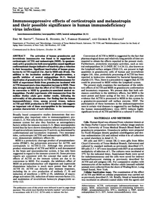
FIG. 5. Expression of ACTH and HIV nucleocapsid (p24) antigens in HIV-infected H9 cells. Infected cells were stained by immunofluo-
rescence with normal rabbit serum at 45 days after infection (A), with anti-ACTH at 25 days after infection (B), with anti-ACTH at 45 days after
infection (C), or with anti-p24 at 45 days after infection (D).
pituitary and adrenal glands can be altered in AIDS, led us to
examine this virus for induction of ACTH and MSH.
Fig. 5 shows that HIV infection of H9 T-lymphoma cells
induced the production of intracellular ACTH. Noninfected
H9 cells or normal rabbit serum-stained controls exhibited
only background staining for ACTH or the HIV p24 antigen.
To determine if ACTH-like peptides are secreted and pro-
cessed to MSH, radioimmunoassays were performed on
supernatant fluids ofcultures. Table 1 shows both ACTH and
MSH to be present. Significant amounts ofMSH were found,
and on a molar basis this suggests that there are approxi-
mately 2 to 3 times more MSH than ACTH molecules.
DISCUSSION
Previous reports have shown that the presence ofACTH and
MSH, individually, can modulate immune responses. The
data presented here provide compelling evidence that the
cellular immunosuppression attributed to ACTH may actu-
ally be a composite ofACTH and MSH actions. Specifically,
we have shown that MSH acts rapidly, within minutes of
application to the cells. Conversely, ACTH requires hours,
which is sufficient time for its processing into MSH. ACTH
also blocks MSH activity, but only when added concomi-
tantly, presumably by competing with MSH for binding to its
receptor. Since there appears to be some tolerance of the
MSH receptor for ACTH (27), the data are consistent with
the view of competition.
Alternatively, ACTH receptors could be present on the
cells in addition to MSH binding sites. However, they would
Table 1. ACTH and MSH production by HIV-infected H9 cells
Time, Hormone conc., pg/ml
days ACTH MSH
0 ND ND
15 32.0 + 0.1 40.0 ± 4.2
22 47.5 ± 12.0 ND
35 29.0 ± 5.6 42.0 ± 2.8
58 53.5 ± 9.2 38.5 ± 2.1
Supernatant fluids from HIV-infected cultures were collected at
the indicated times after infection and radioimmunoassayed for
ACTH or MSH. Results are mean ± SD. ND, none detected.
have to be slower acting and have lower affinity than the
MSH binding sites and somehow inhibit intracellular action
ofMSH. Thus, processing ofACTH to MSH is supported by
kinetics and similarity of action, detection of MSH subse-
quent to addition ofACTH, plus inhibition ofthe proteolytic
mechanism thought to generate MSH.
Since ACTH does bind with high affinity to lymphocytes
(28, 29) and activates their intracellular pathways (30, 31), it
undoubtedly has its own direct immunomodulatory effects.
The effects of ACTH described here probably are a consol-
idation of ACTH and MSH activities, dependent upon the
cell types and numbers, kinetics, and the presence ofmultiple
other regulatory factors such as lymphokines and cortico-
steroid hormones.
The results suggest that at least part if not all of the
proteolytic processing of ACTH to MSH is phosphoramidon
sensitive. This strongly implicates NEP (13, 14) as one ofthe
proteases involved. This would be a mode of operation for
NEP not previously described and one different from the
classical trypsin-like cleavage generally thought to process
POMC (32). These results do not eliminate the possibility of
other proteolytic enzymes being involved. A carboxypepti-
dase or an endopeptidase plus several newly cloned conver-
tases have been shown to generate an ACTH-(1-16) fragment
and one or all of these enzymes may be phosphoramidon
sensitive (32). Harbour et al. (15) reported an acid-dependent
proteolytic activity associated with B lymphocytes that is
induced by bacterial endotoxin. This enzyme truncates
ACTH-(1-39) to a species approximately 24 residues in
length, a result which indicates that it will be important to
determine the relationships of these enzymes in immuno-
modulation. NEP cleavage occurs at the amino side of
hydrophobic residues (13). A candidate site on ACTH for
cleavage by NEP could be residues 12-13 (Pro-Val), gener-
ating ACTH-(1-12). Such a fragment should retain MSH-like
immunoreactivity and bioactivity. In addition, blockage of
MSH degradation by phosphoramidon (Fig. 3) is consistent
with NEP activity (23). Degradation of MSH by NEP could
represent another immunoregulatory mechanism.
Shipp et al. (14) found a regulation ofenkephalin signals in
lymphoid cells by coexpression of cell surface opioid recep-
tors and the CD10/NEP enzyme. Considered in this context,
Immunology: Smith et al.
5. Proc. Natl. Acad. Sci. USA 89 (1992)
our results showing induction of ACTH by HIV in a T-cell
line illustrate the broad implications of this ACTH -+ MSH
conversion mechanism. Its universality is suggested, since
additional viruses have been shown to induce ACTH forma-
tion in lymphocytes. Our HIV results do contrast with those
ofOates etal. (33), but their failure tofind induction ofPOMC
mRNA after infection may be due to differences in the length
of infection time.
We are not certain what roles may be played by ACTH and
MSH in AIDS or other viral infections. However, alterations
in the functions of the pituitary and adrenal glands have been
reported and may be a common feature of virus infections in
general (24, 25, 33, 34). ACTH from lymphocytes may
contribute to these systemic effects (24) as undoubtedly do
other cytokines such as interleukin 1 (21, 35). Presumably,
the induction of ACTH and MSH formation is adaptive for
either the virus or the host. MSH or other related fragments
might be active in enhancing or inhibiting HIV replication
directly. The ability to inactivate granulocytes means that
neuropeptides debilitate host defense mechanisms, particu-
larly those that might protect against opportunistic infection.
Also, ACTH induces tumor necrosis factor a production in
vitro (11), which if it occurred in vivo might contribute to the
wasting seen in AIDS.
The overall means by which HIV compromises the host's
immune system is not known. There are many incongruous
features ofHIV infection, such as that the number ofinfected
cells is too low to account for the magnitude of immunode-
ficiency (36, 37). The overall effect is probably due to
multiple factors rangingfrom manipulation ofnormal immune
functions to cytotoxic and interfering activities of viral pro-
teins (see ref. 38 for review).
When considered with our findings, it becomes apparent
that the immunosuppression can result from many causes.
MSH has been shown to have multiple immunomodulatory
effects such as antipyresis, inhibition of polymorphonuclear
cell mobilization, and inhibition of cytokine production (4,
39). ACTH inhibits production of IFN-y (7) and activation of
macrophages by IFN-y (10). ACTH is induced by a number
of stimuli, either at the pituitary gland or lymphoid sites,
ranging from "stress" and circadian rhythms to pathogenic
stimuli (viruses, bacterial lipopolysaccharide, tumor cells)
(1). Since suppressed immune responses are associated with
many of these conditions, ACTH's processing into MSH,
which has a stronger and faster effect, may be a fundamental
mechanism for debilitating host defenses. Additionally, since
the conversion ofACTH to MSH may require NEP found on
the surface of only certain lymphoid cells, the immunosup-
pressive phenomenon may initially reside at the local level
and then be followed by a broader suppression of other cells
in the vicinity of the MSH.
The authors thank Ms. Anne Millard for excellent technical
assistance and Dr. Berta Scharrer for excellent conceptual and
editorial input. This research was supported in part by grants from
the National Institutes of Health (DK41034-03 and MH08180), the
Office of Naval Research (N00014-J-1095 and N00014J-89-J1%2),
and the Alcohol, Drug Abuse, and Mental Health Administration
(MARC 17138).
1. Smith, E. M., Hughes, T. K., Leung, M. K. & Stefano, G. B.
(1991) Adv. Neuroimmunol. 1, 7-16.
2. Stefano, G. B., Leung, M. K., Zhao, X. & Scharrer, B. (1989)
Proc. Natl. Acad. Sci. USA 86, 626-630.
3. Smith, E. M., Galin, F. S., LeBoeuf, R. D., Coppenhaver,
D. H., Harbour, D. V. & Blalock, J. E. (1990) Proc. Natl.
Acad. Sci. USA 87, 1057-1060.
4. Van Epps, D. E. & Mason, M. M. (1991) in Comparative
Aspects ofNeuropeptide Function, eds. Florey, E. & Stefano,
G. B. (Manchester Univ., Manchester, U.K.), pp. 335-345.
5. Stefano, G. B., Cadet, P. & Scharrer, B. (1989) Proc. Nat!.
Acad. Sci. USA 86, 6307-6311.
6. Stefano, G. B., Smith, D. M., Smith, E. M. & Hughes, T. K.
(1991) in Molluscan Neurobiology, eds. Boer, H., Garearts, G.
& Joosse, J. (Elsevier/North Holland, Amsterdam), in press.
7. Johnson, H. M., Smith, E. M., Torres, B. A. & Blalock, J. E.
(1982) Proc. Nat!. Acad. Sci. USA 79, 4171-4174.
8. Bost, K. L., Clarke, B. L., Xu, J., Kiyono, H., McGhee, J. R.
& Pascual, D. (1990) J. Immunol. 145, 4326-4331.
9. Johnson, H. M., Torres, B. A., Smith, E. M., Dion, L. D. &
Blalock, J. E. (1984) J. Immunol. 132, 246-250.
10. Koff, W. C. & Dunegan, M. A. (1985) J. Immunol. 135, 350-
354.
11. Hughes, T. K. & Smith, E. M. (1989) J. Biol. Regul. Homeo-
static Agents 3, 163-166.
12. Ottaviani, E., Caselgrandi, E., Bondi, M., Cossarizza, A.,
Monti, D. & Franceschi, C. (1991) Adv. Neuroimmunol. 1,
27-39.
13. Turner, A. J., Matsas, R. & Kenny, A. J. (1985) Biochem.
Pharmacol. 34, 1347-1356.
14. Shipp, M. A., Stefano, G. B., D'Adamio, L., Switzer, S. N.,
Howard, F. D., Sinisterra, J., Scharrer, B. & Reinherz, E. L.
(1990) Nature (London) 347, 394-3%.
15. Harbour, D. V., Smith, E. M. & Blalock, J. E. (1987) J.
Neurosci. Res. 18, 95-101.
16. Boyum, A. (1968) Scand. J. Clin. Lab. Invest. 21, 77-89.
17. Cloyd, M. W. & Moore, B. E. (1990) Virology 174, 103-116.
18. Smith, E. M. & Blalock, J. E. (1981) Proc. Natl. Acad. Sci.
USA 78, 7530-7534.
19. Cushman, D. W., Cheung, H. S., Sabo, E. F. & Ondetti,
M. A. (1977) Biochemistry 16, 5484-5491.
20. Hughes, T. K., Smith, E. M., Chin, R., Cadet, P., Sinisterra,
J., Leung, M. K., Shipp, M. A., Scharrer, B. A. & Stefano,
G. B. (1990) Proc. Natl. Acad. Sci. USA 87, 4426-4429.
21. Woloski, B. M. R. N. J., Smith, E. M., Meyer, W. J., III,
Fuller, G. M. & Blalock, J. E. (1985) Science 230, 1035-1037.
22. Hughes, T. K., Smith, E. M., Barnett, J. A., Charles, R. &
Stefano, G. B. (1991) Dev. Comp. Immunol. 15, 117-122.
23. Deschodt-Lanckman, M., Vanneste, Y., Loir, B., Michel, A.,
Libert, A., Ghanem, G. & Lejeune, F. (1990) Int. J. Cancer 46,
1124-1130.
24. Smith, E. M., Meyer, W. J., II, & Blalock, J. E. (1982)
Science 218, 1311-1312.
25. Westly, H. J., Kleiss, A. J., Kelley, K. W., Wong, P. K. Y. &
Yuen, P. H. (1986) J. Exp. Med. 163, 1589-1594.
26. Smith, E. M., Hashemi, F. & Hughes, T. K. (1991) FASEB J.
5, 1486 (abstr.).
27. Sayers, G., Seelig, S. & Kumar, S. (1975) J. Steroid Biochem.
6, 371-375.
28. Smith, E. M., Brosnan, P., Meyer, W. J., III, & Blalock, J. E.
(1987) N. Engl. J. Med. 317, 1266-1269.
29. Clarke, B. & Bost, K. L. (1989) J. Immunol. 143, 464-469.
30. Johnson, E. W., Blalock, J. E. & Smith, E. M. (1988) Bio-
chem. Biophys. Res. Commun. 157, 1205-1211.
31. Kavelaars, A., Ballieux, R. E. & Heijnen, C. (1988) Brain
Behav. Immun. 2, 57-66.
32. Scott, A. P., Ratcliffe, J. G., Rees, L. H., Landon, J., Bennett,
H. P. J., Lowry, P. J. &McMartin, C. (1973) Nature NewBiol.
244, 65-67.
33. Oates, E. L., Allaway, G. P., Armstrong, G. R., Boyajian,
R. A., Kehrl, J. H. & Prabhakar, B. S. (1988) J. Biol. Chem.
263, 10041-10044.
34. Dunn, A. J., Powell, M. L., Meitin, C. & Small, P. A., Jr.
(1989) Physiol. Behav. 45, 591-594.
35. Smith, E. M. (1988) Prog. Allergy 43, 121-139.
36. Harper, M., Marselle, L., Gallo, R. & Wong-Staal, F. (1986)
Proc. Natl. Acad. Sci. USA 83, 772-776.
37. Duesberg, P. (1988) Science 241, 514.
38. Rosenberg, Z. F. & Fauci, A. S. (1990) Immunol. Today 11,
176-180.
39. Lipton, J. M. (1990) Yale J. Biol. Med. 63, 173-182.
786 Immunology: Smith et al.