SlideShare a Scribd company logo
1 of 94
DR. FARRUKH JAVEED
CONSULTANT NEUROSURGEON
JPMC, Karachi.
LOBES OF THE
CEREBRUM
PARIETAL
LOBE
TEMPORAL LOBE
FRONTAL LOBE
LIMBIC LOBE
OCCIPITAL LOBE
FRONTAL LOBE
The frontal lobe is the area of
the brain responsible for higher
cognitive functions.
Problem solving
• Memory
• Language
• Motivation
• Judgment
• Impulse control
• Social and sexual behavior.
TEMPORAL LOBE
The temporal lobe plays
a role in emotions, and
is also responsible for
smelling, tasting,
perception, memory,
understanding music,
aggressiveness, and
sexual behavior.
The temporal lobe also
contains the language
area of the brain.
PARIETAL LOBE
The parietal lobe plays a role in
our sensations of touch, smell,
and taste.
It also processes sensory and
spatial awareness, and is a key
component in eye-hand co-
ordination and arm movement.
The parietal lobe also contains a
specialized area called
Wernicke’s area that is
responsible for matching written
words with the sound of spoken
speech.
OCCIPITAL LOBE
The occipital
lobe is at the
rear of the brain
and controls
vision and
recognition.
LANGUAGE
Wernicke’s area is a
specialized portion of the
parietal lobe that recognizes
and understands written and
spoken language.
Wernicke’s area surrounds
the auditory association
area.
Damage to this part of the
brain can result in someone
hearing speech, but not
understanding it.
Wernicke’s
Area
Auditory Association
Area
HEARING
There are two auditory
areas of the brain:
• The primary auditory area
(brown circle) is what
detects sounds that are
transmitted from the ear. It
is located in the sensory
cortex.
• The auditory association
area (purple circle) is the
part of the brain that is
used to recognize the
sounds as speech, music,
or noise.
SPEECH
Broca’s area is where we
formulate speech and the
area of the brain that
sends motor instructions
to the motor cortex.
Injury to Broca’s area can
cause difficulty in
speaking. The individual
may know what words he
or she wishes to speak,
but will be unable to do
so.
Broca’s Area
MOTOR CORTEX
The motor portion of the
cerebrum is illustrated here. The
light red area is the premotor
cortex, which is responsible for
repetitive motions of learned
motor skills. The dark red area is
the primary motor area, and is
responsible for control of skeletal
muscles.
Different areas of the brain are
associated with different parts of
the body.
Injury to the motor cortex can
result in motor disturbance in the
associated body part.
SENSORY CORTEX
The sensory portion of the
cerebrum is illustrated here.
Different areas of the brain
are associated with different
parts of the body, as can be
seen below.
Injury to the sensory cortex
can result in sensory
disturbance in the associated
body part.
CEREBELLUM
The cerebellum is connected
to the brainstem, and is the
center for body movement
and balance.
INCIDENCE OF CNS
TUMORS
 One-third of CNS tumors
are metastatic lesions
 One third are gliomas
and
 One-third is of nonglial
origin.
World Health Organization
(WHO) Classification
Categorized & considered by cell layer of origin
 Tumors of Neuroepithelial tissue
 Tumors of Meninges
 Tumors of Craniospinal Nerves
 Hematopoietic Neoplasms
 Germ Cell Tumors
 Sellar Tumors
 Metastatic Tumors
Brain Tumors in General
 Presentation – Headache, seizure, neurological
deficit, visual loss, personality changes, etc
 History – Progressive onset of symptoms
(weeks to months)
 Physical Exam – Depends upon brain region
affected
HEADACHE
ONSET
• ABRUPT(waking from sleep)
• Triggered by exertion or Valsalva
CHANGE
• First or worst
• Change or progression in pattern
• New headache >50yrs
• New headache in cancer or AIDS pts
ASSOCIATED
• Abnormal neurological exam
• Neurological symptoms > 1 hours
• Associated with altered / loss of consciousness
TYPES OF BRAIN
HERNIATION
DIAGNOSIS
 Imaging Studies – CT, MRI, PET
 Laboratory Studies – Important for
systemic tumors and metastasis, e.g.
leukemia •
Treatment & Prognosis
 Varies per histology of specific tumors
CARE OF THE BRAIN TUMOR
PATIENT
Steroids
 Help resolve edema and symptoms
associated with it
 Can confuse the issue of lymphoma,
because rapid “disappearance” is seen for
lymphoma, followed by recurrence
Anti-convulsants
 Recommended particularly for supra
tentorial & epileptogenic areas, e.g. mesial
temporal lobe, and with past h/o seizures
Treatment Options in general
for Intracranial Tumors
 Need Biopsy for definitve identification…
then one or combination of the following:
 Surgery
 Radiation Therapy
 Whole brain
 Focused beam
 Gamma Knife Stereotactic Radiosurgery
 Chemotherapy
 Systemic
 Local
COMMON INTRAAXIAL TUMORS IN
ADULTS
Supratentorial Infratentorial
Metastasis Metastasis
Gliomas
Fibrillary Astrocytoma
Anaplastic Astrocytoma
Glioblastoma Multiforme
Oligodendroglioma
Hemangioblastoma
COMMON INTRAAXIAL TUMORS
IN PEDIATRICS
Supratentorial Infratentorial
Astrocytoma Juvenile Pilocytic Astrocytoma
Pleomorphic Xanthoastrocytoma PNET (Medulloblastoma)
PNET Ependymoma
DNET Brainstem Astrocytoma
Ganglioglioma
Although cancer is rare in children, brain tumors are the most
common type of childhood cancer after leukemia and
lymphoma.
SIGNS OF EXTRA AXIAL
LOCATION
 CSF cleft
 Displaced subarachnoid vessels
 Cortical gray between mass and white matter
 Displaced and expanded subarachnoid
spaces
 Broad dural base
 Bony reaction
 The T2W-images show a schwannoma located
in the cerebellopontine angle (CPA).
 CSF Cleft (yellow arrow)
 Displaced subarachnoid Space (blue arrow)
 Gray Matter between mass and White matter
(curved arrow)
 Wide CSF spaces (long arrow)
Meningioma (Enplaque variety)
 broad dural base and a dural tail of enhancement(blue
arrow)
 There is hyperostosis in the adjacent bone (yellow arrow)
 lesion enhances homogeneously
 A hyperintense contrast enhancing lesion
with extension to the prepontine area (blue
arrows) and into the foramen magnum (red
arrow).
 This Lesion proved to be Ependymoma
 Above image shows a diffusely infiltrating intra-axial tumor
occupying most of the right hemisphere with only a minimal
mass effect.
 Low-grade astrocytoma.
Midline Crossing Tumors
 Glioblastoma multiforme (GBM) frequently
crosses the midline by infiltrating the white
matter tracts of the corpus callosum.
 Radiation necrosis can look like recurrent GBM
and can sometimes cross the midline.
 Meningioma is an extra-axial tumor and can
spread along the meninges to the contralateral
side.
Continued
GBM Radiation Necrosis Meningioma
CALCIFICATIONS
Intraaxial
Tumors
Extraaxial
Tumors
Astrocytomas (20%) Meningiomas (25%)
Craniopharyngiomas (90%)
Oligodendrogliomas (80%)
Chordomas
Metastasis
Chondrosarcomas
Ependymomas (50%)
Choroid plexus papilloma
(25%)
Ganglioglioma (40%)
 The CT shows a
mass with
calcifications, which
extends all the way
to the cortex.
 The most likely
diagnosis is
Oligodendroglioma.
 A calcified mass seen in the
suprasellar region, causing
obstructive hydrocephalus.
 Craniopharyngiomas are slow
growing,calcified, cystic
tumors arising from remnants
of Rathke's cleft.
 They are located in the
suprasellar region and
primarily seen in children with
a small second peak incidence
in older adults.
 On the coronal and sagittal TW1I there is a large
mass centered around the sella with a broad dural
base. & extension into the sella.
 CT shows densely calcified tumor.
Melanoma GBM PNET
DIFFUSION WEIGHTED
IMAGING
 Normally water protons have the ability to diffuse
extracellularly and loose signal.
 High intensity on DWI indicates restriction of the
ability of water protons to diffuse extracellularly.
 Restricted diffusion is seen in abscesses,
epidermoid cysts and acute infarction
 In most tumors there is no restricted diffusion -
even in necrotic or cystic components. This
results in a normal, low signal on DWI.
 We see restriction in above left extreme image
as it is hyperintense, while lower extreme left
image shows no restriction.
 Schwannoma extending into the middle cranial
fossa with homogeneous enhancement (right).
 Primary Lymphoma shows vivid enhancement (left).
Contrast Enhancement
Meningioma Schwannoma Lymphoma Choroid Plexus
Papilloma
Contrast Enhancement
Hemangioblastoma Pilocytic Astrocytoma Ganglioglioma
 Above image shows glioblastoma multiforme (GBM).
 The enhancement indicates that this is a high-grade tumor
 There is also a cystic component with ring enhancement..
RING ENHANCEMENT
1. Metastasis
2. Glioblastoma Multiforme
3. Tuberculoma
4. Abscess,
5. Infectious disease Toxoplasma
6. Multiple Sclerosis
7. Chronic hematoma
Contrast Enhancement
 Many non-
tumorous
lesions can
mimic a
brain
tumor.
 Infections and vascular lesions can also
mimic a CNS tumor.
METASTATIC BRAIN
TUMOR
 History – Rapidly
progressive onset of
symptoms (weeks vs.
months)
 Most common sources
are LUNG, BREAST (in
women), Thyroid,
RENAL, & G.I. tract
DIAGNOSIS
Imaging Studies –
 MRI: Tumor at grey-white
matter junction, usually
associated with edema
Systemic work-up includes
 CT scan of chest, abdomen
and pelvis
 Mammography
 Bone scan
Diagnostic Laboratory Studies
 CBC, ESR, LFTs,
TREATMENT
 For solitary lesion or less
than 4 lesions all < 3 cm. –
biopsy if undiagnosed, plus
Gamma Knife
 For > 3 cm. tumor, surgery
followed by WBRT
 For > 4 lesions, biopsy for
diagnosis, plus whole brain
radiation therapy
 Prognosis: 7 – 12 mos.
GLIOMAS
Presentation
 Depends upon brain
location, usually H/A over
several weeks, seizure,
vomiting, visual chages or
word finding difficulty
Physical Exam –
 Depends upon brain region
affected
Diagnostic Imaging Studies –
 CT, MRI
Treatment
 Surgery, Radiation, Chemotherapy (varies with
age group & type)
Survival depends on pathology: –
 Pilocytic astrocytoma: resection Æ cure; Grade
1 Astrocytoma (nuclear atypia) – 8-10 yrs;
 Grade 2 Anaplastic astrocytoma (+endothelial
proliferation) – 2 yrs
 Grade 3 (+necrosis) GBM – 11 months
MENINGIOMA
Presentation
 Meningiomas occur in
middle aged females more
frequently
History
 Meningiomas are usually
slow-growing (months to
years)
 CT/MRI: Show meninges-
based tumor (usually), with
variable amount of edema
TREATMENT
 Meninigiomas: Gross total resection curative
 Need for radiation therapy depends on grade
Prognosis
 – Meningiomas: Can be cured with gross
total resection with favorable prognosis
PITUITARY ADENOMA
Presentation
 Headache, visual defect
(classically bitemporal
hemianopsia), endocrine
abnormalities (dependent
upon hormone secreted) –
 Prolactinoma most common
 ACTH with Cushing’s
disease (w/hypertension,
stria, buffalo hump, morbid
obesity);
 GH with acromegaly
History
 Slow-growing, over months to
years
 Headache with progressive
visual loss
 Most common presentation is
bitemporal hemi anopia
Diagnostic Laboratory Studies
 Endocrine panel
 Test IGF-1 for GH-secreting
tumor;
 Use dexamethasone
suppression test for Cushing’s
disease vs. ectopic Cushing’s
syndrome
Treatment
 Medical treatment for Prolactinoma only
with dopamine antagonists
 Surgical excision usually initial treatment
(usually transnasal approach), with
Gamma Knife for recurrence vs. local
focused radiation
Prognosis
 Generally benign, so proportional to gross
total resection & control of endocrine
effects;
Vestibular schwannoma
Presentation
 Hearing difficulty in
affected ear, vertigo,
headache/facial
weakness/facial
numbness if large
tumor compressing
trigeminal nerve
Physical Exam
 Evaluate facial
weakness & hearing
ability
Diagnostic Imaging
Studies
 MRI Brain with
MRA/MRV
OTHERS
 Audiometry
NEURO FIBROMOTOSIS
Treatment
 Surgical resection if >3 cm. or causing
excessive mass effect, brainstem
compression; GK for smaller lesions (<3 cm.)
Prognosis
 Benign tumor, so good with total excision and
lack of recurrence
Lymphoma
Presentation
 Sporadic, more common in
immunosuppresed or
immunocompromised
patients (e.g. HIV+); may or
may not be systemic @
presentation.
 Spinal cord (epidural)
compression or
carcinomatous meningitis
 Multiple cranial nerve deficits
Diagnostic Imaging
Studies
 CT / MRI Brain
Treatment
 Biopsy for establishing
diagnosis, followed by
radiation; chemotherapy
(intra thecal
methotrexate) has
shown increased
survival
PROGNOSIS
 No treatment 2.5 months.;
 XRT 10 months
 Intraventricular methotrexate 41 months.
 Prognosis worse in AIDS pts. (4 months)
Pineal Region Tumors
Presentation
 Headache,
Hydrocephalus (if large)
Physical Exam
 May develop Perinaud’s
syndrome (up gaze
palsy, convergence, and
accomodation
impairment)
Diagnostic Imaging
Studies
 MRI
Diagnostic Laboratory
Studies
 CSF can be sent for
markers – AFP, HCG,
PLAP
 Tumor located in the pineal region with calcifications.
 This is most likely a germinoma.
Treatment
 Depends upon histology:
 Germinoma – biopsy & irradiate;
 Pineoblastoma: resect & irradiate
Prognosis
 Depends upon histology: Pineocytoma
better than germinoma, pineoblastoma
Lymphoma Meningioma GBM
Meningioma Lymphoma GBM
Multifocal Disease
 Multiple tumors in the brain usually indicate
metastatic disease.
 Primary brain tumors are typically seen in a
single region
 Some tumors can be multifocal as a
result of seeding metastases: this can
occur in medulloblastomas (PNET-MB),
ependymomas, GBMs and
oligodendrogliomas.
 Meningiomas and schwannomas can be
multiple, especially in neurofibromatosis
type II.
Metastasis Neurofibromatosis II
COMMON CP ANGLE
TUMORS
Schwannoma
Meningioma
Epidermoid
Arachnoid cyst
Paraganglioma
Metastasis
 Cystic schwannoma of Left CP Angle.
LOW GRADE
ASTROCYTOMA
CYSTIC TUMORS WITH MURAL
NODULE
Brain tumors for Undergraduate Students

More Related Content

What's hot

Presentation2.pptx. posterior fossa tumour
Presentation2.pptx. posterior fossa tumourPresentation2.pptx. posterior fossa tumour
Presentation2.pptx. posterior fossa tumourAbdellah Nazeer
 
Brain tumors 2020 by Assist.Prof. Ines Strenja MD, PhD Clinical hospital R...
Brain tumors  2020 by Assist.Prof. Ines Strenja  MD, PhD  Clinical hospital R...Brain tumors  2020 by Assist.Prof. Ines Strenja  MD, PhD  Clinical hospital R...
Brain tumors 2020 by Assist.Prof. Ines Strenja MD, PhD Clinical hospital R...Neurology Dpt, Clinical Hospital Rijeka
 
Imaging of white matter diseases
Imaging of white matter diseasesImaging of white matter diseases
Imaging of white matter diseasesNavni Garg
 
Radionuclide neuroendocrine tumors functional imaging
Radionuclide neuroendocrine tumors functional imagingRadionuclide neuroendocrine tumors functional imaging
Radionuclide neuroendocrine tumors functional imagingHussein Farghaly
 
Presentation1.pptx, radiological imaging of intra cranial calcification.
Presentation1.pptx, radiological imaging of intra cranial calcification.Presentation1.pptx, radiological imaging of intra cranial calcification.
Presentation1.pptx, radiological imaging of intra cranial calcification.Abdellah Nazeer
 
Imaging in stroke
Imaging in stroke Imaging in stroke
Imaging in stroke Deepak Garg
 
Neuroradiology craniopharyngioma
Neuroradiology craniopharyngiomaNeuroradiology craniopharyngioma
Neuroradiology craniopharyngiomaRoopchand Ps
 
MRI imaging of brain tumors. A practical approach.
MRI imaging of brain tumors. A practical approach. MRI imaging of brain tumors. A practical approach.
MRI imaging of brain tumors. A practical approach. hazem youssef
 
Liver lesions
Liver lesionsLiver lesions
Liver lesionsairwave12
 
Neuro radiology neuroimaging
Neuro radiology   neuroimagingNeuro radiology   neuroimaging
Neuro radiology neuroimagingMarwa Khalifa
 
Lytic leisons of the skull
Lytic leisons of the skullLytic leisons of the skull
Lytic leisons of the skullMilan Silwal
 
Radiological Evaluation of CNS Tumors
Radiological Evaluation of CNS TumorsRadiological Evaluation of CNS Tumors
Radiological Evaluation of CNS TumorsSubhash Thakur
 
42925901 brain-tumor
42925901 brain-tumor42925901 brain-tumor
42925901 brain-tumorMuhammad Adi
 

What's hot (20)

Presentation2.pptx. posterior fossa tumour
Presentation2.pptx. posterior fossa tumourPresentation2.pptx. posterior fossa tumour
Presentation2.pptx. posterior fossa tumour
 
Intra axial posterior fossa tumor
Intra axial posterior fossa tumorIntra axial posterior fossa tumor
Intra axial posterior fossa tumor
 
Cranial Ultrasound of neonate
Cranial Ultrasound of neonateCranial Ultrasound of neonate
Cranial Ultrasound of neonate
 
High Grade Glioma
High Grade GliomaHigh Grade Glioma
High Grade Glioma
 
Brain tumors 2020 by Assist.Prof. Ines Strenja MD, PhD Clinical hospital R...
Brain tumors  2020 by Assist.Prof. Ines Strenja  MD, PhD  Clinical hospital R...Brain tumors  2020 by Assist.Prof. Ines Strenja  MD, PhD  Clinical hospital R...
Brain tumors 2020 by Assist.Prof. Ines Strenja MD, PhD Clinical hospital R...
 
Imaging of white matter diseases
Imaging of white matter diseasesImaging of white matter diseases
Imaging of white matter diseases
 
Radionuclide neuroendocrine tumors functional imaging
Radionuclide neuroendocrine tumors functional imagingRadionuclide neuroendocrine tumors functional imaging
Radionuclide neuroendocrine tumors functional imaging
 
Presentation1.pptx, radiological imaging of intra cranial calcification.
Presentation1.pptx, radiological imaging of intra cranial calcification.Presentation1.pptx, radiological imaging of intra cranial calcification.
Presentation1.pptx, radiological imaging of intra cranial calcification.
 
Imaging in CNS Infections
Imaging in CNS InfectionsImaging in CNS Infections
Imaging in CNS Infections
 
Imaging in stroke
Imaging in stroke Imaging in stroke
Imaging in stroke
 
Neuroradiology craniopharyngioma
Neuroradiology craniopharyngiomaNeuroradiology craniopharyngioma
Neuroradiology craniopharyngioma
 
MRI imaging of brain tumors. A practical approach.
MRI imaging of brain tumors. A practical approach. MRI imaging of brain tumors. A practical approach.
MRI imaging of brain tumors. A practical approach.
 
Liver lesions
Liver lesionsLiver lesions
Liver lesions
 
Neuro radiology neuroimaging
Neuro radiology   neuroimagingNeuro radiology   neuroimaging
Neuro radiology neuroimaging
 
Medulloblastoma
MedulloblastomaMedulloblastoma
Medulloblastoma
 
Gliomas - Brain Tumor
Gliomas - Brain TumorGliomas - Brain Tumor
Gliomas - Brain Tumor
 
Lytic leisons of the skull
Lytic leisons of the skullLytic leisons of the skull
Lytic leisons of the skull
 
Brain Tumors
Brain TumorsBrain Tumors
Brain Tumors
 
Radiological Evaluation of CNS Tumors
Radiological Evaluation of CNS TumorsRadiological Evaluation of CNS Tumors
Radiological Evaluation of CNS Tumors
 
42925901 brain-tumor
42925901 brain-tumor42925901 brain-tumor
42925901 brain-tumor
 

Similar to Brain tumors for Undergraduate Students

Spinal cord lesions and its radiological imaging finding.
Spinal cord lesions and its radiological imaging finding.Spinal cord lesions and its radiological imaging finding.
Spinal cord lesions and its radiological imaging finding.Navneet Ranjan
 
Brain tumor imaginig 3 10th may 02
Brain tumor imaginig   3  10th may 02Brain tumor imaginig   3  10th may 02
Brain tumor imaginig 3 10th may 02PS Deb
 
Primitive Neuroectodermal Tumor.pptx
Primitive Neuroectodermal Tumor.pptxPrimitive Neuroectodermal Tumor.pptx
Primitive Neuroectodermal Tumor.pptxMedhatMoustafa3
 
Spinal tumour lecture - copy
Spinal tumour   lecture - copySpinal tumour   lecture - copy
Spinal tumour lecture - copywasek_bd
 
Essentials of gliomas
Essentials of gliomas Essentials of gliomas
Essentials of gliomas NeurologyKota
 
INTRAMEDULLARY TUMOR OF SPINAL CORD
INTRAMEDULLARY TUMOR OF SPINAL CORDINTRAMEDULLARY TUMOR OF SPINAL CORD
INTRAMEDULLARY TUMOR OF SPINAL CORDsuresh Bishokarma
 
imaging of itraconal mass EOS 2015.pptx
imaging of itraconal mass EOS 2015.pptximaging of itraconal mass EOS 2015.pptx
imaging of itraconal mass EOS 2015.pptxayman972811
 
Approach to CNS tumors Dr. Muhammad Bin Zulfiqar
Approach to CNS tumors Dr. Muhammad Bin ZulfiqarApproach to CNS tumors Dr. Muhammad Bin Zulfiqar
Approach to CNS tumors Dr. Muhammad Bin ZulfiqarDr. Muhammad Bin Zulfiqar
 
Supratentorial brain tumours
Supratentorial brain tumoursSupratentorial brain tumours
Supratentorial brain tumourstrial4neha
 
Brain and spinal cord tumours.pptx
Brain and spinal cord tumours.pptxBrain and spinal cord tumours.pptx
Brain and spinal cord tumours.pptxMohammedAbdela7
 
Oncologic conditions of brain and spinal cord
Oncologic conditions of brain and spinal cordOncologic conditions of brain and spinal cord
Oncologic conditions of brain and spinal cordBobby Abraham
 
Parapharyngeal swellings
Parapharyngeal swellingsParapharyngeal swellings
Parapharyngeal swellingsMai Hatem
 
Spinal tumors lecture
Spinal tumors lectureSpinal tumors lecture
Spinal tumors lecturetest
 
PINEAL REGION TUMORS DIAGNOSIS & SURGICAL APPROACHES.pptx
PINEAL REGION TUMORS DIAGNOSIS & SURGICAL APPROACHES.pptxPINEAL REGION TUMORS DIAGNOSIS & SURGICAL APPROACHES.pptx
PINEAL REGION TUMORS DIAGNOSIS & SURGICAL APPROACHES.pptxMedhatMoustafa3
 
Pleomorphic adenoma
Pleomorphic adenomaPleomorphic adenoma
Pleomorphic adenomaAhmed Shoeeb
 

Similar to Brain tumors for Undergraduate Students (20)

Spinal cord lesions and its radiological imaging finding.
Spinal cord lesions and its radiological imaging finding.Spinal cord lesions and its radiological imaging finding.
Spinal cord lesions and its radiological imaging finding.
 
Brain tumours
Brain tumoursBrain tumours
Brain tumours
 
Harbor UCLA Neuro-Radiology Case 6
Harbor UCLA Neuro-Radiology Case 6Harbor UCLA Neuro-Radiology Case 6
Harbor UCLA Neuro-Radiology Case 6
 
Brain tumor imaginig 3 10th may 02
Brain tumor imaginig   3  10th may 02Brain tumor imaginig   3  10th may 02
Brain tumor imaginig 3 10th may 02
 
Primitive Neuroectodermal Tumor.pptx
Primitive Neuroectodermal Tumor.pptxPrimitive Neuroectodermal Tumor.pptx
Primitive Neuroectodermal Tumor.pptx
 
Spinal tumour lecture - copy
Spinal tumour   lecture - copySpinal tumour   lecture - copy
Spinal tumour lecture - copy
 
Essentials of gliomas
Essentials of gliomas Essentials of gliomas
Essentials of gliomas
 
INTRAMEDULLARY TUMOR OF SPINAL CORD
INTRAMEDULLARY TUMOR OF SPINAL CORDINTRAMEDULLARY TUMOR OF SPINAL CORD
INTRAMEDULLARY TUMOR OF SPINAL CORD
 
Medulloblastomas
MedulloblastomasMedulloblastomas
Medulloblastomas
 
imaging of itraconal mass EOS 2015.pptx
imaging of itraconal mass EOS 2015.pptximaging of itraconal mass EOS 2015.pptx
imaging of itraconal mass EOS 2015.pptx
 
Approach to CNS tumors Dr. Muhammad Bin Zulfiqar
Approach to CNS tumors Dr. Muhammad Bin ZulfiqarApproach to CNS tumors Dr. Muhammad Bin Zulfiqar
Approach to CNS tumors Dr. Muhammad Bin Zulfiqar
 
Supratentorial brain tumours
Supratentorial brain tumoursSupratentorial brain tumours
Supratentorial brain tumours
 
Brain and spinal cord tumours.pptx
Brain and spinal cord tumours.pptxBrain and spinal cord tumours.pptx
Brain and spinal cord tumours.pptx
 
Meningioma
MeningiomaMeningioma
Meningioma
 
Oncologic conditions of brain and spinal cord
Oncologic conditions of brain and spinal cordOncologic conditions of brain and spinal cord
Oncologic conditions of brain and spinal cord
 
Parapharyngeal swellings
Parapharyngeal swellingsParapharyngeal swellings
Parapharyngeal swellings
 
Spinal tumors lecture
Spinal tumors lectureSpinal tumors lecture
Spinal tumors lecture
 
PINEAL REGION TUMORS DIAGNOSIS & SURGICAL APPROACHES.pptx
PINEAL REGION TUMORS DIAGNOSIS & SURGICAL APPROACHES.pptxPINEAL REGION TUMORS DIAGNOSIS & SURGICAL APPROACHES.pptx
PINEAL REGION TUMORS DIAGNOSIS & SURGICAL APPROACHES.pptx
 
Testicular tumours
Testicular tumoursTesticular tumours
Testicular tumours
 
Pleomorphic adenoma
Pleomorphic adenomaPleomorphic adenoma
Pleomorphic adenoma
 

More from Farrukh Javeed

Understanding CT Scan and MRI Brain for Undergraduate Medical Students
Understanding CT Scan and MRI Brain for Undergraduate Medical StudentsUnderstanding CT Scan and MRI Brain for Undergraduate Medical Students
Understanding CT Scan and MRI Brain for Undergraduate Medical StudentsFarrukh Javeed
 
Head injury for Undergraduate Medical Students
Head injury for Undergraduate Medical StudentsHead injury for Undergraduate Medical Students
Head injury for Undergraduate Medical StudentsFarrukh Javeed
 
Hydrocephalus for Undergraduate Medical Students
Hydrocephalus for Undergraduate Medical StudentsHydrocephalus for Undergraduate Medical Students
Hydrocephalus for Undergraduate Medical StudentsFarrukh Javeed
 
Neural Tube Defects for Undergraduate Medical Students
Neural Tube Defects for Undergraduate Medical StudentsNeural Tube Defects for Undergraduate Medical Students
Neural Tube Defects for Undergraduate Medical StudentsFarrukh Javeed
 
Farrukh neurosurgery long case history &amp; examination technique
Farrukh   neurosurgery long case history &amp; examination techniqueFarrukh   neurosurgery long case history &amp; examination technique
Farrukh neurosurgery long case history &amp; examination techniqueFarrukh Javeed
 
Radiology quiz - Oligodendroglioma
Radiology quiz - OligodendrogliomaRadiology quiz - Oligodendroglioma
Radiology quiz - OligodendrogliomaFarrukh Javeed
 
Principles of Patient Counseling
Principles of Patient CounselingPrinciples of Patient Counseling
Principles of Patient CounselingFarrukh Javeed
 
Mechanical Ventilation
Mechanical VentilationMechanical Ventilation
Mechanical VentilationFarrukh Javeed
 
Cervical Fusion: C1-C2 Fusion
Cervical Fusion: C1-C2 FusionCervical Fusion: C1-C2 Fusion
Cervical Fusion: C1-C2 FusionFarrukh Javeed
 
Nasal Cavity and Sphenoid Sinus Anatomy
Nasal Cavity and Sphenoid Sinus AnatomyNasal Cavity and Sphenoid Sinus Anatomy
Nasal Cavity and Sphenoid Sinus AnatomyFarrukh Javeed
 
Foramen Magnum Meningioma
Foramen Magnum MeningiomaForamen Magnum Meningioma
Foramen Magnum MeningiomaFarrukh Javeed
 
Meningeal Hemangiopericytoma
Meningeal HemangiopericytomaMeningeal Hemangiopericytoma
Meningeal HemangiopericytomaFarrukh Javeed
 
Intraoperative Neurophysiological Monitoring Brain
Intraoperative Neurophysiological Monitoring BrainIntraoperative Neurophysiological Monitoring Brain
Intraoperative Neurophysiological Monitoring BrainFarrukh Javeed
 
Parasagittal Meningioma
Parasagittal MeningiomaParasagittal Meningioma
Parasagittal MeningiomaFarrukh Javeed
 
Petroclival Meningioma
Petroclival MeningiomaPetroclival Meningioma
Petroclival MeningiomaFarrukh Javeed
 

More from Farrukh Javeed (20)

Understanding CT Scan and MRI Brain for Undergraduate Medical Students
Understanding CT Scan and MRI Brain for Undergraduate Medical StudentsUnderstanding CT Scan and MRI Brain for Undergraduate Medical Students
Understanding CT Scan and MRI Brain for Undergraduate Medical Students
 
Head injury for Undergraduate Medical Students
Head injury for Undergraduate Medical StudentsHead injury for Undergraduate Medical Students
Head injury for Undergraduate Medical Students
 
Hydrocephalus for Undergraduate Medical Students
Hydrocephalus for Undergraduate Medical StudentsHydrocephalus for Undergraduate Medical Students
Hydrocephalus for Undergraduate Medical Students
 
Neural Tube Defects for Undergraduate Medical Students
Neural Tube Defects for Undergraduate Medical StudentsNeural Tube Defects for Undergraduate Medical Students
Neural Tube Defects for Undergraduate Medical Students
 
Farrukh neurosurgery long case history &amp; examination technique
Farrukh   neurosurgery long case history &amp; examination techniqueFarrukh   neurosurgery long case history &amp; examination technique
Farrukh neurosurgery long case history &amp; examination technique
 
Radiology quiz - Oligodendroglioma
Radiology quiz - OligodendrogliomaRadiology quiz - Oligodendroglioma
Radiology quiz - Oligodendroglioma
 
Urinary Incontinence
Urinary IncontinenceUrinary Incontinence
Urinary Incontinence
 
Principles of Patient Counseling
Principles of Patient CounselingPrinciples of Patient Counseling
Principles of Patient Counseling
 
Mechanical Ventilation
Mechanical VentilationMechanical Ventilation
Mechanical Ventilation
 
Cervical Fusion: C1-C2 Fusion
Cervical Fusion: C1-C2 FusionCervical Fusion: C1-C2 Fusion
Cervical Fusion: C1-C2 Fusion
 
Aneurysmal Bone Cyst
Aneurysmal Bone CystAneurysmal Bone Cyst
Aneurysmal Bone Cyst
 
Nasal Cavity and Sphenoid Sinus Anatomy
Nasal Cavity and Sphenoid Sinus AnatomyNasal Cavity and Sphenoid Sinus Anatomy
Nasal Cavity and Sphenoid Sinus Anatomy
 
Foramen Magnum Meningioma
Foramen Magnum MeningiomaForamen Magnum Meningioma
Foramen Magnum Meningioma
 
Hemangioblastoma
HemangioblastomaHemangioblastoma
Hemangioblastoma
 
Meningeal Hemangiopericytoma
Meningeal HemangiopericytomaMeningeal Hemangiopericytoma
Meningeal Hemangiopericytoma
 
Hypothalamic Gliomas
Hypothalamic GliomasHypothalamic Gliomas
Hypothalamic Gliomas
 
Intraoperative Neurophysiological Monitoring Brain
Intraoperative Neurophysiological Monitoring BrainIntraoperative Neurophysiological Monitoring Brain
Intraoperative Neurophysiological Monitoring Brain
 
Parasagittal Meningioma
Parasagittal MeningiomaParasagittal Meningioma
Parasagittal Meningioma
 
Petroclival Meningioma
Petroclival MeningiomaPetroclival Meningioma
Petroclival Meningioma
 
Stereotaxy Brain
Stereotaxy BrainStereotaxy Brain
Stereotaxy Brain
 

Recently uploaded

Lippincott Microcards_ Microbiology Flash Cards-LWW (2015).pdf
Lippincott Microcards_ Microbiology Flash Cards-LWW (2015).pdfLippincott Microcards_ Microbiology Flash Cards-LWW (2015).pdf
Lippincott Microcards_ Microbiology Flash Cards-LWW (2015).pdfSreeja Cherukuru
 
Presentació "Real-Life VR Integration for Mild Cognitive Impairment Rehabilit...
Presentació "Real-Life VR Integration for Mild Cognitive Impairment Rehabilit...Presentació "Real-Life VR Integration for Mild Cognitive Impairment Rehabilit...
Presentació "Real-Life VR Integration for Mild Cognitive Impairment Rehabilit...Badalona Serveis Assistencials
 
Nutrition of OCD for my Nutritional Neuroscience Class
Nutrition of OCD for my Nutritional Neuroscience ClassNutrition of OCD for my Nutritional Neuroscience Class
Nutrition of OCD for my Nutritional Neuroscience Classmanuelazg2001
 
COVID-19 (NOVEL CORONA VIRUS DISEASE PANDEMIC ).pptx
COVID-19  (NOVEL CORONA  VIRUS DISEASE PANDEMIC ).pptxCOVID-19  (NOVEL CORONA  VIRUS DISEASE PANDEMIC ).pptx
COVID-19 (NOVEL CORONA VIRUS DISEASE PANDEMIC ).pptxBibekananda shah
 
Case Report Peripartum Cardiomyopathy.pptx
Case Report Peripartum Cardiomyopathy.pptxCase Report Peripartum Cardiomyopathy.pptx
Case Report Peripartum Cardiomyopathy.pptxNiranjan Chavan
 
Glomerular Filtration and determinants of glomerular filtration .pptx
Glomerular Filtration and  determinants of glomerular filtration .pptxGlomerular Filtration and  determinants of glomerular filtration .pptx
Glomerular Filtration and determinants of glomerular filtration .pptxDr.Nusrat Tariq
 
Hematology and Immunology - Leukocytes Functions
Hematology and Immunology - Leukocytes FunctionsHematology and Immunology - Leukocytes Functions
Hematology and Immunology - Leukocytes FunctionsMedicoseAcademics
 
SGK HÓA SINH NĂNG LƯỢNG SINH HỌC 2006.pdf
SGK HÓA SINH NĂNG LƯỢNG SINH HỌC 2006.pdfSGK HÓA SINH NĂNG LƯỢNG SINH HỌC 2006.pdf
SGK HÓA SINH NĂNG LƯỢNG SINH HỌC 2006.pdfHongBiThi1
 
Giftedness: Understanding Everyday Neurobiology for Self-Knowledge
Giftedness: Understanding Everyday Neurobiology for Self-KnowledgeGiftedness: Understanding Everyday Neurobiology for Self-Knowledge
Giftedness: Understanding Everyday Neurobiology for Self-Knowledgeassessoriafabianodea
 
April 2024 ONCOLOGY CARTOON by DR KANHU CHARAN PATRO
April 2024 ONCOLOGY CARTOON by  DR KANHU CHARAN PATROApril 2024 ONCOLOGY CARTOON by  DR KANHU CHARAN PATRO
April 2024 ONCOLOGY CARTOON by DR KANHU CHARAN PATROKanhu Charan
 
SYNDESMOTIC INJURY- ANATOMICAL REPAIR.pptx
SYNDESMOTIC INJURY- ANATOMICAL REPAIR.pptxSYNDESMOTIC INJURY- ANATOMICAL REPAIR.pptx
SYNDESMOTIC INJURY- ANATOMICAL REPAIR.pptxdrashraf369
 
maternal mortality and its causes and how to reduce maternal mortality
maternal mortality and its causes and how to reduce maternal mortalitymaternal mortality and its causes and how to reduce maternal mortality
maternal mortality and its causes and how to reduce maternal mortalityhardikdabas3
 
Wessex Health Partners Wessex Integrated Care, Population Health, Research & ...
Wessex Health Partners Wessex Integrated Care, Population Health, Research & ...Wessex Health Partners Wessex Integrated Care, Population Health, Research & ...
Wessex Health Partners Wessex Integrated Care, Population Health, Research & ...Wessex Health Partners
 
PNEUMOTHORAX AND ITS MANAGEMENTS.pdf
PNEUMOTHORAX   AND  ITS  MANAGEMENTS.pdfPNEUMOTHORAX   AND  ITS  MANAGEMENTS.pdf
PNEUMOTHORAX AND ITS MANAGEMENTS.pdfDolisha Warbi
 
PULMONARY EMBOLISM AND ITS MANAGEMENTS.pdf
PULMONARY EMBOLISM AND ITS MANAGEMENTS.pdfPULMONARY EMBOLISM AND ITS MANAGEMENTS.pdf
PULMONARY EMBOLISM AND ITS MANAGEMENTS.pdfDolisha Warbi
 
Apiculture Chapter 1. Introduction 2.ppt
Apiculture Chapter 1. Introduction 2.pptApiculture Chapter 1. Introduction 2.ppt
Apiculture Chapter 1. Introduction 2.pptkedirjemalharun
 
epilepsy and status epilepticus for undergraduate.pptx
epilepsy and status epilepticus  for undergraduate.pptxepilepsy and status epilepticus  for undergraduate.pptx
epilepsy and status epilepticus for undergraduate.pptxMohamed Rizk Khodair
 
The next social challenge to public health: the information environment.pptx
The next social challenge to public health:  the information environment.pptxThe next social challenge to public health:  the information environment.pptx
The next social challenge to public health: the information environment.pptxTina Purnat
 
Introduction to Sports Injuries by- Dr. Anjali Rai
Introduction to Sports Injuries by- Dr. Anjali RaiIntroduction to Sports Injuries by- Dr. Anjali Rai
Introduction to Sports Injuries by- Dr. Anjali RaiGoogle
 

Recently uploaded (20)

Lippincott Microcards_ Microbiology Flash Cards-LWW (2015).pdf
Lippincott Microcards_ Microbiology Flash Cards-LWW (2015).pdfLippincott Microcards_ Microbiology Flash Cards-LWW (2015).pdf
Lippincott Microcards_ Microbiology Flash Cards-LWW (2015).pdf
 
Presentació "Real-Life VR Integration for Mild Cognitive Impairment Rehabilit...
Presentació "Real-Life VR Integration for Mild Cognitive Impairment Rehabilit...Presentació "Real-Life VR Integration for Mild Cognitive Impairment Rehabilit...
Presentació "Real-Life VR Integration for Mild Cognitive Impairment Rehabilit...
 
Nutrition of OCD for my Nutritional Neuroscience Class
Nutrition of OCD for my Nutritional Neuroscience ClassNutrition of OCD for my Nutritional Neuroscience Class
Nutrition of OCD for my Nutritional Neuroscience Class
 
COVID-19 (NOVEL CORONA VIRUS DISEASE PANDEMIC ).pptx
COVID-19  (NOVEL CORONA  VIRUS DISEASE PANDEMIC ).pptxCOVID-19  (NOVEL CORONA  VIRUS DISEASE PANDEMIC ).pptx
COVID-19 (NOVEL CORONA VIRUS DISEASE PANDEMIC ).pptx
 
Case Report Peripartum Cardiomyopathy.pptx
Case Report Peripartum Cardiomyopathy.pptxCase Report Peripartum Cardiomyopathy.pptx
Case Report Peripartum Cardiomyopathy.pptx
 
Glomerular Filtration and determinants of glomerular filtration .pptx
Glomerular Filtration and  determinants of glomerular filtration .pptxGlomerular Filtration and  determinants of glomerular filtration .pptx
Glomerular Filtration and determinants of glomerular filtration .pptx
 
Hematology and Immunology - Leukocytes Functions
Hematology and Immunology - Leukocytes FunctionsHematology and Immunology - Leukocytes Functions
Hematology and Immunology - Leukocytes Functions
 
SGK HÓA SINH NĂNG LƯỢNG SINH HỌC 2006.pdf
SGK HÓA SINH NĂNG LƯỢNG SINH HỌC 2006.pdfSGK HÓA SINH NĂNG LƯỢNG SINH HỌC 2006.pdf
SGK HÓA SINH NĂNG LƯỢNG SINH HỌC 2006.pdf
 
Giftedness: Understanding Everyday Neurobiology for Self-Knowledge
Giftedness: Understanding Everyday Neurobiology for Self-KnowledgeGiftedness: Understanding Everyday Neurobiology for Self-Knowledge
Giftedness: Understanding Everyday Neurobiology for Self-Knowledge
 
Epilepsy
EpilepsyEpilepsy
Epilepsy
 
April 2024 ONCOLOGY CARTOON by DR KANHU CHARAN PATRO
April 2024 ONCOLOGY CARTOON by  DR KANHU CHARAN PATROApril 2024 ONCOLOGY CARTOON by  DR KANHU CHARAN PATRO
April 2024 ONCOLOGY CARTOON by DR KANHU CHARAN PATRO
 
SYNDESMOTIC INJURY- ANATOMICAL REPAIR.pptx
SYNDESMOTIC INJURY- ANATOMICAL REPAIR.pptxSYNDESMOTIC INJURY- ANATOMICAL REPAIR.pptx
SYNDESMOTIC INJURY- ANATOMICAL REPAIR.pptx
 
maternal mortality and its causes and how to reduce maternal mortality
maternal mortality and its causes and how to reduce maternal mortalitymaternal mortality and its causes and how to reduce maternal mortality
maternal mortality and its causes and how to reduce maternal mortality
 
Wessex Health Partners Wessex Integrated Care, Population Health, Research & ...
Wessex Health Partners Wessex Integrated Care, Population Health, Research & ...Wessex Health Partners Wessex Integrated Care, Population Health, Research & ...
Wessex Health Partners Wessex Integrated Care, Population Health, Research & ...
 
PNEUMOTHORAX AND ITS MANAGEMENTS.pdf
PNEUMOTHORAX   AND  ITS  MANAGEMENTS.pdfPNEUMOTHORAX   AND  ITS  MANAGEMENTS.pdf
PNEUMOTHORAX AND ITS MANAGEMENTS.pdf
 
PULMONARY EMBOLISM AND ITS MANAGEMENTS.pdf
PULMONARY EMBOLISM AND ITS MANAGEMENTS.pdfPULMONARY EMBOLISM AND ITS MANAGEMENTS.pdf
PULMONARY EMBOLISM AND ITS MANAGEMENTS.pdf
 
Apiculture Chapter 1. Introduction 2.ppt
Apiculture Chapter 1. Introduction 2.pptApiculture Chapter 1. Introduction 2.ppt
Apiculture Chapter 1. Introduction 2.ppt
 
epilepsy and status epilepticus for undergraduate.pptx
epilepsy and status epilepticus  for undergraduate.pptxepilepsy and status epilepticus  for undergraduate.pptx
epilepsy and status epilepticus for undergraduate.pptx
 
The next social challenge to public health: the information environment.pptx
The next social challenge to public health:  the information environment.pptxThe next social challenge to public health:  the information environment.pptx
The next social challenge to public health: the information environment.pptx
 
Introduction to Sports Injuries by- Dr. Anjali Rai
Introduction to Sports Injuries by- Dr. Anjali RaiIntroduction to Sports Injuries by- Dr. Anjali Rai
Introduction to Sports Injuries by- Dr. Anjali Rai
 

Brain tumors for Undergraduate Students

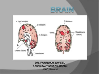
  • 1. DR. FARRUKH JAVEED CONSULTANT NEUROSURGEON JPMC, Karachi.
  • 2. LOBES OF THE CEREBRUM PARIETAL LOBE TEMPORAL LOBE FRONTAL LOBE LIMBIC LOBE OCCIPITAL LOBE
  • 3. FRONTAL LOBE The frontal lobe is the area of the brain responsible for higher cognitive functions. Problem solving • Memory • Language • Motivation • Judgment • Impulse control • Social and sexual behavior.
  • 4. TEMPORAL LOBE The temporal lobe plays a role in emotions, and is also responsible for smelling, tasting, perception, memory, understanding music, aggressiveness, and sexual behavior. The temporal lobe also contains the language area of the brain.
  • 5. PARIETAL LOBE The parietal lobe plays a role in our sensations of touch, smell, and taste. It also processes sensory and spatial awareness, and is a key component in eye-hand co- ordination and arm movement. The parietal lobe also contains a specialized area called Wernicke’s area that is responsible for matching written words with the sound of spoken speech.
  • 6. OCCIPITAL LOBE The occipital lobe is at the rear of the brain and controls vision and recognition.
  • 7. LANGUAGE Wernicke’s area is a specialized portion of the parietal lobe that recognizes and understands written and spoken language. Wernicke’s area surrounds the auditory association area. Damage to this part of the brain can result in someone hearing speech, but not understanding it. Wernicke’s Area Auditory Association Area
  • 8. HEARING There are two auditory areas of the brain: • The primary auditory area (brown circle) is what detects sounds that are transmitted from the ear. It is located in the sensory cortex. • The auditory association area (purple circle) is the part of the brain that is used to recognize the sounds as speech, music, or noise.
  • 9. SPEECH Broca’s area is where we formulate speech and the area of the brain that sends motor instructions to the motor cortex. Injury to Broca’s area can cause difficulty in speaking. The individual may know what words he or she wishes to speak, but will be unable to do so. Broca’s Area
  • 10. MOTOR CORTEX The motor portion of the cerebrum is illustrated here. The light red area is the premotor cortex, which is responsible for repetitive motions of learned motor skills. The dark red area is the primary motor area, and is responsible for control of skeletal muscles. Different areas of the brain are associated with different parts of the body. Injury to the motor cortex can result in motor disturbance in the associated body part.
  • 11. SENSORY CORTEX The sensory portion of the cerebrum is illustrated here. Different areas of the brain are associated with different parts of the body, as can be seen below. Injury to the sensory cortex can result in sensory disturbance in the associated body part.
  • 12. CEREBELLUM The cerebellum is connected to the brainstem, and is the center for body movement and balance.
  • 13. INCIDENCE OF CNS TUMORS  One-third of CNS tumors are metastatic lesions  One third are gliomas and  One-third is of nonglial origin.
  • 14. World Health Organization (WHO) Classification Categorized & considered by cell layer of origin  Tumors of Neuroepithelial tissue  Tumors of Meninges  Tumors of Craniospinal Nerves  Hematopoietic Neoplasms  Germ Cell Tumors  Sellar Tumors  Metastatic Tumors
  • 15. Brain Tumors in General  Presentation – Headache, seizure, neurological deficit, visual loss, personality changes, etc  History – Progressive onset of symptoms (weeks to months)  Physical Exam – Depends upon brain region affected
  • 16. HEADACHE ONSET • ABRUPT(waking from sleep) • Triggered by exertion or Valsalva CHANGE • First or worst • Change or progression in pattern • New headache >50yrs • New headache in cancer or AIDS pts ASSOCIATED • Abnormal neurological exam • Neurological symptoms > 1 hours • Associated with altered / loss of consciousness
  • 17.
  • 19. DIAGNOSIS  Imaging Studies – CT, MRI, PET  Laboratory Studies – Important for systemic tumors and metastasis, e.g. leukemia • Treatment & Prognosis  Varies per histology of specific tumors
  • 20. CARE OF THE BRAIN TUMOR PATIENT Steroids  Help resolve edema and symptoms associated with it  Can confuse the issue of lymphoma, because rapid “disappearance” is seen for lymphoma, followed by recurrence Anti-convulsants  Recommended particularly for supra tentorial & epileptogenic areas, e.g. mesial temporal lobe, and with past h/o seizures
  • 21. Treatment Options in general for Intracranial Tumors  Need Biopsy for definitve identification… then one or combination of the following:  Surgery  Radiation Therapy  Whole brain  Focused beam  Gamma Knife Stereotactic Radiosurgery  Chemotherapy  Systemic  Local
  • 22. COMMON INTRAAXIAL TUMORS IN ADULTS Supratentorial Infratentorial Metastasis Metastasis Gliomas Fibrillary Astrocytoma Anaplastic Astrocytoma Glioblastoma Multiforme Oligodendroglioma Hemangioblastoma
  • 23. COMMON INTRAAXIAL TUMORS IN PEDIATRICS Supratentorial Infratentorial Astrocytoma Juvenile Pilocytic Astrocytoma Pleomorphic Xanthoastrocytoma PNET (Medulloblastoma) PNET Ependymoma DNET Brainstem Astrocytoma Ganglioglioma Although cancer is rare in children, brain tumors are the most common type of childhood cancer after leukemia and lymphoma.
  • 24.
  • 25. SIGNS OF EXTRA AXIAL LOCATION  CSF cleft  Displaced subarachnoid vessels  Cortical gray between mass and white matter  Displaced and expanded subarachnoid spaces  Broad dural base  Bony reaction
  • 26.  The T2W-images show a schwannoma located in the cerebellopontine angle (CPA).  CSF Cleft (yellow arrow)  Displaced subarachnoid Space (blue arrow)  Gray Matter between mass and White matter (curved arrow)  Wide CSF spaces (long arrow)
  • 27. Meningioma (Enplaque variety)  broad dural base and a dural tail of enhancement(blue arrow)  There is hyperostosis in the adjacent bone (yellow arrow)  lesion enhances homogeneously
  • 28.  A hyperintense contrast enhancing lesion with extension to the prepontine area (blue arrows) and into the foramen magnum (red arrow).  This Lesion proved to be Ependymoma
  • 29.  Above image shows a diffusely infiltrating intra-axial tumor occupying most of the right hemisphere with only a minimal mass effect.  Low-grade astrocytoma.
  • 30. Midline Crossing Tumors  Glioblastoma multiforme (GBM) frequently crosses the midline by infiltrating the white matter tracts of the corpus callosum.  Radiation necrosis can look like recurrent GBM and can sometimes cross the midline.  Meningioma is an extra-axial tumor and can spread along the meninges to the contralateral side. Continued
  • 32. CALCIFICATIONS Intraaxial Tumors Extraaxial Tumors Astrocytomas (20%) Meningiomas (25%) Craniopharyngiomas (90%) Oligodendrogliomas (80%) Chordomas Metastasis Chondrosarcomas Ependymomas (50%) Choroid plexus papilloma (25%) Ganglioglioma (40%)
  • 33.  The CT shows a mass with calcifications, which extends all the way to the cortex.  The most likely diagnosis is Oligodendroglioma.
  • 34.  A calcified mass seen in the suprasellar region, causing obstructive hydrocephalus.  Craniopharyngiomas are slow growing,calcified, cystic tumors arising from remnants of Rathke's cleft.  They are located in the suprasellar region and primarily seen in children with a small second peak incidence in older adults.
  • 35.  On the coronal and sagittal TW1I there is a large mass centered around the sella with a broad dural base. & extension into the sella.  CT shows densely calcified tumor.
  • 37. DIFFUSION WEIGHTED IMAGING  Normally water protons have the ability to diffuse extracellularly and loose signal.  High intensity on DWI indicates restriction of the ability of water protons to diffuse extracellularly.  Restricted diffusion is seen in abscesses, epidermoid cysts and acute infarction  In most tumors there is no restricted diffusion - even in necrotic or cystic components. This results in a normal, low signal on DWI.
  • 38.  We see restriction in above left extreme image as it is hyperintense, while lower extreme left image shows no restriction.
  • 39.  Schwannoma extending into the middle cranial fossa with homogeneous enhancement (right).  Primary Lymphoma shows vivid enhancement (left).
  • 40. Contrast Enhancement Meningioma Schwannoma Lymphoma Choroid Plexus Papilloma
  • 42.  Above image shows glioblastoma multiforme (GBM).  The enhancement indicates that this is a high-grade tumor  There is also a cystic component with ring enhancement..
  • 43. RING ENHANCEMENT 1. Metastasis 2. Glioblastoma Multiforme 3. Tuberculoma 4. Abscess, 5. Infectious disease Toxoplasma 6. Multiple Sclerosis 7. Chronic hematoma
  • 45.  Many non- tumorous lesions can mimic a brain tumor.
  • 46.  Infections and vascular lesions can also mimic a CNS tumor.
  • 47. METASTATIC BRAIN TUMOR  History – Rapidly progressive onset of symptoms (weeks vs. months)  Most common sources are LUNG, BREAST (in women), Thyroid, RENAL, & G.I. tract
  • 48. DIAGNOSIS Imaging Studies –  MRI: Tumor at grey-white matter junction, usually associated with edema Systemic work-up includes  CT scan of chest, abdomen and pelvis  Mammography  Bone scan Diagnostic Laboratory Studies  CBC, ESR, LFTs,
  • 49.
  • 50. TREATMENT  For solitary lesion or less than 4 lesions all < 3 cm. – biopsy if undiagnosed, plus Gamma Knife  For > 3 cm. tumor, surgery followed by WBRT  For > 4 lesions, biopsy for diagnosis, plus whole brain radiation therapy  Prognosis: 7 – 12 mos.
  • 51. GLIOMAS Presentation  Depends upon brain location, usually H/A over several weeks, seizure, vomiting, visual chages or word finding difficulty Physical Exam –  Depends upon brain region affected Diagnostic Imaging Studies –  CT, MRI
  • 52.
  • 53. Treatment  Surgery, Radiation, Chemotherapy (varies with age group & type) Survival depends on pathology: –  Pilocytic astrocytoma: resection Æ cure; Grade 1 Astrocytoma (nuclear atypia) – 8-10 yrs;  Grade 2 Anaplastic astrocytoma (+endothelial proliferation) – 2 yrs  Grade 3 (+necrosis) GBM – 11 months
  • 54. MENINGIOMA Presentation  Meningiomas occur in middle aged females more frequently History  Meningiomas are usually slow-growing (months to years)  CT/MRI: Show meninges- based tumor (usually), with variable amount of edema
  • 55.
  • 56.
  • 57. TREATMENT  Meninigiomas: Gross total resection curative  Need for radiation therapy depends on grade Prognosis  – Meningiomas: Can be cured with gross total resection with favorable prognosis
  • 58. PITUITARY ADENOMA Presentation  Headache, visual defect (classically bitemporal hemianopsia), endocrine abnormalities (dependent upon hormone secreted) –  Prolactinoma most common  ACTH with Cushing’s disease (w/hypertension, stria, buffalo hump, morbid obesity);  GH with acromegaly
  • 59.
  • 60. History  Slow-growing, over months to years  Headache with progressive visual loss  Most common presentation is bitemporal hemi anopia Diagnostic Laboratory Studies  Endocrine panel  Test IGF-1 for GH-secreting tumor;  Use dexamethasone suppression test for Cushing’s disease vs. ectopic Cushing’s syndrome
  • 61.
  • 62. Treatment  Medical treatment for Prolactinoma only with dopamine antagonists  Surgical excision usually initial treatment (usually transnasal approach), with Gamma Knife for recurrence vs. local focused radiation Prognosis  Generally benign, so proportional to gross total resection & control of endocrine effects;
  • 63. Vestibular schwannoma Presentation  Hearing difficulty in affected ear, vertigo, headache/facial weakness/facial numbness if large tumor compressing trigeminal nerve
  • 64. Physical Exam  Evaluate facial weakness & hearing ability Diagnostic Imaging Studies  MRI Brain with MRA/MRV OTHERS  Audiometry
  • 66. Treatment  Surgical resection if >3 cm. or causing excessive mass effect, brainstem compression; GK for smaller lesions (<3 cm.) Prognosis  Benign tumor, so good with total excision and lack of recurrence
  • 67. Lymphoma Presentation  Sporadic, more common in immunosuppresed or immunocompromised patients (e.g. HIV+); may or may not be systemic @ presentation.  Spinal cord (epidural) compression or carcinomatous meningitis  Multiple cranial nerve deficits
  • 68.
  • 69. Diagnostic Imaging Studies  CT / MRI Brain Treatment  Biopsy for establishing diagnosis, followed by radiation; chemotherapy (intra thecal methotrexate) has shown increased survival
  • 70. PROGNOSIS  No treatment 2.5 months.;  XRT 10 months  Intraventricular methotrexate 41 months.  Prognosis worse in AIDS pts. (4 months)
  • 71. Pineal Region Tumors Presentation  Headache, Hydrocephalus (if large) Physical Exam  May develop Perinaud’s syndrome (up gaze palsy, convergence, and accomodation impairment)
  • 72. Diagnostic Imaging Studies  MRI Diagnostic Laboratory Studies  CSF can be sent for markers – AFP, HCG, PLAP
  • 73.  Tumor located in the pineal region with calcifications.  This is most likely a germinoma.
  • 74. Treatment  Depends upon histology:  Germinoma – biopsy & irradiate;  Pineoblastoma: resect & irradiate Prognosis  Depends upon histology: Pineocytoma better than germinoma, pineoblastoma
  • 77. Multifocal Disease  Multiple tumors in the brain usually indicate metastatic disease.  Primary brain tumors are typically seen in a single region
  • 78.  Some tumors can be multifocal as a result of seeding metastases: this can occur in medulloblastomas (PNET-MB), ependymomas, GBMs and oligodendrogliomas.  Meningiomas and schwannomas can be multiple, especially in neurofibromatosis type II.
  • 81.  Cystic schwannoma of Left CP Angle.
  • 82.
  • 83.
  • 85.
  • 86.
  • 87.
  • 88.
  • 89.
  • 90.
  • 91.
  • 92.
  • 93. CYSTIC TUMORS WITH MURAL NODULE

Editor's Notes

  1. The most common tumors in adults are listed in the table on the left. Note that metastases are by far the most common.  It is important to realize that 50% of metastases are solitary. Particularly in the posterior fossa, metastases should be in the top 3 of the differential diagnostic list. Hemangioblastoma is an uncommon tumor, but it is the most common primary intra-axial tumor in the adult. Supratentorially, metastases are also the most common tumors, followed by gliomas.
  2. Bony changes are seen in bone tumors like chordomas, chondrosarcomas and metastases.  They can also be secondary, as is seen in meningiomas and other tumors.