1. LUPUS ERYTHEMATOSUS CELL
Systemic Lupus Erythematosus (SLE) is a chronic rheumatic disease which affects
joints, muscles and other parts of the body. Lupus involves inflammation (the
immune system's response to kill foreign agents, virus, and bacteria). Systemic
lupus erythematosus involves chronic inflammation that can affect many parts of
the body, including: Heart, lungs, skin, joints, blood-forming organs, kidneys,
nervous system. It is a connective tissue disease that affects most commonly
women of child bearing age and is characterized by skin rash, arthralgia, fever,
renal, cardiac and vascular lesions, anemia, leucopenia and often
thrombocytopenia.
There is a factor in the serum (an immunoglobulin of the IgG, IgM or IgA class)
that has the ability to cause depolymerization of the nuclear chromatin of
polymorphonuclear leucocytes and this depolymerized material is subsequently
phagocytosed by an intact polymorph giving rise to the Lupus erythematosus (LE)
cell.
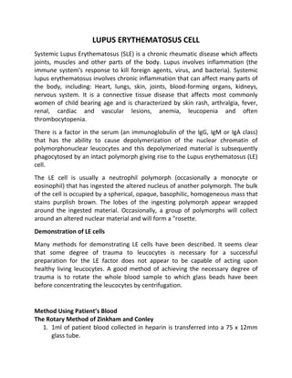
The LE cell is usually a neutrophil polymorph (occasionally a monocyte or
eosinophil) that has ingested the altered nucleus of another polymorph. The bulk
of the cell is occupied by a spherical, opaque, basophilic, homogeneous mass that
stains purplish brown. The lobes of the ingesting polymorph appear wrapped
around the ingested material. Occasionally, a group of polymorphs will collect
around an altered nuclear material and will form a "rosette.
Demonstration of LE cells
Many methods for demonstrating LE cells have been described. It seems clear
that some degree of trauma to leucocytes is necessary for a successful
preparation for the LE factor does not appear to be capable of acting upon
healthy living leucocytes. A good method of achieving the necessary degree of
trauma is to rotate the whole blood sample to which glass beads have been
before concentrating the leucocytes by centrifugation.
Method Using Patient’s Blood
The Rotary Method of Zinkham and Conley
1. 1ml of patient blood collected in heparin is transferred into a 75 x 12mm
glass tube.
2. 2. Four glass beads are added and the tube is sealed with a tightly fitting
rubber bung.
3. The preparation is rotated at 33 rpm at room temperature for30 minutes
and placed at 37oC for 10-15 minutes.
4. The contents of the tube are transferred to a Wintrobe tube and
centrifuged at 200g for 10 minutes.
5. Buffy coat smears are prepared, dried in the air, fixed in methanol and are
stained with Romanowsky stain in the usual manner.
Examination of Films
The films, especially their edges and tails are searched for a minimum of 10
minutes (a minimum of 500 polymorphs should be counted) before a negative
report is given. Frequently, dead nuclei will be seen lying freely; if numerous,
these may heighten suspicions but they are never diagnostic. LE cells must be
differentiated from "tart cells" which are usually monocytes that have
phagocytosed the nucleus of a lymphocyte. The ingested nuclear material is well
preserved in contrast to the LE cell inclusion body. Tart cells are often associated
with leucoagglutinins and may occasionally occur in patients on drug therapy.
Interpretation:
A positive LE cell test is very suggestive of SLE and the test is a very useful
diagnostic test. The test is positivein 75% of patients with SLE. However, false
positive results have been reported in lupoid hepatitis, patients with severe and
highly active rheumatoid arthritis and patients on drug therapy