SlideShare a Scribd company logo
1 of 56
Dr. MAMTA
Dept of ophthalmology
RNT Medical college, Udaipur
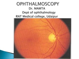
 Ophthalmoscopy is a clinical examination of
the interior of the eye by means of an
ophthalmoscope.
 It is primarily done to assess the state of
fundus and detect the opacities of ocular
media.
 The ophthalmoscope was invented by von
Helmholtz in 1850.
nasal
temporal
DISC: LOCATION –nasal to geometric axis
DIAMETER – 1.5mm [1 disc diameter]
COLOR – Pale pink
SHAPE – Circular
EDGES – Regular
TERMINATION OF ALL
LAYERS EXCEPT NFL
CUP: C/D ratio – 0.3 to 0.5
 RETINAL SYSTEM :
CENTRAL RETINAL ARTERY
AND
CENTRAL RETINAL VEIN
Arterioles
Venules
Capillaries
 CILIARY SYSTEM : POST.CILIARY ARTERIES
Choriocapillaries
 Specialised region of retina
 Diameter – 5.5 mm
 Location – 2 DD - temporal margin of disc
 Color – Yellow; deep pigmented
 4 zones : Foveola -0.35 mm
Fovea -1.50 mm
Parafovea
Perifovea
 Retinal vessels
 Cilioretinal artery
 Ophthalmoscopic methods of examination
are-
(1)Distant direct ophthalmoscopy
(2)Direct ophthalmoscopy
(3) Indirect ophthalmoscopy
Parts-
◦ Light source
◦ Eyepiece
◦ Lens rack and power dial
◦ Aperture selector
◦ Filter selector
◦ On/off and brightness control
◦ Power handle
 For a good view of fundus the pupil should be
dilated by instilling few drops of short acting
mydriatic drug (e.g. combination of
tropicamide and phenylephrine )
 The subject should be examined in sitting or
lying down position.
 Examination room should be semidark.
 keep the eye as still as possible.
For examining right eye of the patient,
 Examiner should stand on right side of the
patient.
 Hold the instrument in his right hand.
 Use examiner’s right eye.
If examining left eye, stand on left side, hold
instrument in left hand use left eye.
 Viewing should begin about half meter away
from the eye.
 First see the “Red reflex”
 Initially the lens power in the instrument
should be set to zero, and if refractive error
present in patient or examiner, e.g. if the
patient is myopic then set the (-ve )lens, if
the examiner or patient is hypermetropic
then set the lens to (+ve) lens. If both
patient & examiner have refractive error
then sum together their powers.
 Rotate the lens dial until the optic disc is focused
clearly.
(Red numbers/negative numbers in myopia or
short-sightedness.
Green numbers/positive numbers in hyperopia or
far-sightedness.)
 Examine the optic disc for:
Shape – normally round or slightly oval
Clarity of the outline-clear outline or rim
Colour-pale pink contrast to rich red of rest of
fundus.
 Examine the macula last.
 To locate the macula, focus on the disc, then
move 2 disc diameters temporally. The
macula is darker in colour than the
surrounding fundus, and devoid of blood
vessels.
 May also ask the patient to look at the light,
this automatically puts the macula into full
view.
 It should be performed routinely before the
direct ophthalmoscope, as it gives a lot of
useful information.
 It can be performed with the help of a self-
illuminated ophthalmoscope or a simple plain
mirror with a hole at the centre.
Procedure- The light is thrown into patients
eye sitting in a semi-darkroom, from a
distance of 20-25 cm and the features of the
red glow in the pupillary area are noted.
 1. To diagnose opacities in the refractive
media- any opacity in the refractive media is
seen as a black shadow in the red glow.
 2. To differentiate between a mole and a
hole of the iris- a small hole and mole on the
iris appears as a black spot on oblique
illumination
 3. To recognise detached retina or a tumour
arising from the fundus is seen as a greyish
reflex.
 It is the most commonly practised method for
routine fundus examination.
 It works on the basic optical principle of glass
plate ophthalmoscope introduced by von
helmholtz.
 A convergent beam of light is reflected into
the patients pupil. The emergent rays from
any point of the patients fundus reach the
observers retina through the viewing hole in
the ophthalmoscope.
 The emergent rays from the patients eye are
parallel and brought to focus on the retina of
the emmetropic observer when
accommodation is relaxed.
 In hypermetropic patient,the emergent rays
will be divergent and with the help of convex
lens can be brought to focus on the observers
retina.
 In myopic patient,the rays will be convergent
and use concave lens for focus on the
observers retina.
Divergent
light, if subject
hypermetropic
Reflected
light
source
Convergent
light, if subject
myopic
Divergent
light
Convergent
light
Parallel
light
Focussed
on retina
Corrective lens is
placed along pathway
 The image is erect, virtual and about 15 times
magnified in emmetropes.
 Technique- should be performed in a semi-
darkroom with the patient seated and looking
straight ahead , while the observer standing
or seated slightly over to the side of the eye
to be examined by the observer with his or
her right eye and left with the left.
 The observer should reflect beam of light
from the ophthalmoscope into patients pupil.
Once the red reflex is seen the observer
should move as close to the patients eye as
possible.
 Once the retina is focused the details should
be examined systematically starting from
disc,blood vessels, the four quadrant of the
general background and the macula.
Monocular Indirect Ophthalmoscopy
It consists of-
 Illumination rheostat at its base,
 Focusing lever for image refinement,
 Filter dial with red free and yellow filters,
 Forehead rest for proper observer head
positioning, and
 Iris diaphragm lever to adjust the illumination
beam diameter.
 Optics- an internal relay lens system re-
inverts the initially inverted image to a real
erect one, which is then magnified. This
image is focusable using the focusing lever.
Indications are-
 Need for an increased field of view
 Small pupils
 Uncooperative children
 Patients intolerance of bright light of
binocular indirect ophthalmoscope.
 Extent of view- although vitreous base views
are possible with monocular IO, its greatest
effectiveness extends anteriorly to the
peripheral equatorial region.
 Advantage- field of view similar to IO and,
 Erect real image similar to DO.
 Disadvantage –are lack of stereopsis,
 Limited illumination and,
 Fixed magnification.
 It is very popular method for examination of
posterior segment introduced by Nagel in
1864.
 PRINCIPLE- To make the eye highly myopic by
placing a strong convex lens in front of
patients eye so that the emergent rays from
an area of the fundus are brought to the
focus as a real,inverted image between the
lens and the observers eye.
 An inverted reverse real image
 Magnification = 2 to 4 X
 Field of view = 40 to 50 degrees
 Optimal working distance = 40 to 50 cms
 Good illumination & stereopsis
 Ease of use with scleral indentor
 Lenses from 14 to 30 D range
 CHARACTERISTICS- Magnification of image
depends upon the dioptric power of convex
lens, position of the lens in relation of the
eyeball and refractive state of eyeball.
 About 5 times magnification is obtained with
+13 D lens.
 With a stronger lens,image will be smaller,
but brighter and field of vision will be more.
(1) Dark room
(2) source of light and concave mirror or self
illuminated indirect ophthalmoscope
(3) Convex lens
(4) pupils of the patient should be dilated.
 The patient is made to lie in the supine
position, with one pillow on a bed or couch
and instructed to keep both eyes open.
 The examiner throws the light into patients
eye from an arms distance.
 In practise, Binocular ophthalmoscope with
head band or that mounted on the spectacle
frame is employed most frequently.
 Keeping his or her eyes on the reflex, the
examiner then interposes the condensing
lens in the path of beam of light, close to the
patient eye, and then slowly moves the lens
away from eye until the image of retina is
clearly seen.
 The examiner moves around the head of the
patient to examine different quadrants of the
fundus.
 He or she has to stand opposite to clock hour
position to be examined.
 By asking the patient to look in extreme gaze,
and using of scleral indenter, the whole
peripheral retina up to ora serrata can be
examined.
 Scleral indentation- helps in making prominent
the barely perceptible lesions, done with the
depressor placed on patients lid.
 Examiner should move the scleral depressor in a
direction opposite to that in which he or she
wishes the depression to appear.
 Scleral depressor should be rolled gently and
tangentially over the eye surface.
 The temporal part of upper lid is sufficiently lax
so depressor can be placed inferiorly in the
horizontal meridian.
-Thimble scleral
depressor
-Pencil type depressor
-Cotton tipped applicator
To examine periphery between equator and ora serrata by creating a
mound to view.
Start superonasal superior ,superotemporal,
Inferotemporal, inferior, inferonasal
 FUNDUS DRAWING-
 The image seen with IO is vertically inverted and
laterally reversed; top of the retinal chart is
placed towards the foot end of the patient
[i.e.upside down]
 Fundus drawing is made on a special Amsler
chart,which has 12 clock hours marked and has
three concentric circles made on it-
 Innermost circle,middle circle and outermost
circle represents to the equator,the ora serrata
and,the midpoint of pars plana respectively.
-Technique is difficult and can be mastered by
hours of practice.
-Magnification is less compare to DO.
-It is impossible with very small pupil.
-Patient is usually more uncomfortable with
intense light of IO and with scleral
indentation.
-Reflex sneezing can occur on exposure to
bright light.
Advantages of Indirect system
• Image not affected by the patients refractive power
• In eyes with nystagmus
• Delivery of LASER
• Binocular examination of fundus up-to the periphery
• Large field of view allow for the panoramic view
• Better Resolution
• Use in operating room for cryo/scleral buckling
• Better view in presence of media opacities
• Increased illumination
• Reduced distortion
Dropout in glaucoma,
myelination at disc margi
Crossings: nipping in
systemic hypertension
Cupping and notching
in glaucoma
Swelling and blurred
margins in papilloedem
and optic neuritis
Vessel walls: sheathing in
systemic hypertension, leakage
and neovascularisation in diabetes.
Bifurcations: embolisms,
branch occlusions
Pigmentation changes
in retinitis pigmentosa
Haemorrhages
in diabetes,
vessel occlusion,
hypertension
Myopic crescent
in myopia
ARMD, drusen,
macular holes
Retinal tears,
detachments
Anterior ischaemic opti
neuropathy in diabetes
Vessels
Optic disc
Fovea
Background/periphery
Retinal nerve fibre layer
Vitreous
Asteroid hyalosis, floater
haemorrhages
 (1) Hruby lens biomicroscopy
 (2) Contact lens biomicroscopy
 (3) Indirect fundus biomicroscopy
 Hruby lens is a planoconcave lens with
diopteric power 58.6D, which neutralizes the
optical power of normal eye (60D) and forms
a virtual,erect image of the fundus.
 This lens provides a small field with low
magnification and cannot visualize the
fundus beyond equator.
 It combines stereopsis, high illumination and
high magnification with the advantages of slit
beam.
 Modified Koeppe lens, i.e. posterior fundus
contact lens can be used to examine the
posterior segment, provides a virtual and
erect image.
 Goldmanns three-mirror contact lens, consist
of a central contact lens and three mirror
placed in the cone,each with different angles
of inclination.
 With this, the central as well as peripheral parts
of the fundus can be visualized.
 It also provides a virtual and erect image.
 By flat central portion can see posterior
vitreous and pole.
 Angled mirror - 73 deg - Peripheral fundus
67 deg - Pars plana
59 deg - Gonioscopy
 Eliminates total internal reflection by
replacement with cornea – goldmann contact lens
interface.
 Also known as non-contact fundus biomicroscopy,
most commonly used lenses are +78D and +90D,
which are mostly double aspheric lenses, so it does
not matter which side is held towards the patient.
 Optics similar to IO, thus a real, inverted image is
formed between the condensing lens and objective
lens of slit-lamp.
 Magnification provided by fundus non-contact lenses
is calculated by dividing power of the eye by the
power of lens, e.g. +90D lens provides a
magnification of 60/90=0.75X,i.e. a minification of
the image.
However, the Magnified image seen because of
the magnification provided by the slit-lamp.
 Magnifying knob to 10X or 16X.
 Field of view high powered lens provides larger
field of view but lesser magnification.
 Drawing the slit lamp biomicroscopic view:
Done on an inverted fundus chart and paper
is turned as patients gaze direction changes in
respective clock hour meridian.
 Performing indirect slit lamp biomicroscopy
 Evaluating fundus :
Provides wide field 130 degree and
high power lens
A real inverted magnified image is
formed
It is used in both posterior fundus
examination and also
Laser pan retinal photo
Coagulation.
THANK YOU

More Related Content

What's hot (20)

GONIOSCOPY
GONIOSCOPY GONIOSCOPY
GONIOSCOPY
 
Optical coherence tomography(OCT) --macula
Optical coherence tomography(OCT) --maculaOptical coherence tomography(OCT) --macula
Optical coherence tomography(OCT) --macula
 
Presbyopia
PresbyopiaPresbyopia
Presbyopia
 
Visual acuity
Visual acuityVisual acuity
Visual acuity
 
Techniques of tear film evaluation by Raju Kaiti
Techniques of tear film evaluation  by Raju KaitiTechniques of tear film evaluation  by Raju Kaiti
Techniques of tear film evaluation by Raju Kaiti
 
colour vision
colour visioncolour vision
colour vision
 
ROPLAS TEST
ROPLAS TESTROPLAS TEST
ROPLAS TEST
 
Indirect ophthalmoscopy
Indirect ophthalmoscopy Indirect ophthalmoscopy
Indirect ophthalmoscopy
 
Direct ophthalmoscope
Direct ophthalmoscopeDirect ophthalmoscope
Direct ophthalmoscope
 
primary closed angle glaucoma (Acute congestive glaucoma)
primary closed angle glaucoma (Acute congestive glaucoma)primary closed angle glaucoma (Acute congestive glaucoma)
primary closed angle glaucoma (Acute congestive glaucoma)
 
Tests for stereopsis
Tests for stereopsisTests for stereopsis
Tests for stereopsis
 
Maddox Rod test
Maddox Rod testMaddox Rod test
Maddox Rod test
 
Synoptophore
SynoptophoreSynoptophore
Synoptophore
 
Tear film test
Tear film testTear film test
Tear film test
 
Colour vision examination
Colour vision examinationColour vision examination
Colour vision examination
 
Perimetry
PerimetryPerimetry
Perimetry
 
Direct ophthalmoscopy
Direct ophthalmoscopyDirect ophthalmoscopy
Direct ophthalmoscopy
 
Tonometry
TonometryTonometry
Tonometry
 
Pachymetry
PachymetryPachymetry
Pachymetry
 
Keratometry
KeratometryKeratometry
Keratometry
 

Viewers also liked

Retinal diagram dr sabin sahu
Retinal diagram dr sabin sahuRetinal diagram dr sabin sahu
Retinal diagram dr sabin sahuvoveran312
 
Opthalmoscopy Uploaded by Parash
Opthalmoscopy Uploaded by ParashOpthalmoscopy Uploaded by Parash
Opthalmoscopy Uploaded by ParashParash39
 
Looking deep into retina : indirect ophthalmoscopy and fundus drawing
Looking deep into retina : indirect ophthalmoscopy and fundus drawingLooking deep into retina : indirect ophthalmoscopy and fundus drawing
Looking deep into retina : indirect ophthalmoscopy and fundus drawingPrachir Agarwal
 

Viewers also liked (7)

Corneal drawings
Corneal drawingsCorneal drawings
Corneal drawings
 
Retinal diagram dr sabin sahu
Retinal diagram dr sabin sahuRetinal diagram dr sabin sahu
Retinal diagram dr sabin sahu
 
Normal fundus
Normal fundusNormal fundus
Normal fundus
 
Fundus examination
Fundus examinationFundus examination
Fundus examination
 
Opthalmoscopy Uploaded by Parash
Opthalmoscopy Uploaded by ParashOpthalmoscopy Uploaded by Parash
Opthalmoscopy Uploaded by Parash
 
Looking deep into retina : indirect ophthalmoscopy and fundus drawing
Looking deep into retina : indirect ophthalmoscopy and fundus drawingLooking deep into retina : indirect ophthalmoscopy and fundus drawing
Looking deep into retina : indirect ophthalmoscopy and fundus drawing
 
Fundus examination
Fundus examinationFundus examination
Fundus examination
 

Similar to Ophthalmoscopy

Presentation Retina.pptx
Presentation Retina.pptxPresentation Retina.pptx
Presentation Retina.pptxmahendra singh
 
Presentation Retina.pptx
Presentation Retina.pptxPresentation Retina.pptx
Presentation Retina.pptxmahendra singh
 
Ophthalmoscopy
OphthalmoscopyOphthalmoscopy
OphthalmoscopySalalKhan5
 
directophthalmoscope-180730142501 (2).pdf
directophthalmoscope-180730142501 (2).pdfdirectophthalmoscope-180730142501 (2).pdf
directophthalmoscope-180730142501 (2).pdfanju468752
 
ilovepdf_merged.pdf
ilovepdf_merged.pdfilovepdf_merged.pdf
ilovepdf_merged.pdfanju468752
 
directophthalmoscope-180730142501 (2).pptx
directophthalmoscope-180730142501 (2).pptxdirectophthalmoscope-180730142501 (2).pptx
directophthalmoscope-180730142501 (2).pptxanju468752
 
Fundoscopy ppt 2012
Fundoscopy ppt 2012Fundoscopy ppt 2012
Fundoscopy ppt 2012Reina Ramesh
 
ppt on direct opthalmoscopy
 ppt on direct opthalmoscopy ppt on direct opthalmoscopy
ppt on direct opthalmoscopymanisaikoduri
 
Direct ophthalmoscope.pptx
Direct ophthalmoscope.pptxDirect ophthalmoscope.pptx
Direct ophthalmoscope.pptxAnamSehreen
 
Binocular Indirect OPHTHALMOSCOPY
Binocular Indirect OPHTHALMOSCOPYBinocular Indirect OPHTHALMOSCOPY
Binocular Indirect OPHTHALMOSCOPYKh.Towkir-Ul- Islam
 
Ophthalmoscope direct and indirect
Ophthalmoscope direct and indirectOphthalmoscope direct and indirect
Ophthalmoscope direct and indirectAnisur Rahman
 
Direct ophthalmoscopy final
Direct ophthalmoscopy finalDirect ophthalmoscopy final
Direct ophthalmoscopy finalshovon2026
 
Retinoscope theory.pptx
Retinoscope theory.pptxRetinoscope theory.pptx
Retinoscope theory.pptxSHAYRI PILLAI
 
CRANIAL NERVE EXAMINATION.pptx
CRANIAL NERVE EXAMINATION.pptxCRANIAL NERVE EXAMINATION.pptx
CRANIAL NERVE EXAMINATION.pptxanujsharmamla
 
Retinoscopy dr-171026143930 copy
Retinoscopy dr-171026143930 copyRetinoscopy dr-171026143930 copy
Retinoscopy dr-171026143930 copyRitika Sahay
 
Direct ophthalmoscopy final
Direct ophthalmoscopy finalDirect ophthalmoscopy final
Direct ophthalmoscopy finalshovon2026
 
Objective refraction
Objective refractionObjective refraction
Objective refractionsneha_thaps
 

Similar to Ophthalmoscopy (20)

Presentation Retina.pptx
Presentation Retina.pptxPresentation Retina.pptx
Presentation Retina.pptx
 
Presentation Retina.pptx
Presentation Retina.pptxPresentation Retina.pptx
Presentation Retina.pptx
 
Ophthalmoscopy
OphthalmoscopyOphthalmoscopy
Ophthalmoscopy
 
directophthalmoscope-180730142501 (2).pdf
directophthalmoscope-180730142501 (2).pdfdirectophthalmoscope-180730142501 (2).pdf
directophthalmoscope-180730142501 (2).pdf
 
ilovepdf_merged.pdf
ilovepdf_merged.pdfilovepdf_merged.pdf
ilovepdf_merged.pdf
 
directophthalmoscope-180730142501 (2).pptx
directophthalmoscope-180730142501 (2).pptxdirectophthalmoscope-180730142501 (2).pptx
directophthalmoscope-180730142501 (2).pptx
 
Fundoscopy ppt 2012
Fundoscopy ppt 2012Fundoscopy ppt 2012
Fundoscopy ppt 2012
 
ppt on direct opthalmoscopy
 ppt on direct opthalmoscopy ppt on direct opthalmoscopy
ppt on direct opthalmoscopy
 
Direct ophthalmoscope.pptx
Direct ophthalmoscope.pptxDirect ophthalmoscope.pptx
Direct ophthalmoscope.pptx
 
Binocular Indirect Ophthalmoscopy
Binocular Indirect OphthalmoscopyBinocular Indirect Ophthalmoscopy
Binocular Indirect Ophthalmoscopy
 
Binocular Indirect OPHTHALMOSCOPY
Binocular Indirect OPHTHALMOSCOPYBinocular Indirect OPHTHALMOSCOPY
Binocular Indirect OPHTHALMOSCOPY
 
Ophthalmoscope direct and indirect
Ophthalmoscope direct and indirectOphthalmoscope direct and indirect
Ophthalmoscope direct and indirect
 
Direct ophthalmoscopy final
Direct ophthalmoscopy finalDirect ophthalmoscopy final
Direct ophthalmoscopy final
 
Ophthalmoscopy
OphthalmoscopyOphthalmoscopy
Ophthalmoscopy
 
Retinoscope theory.pptx
Retinoscope theory.pptxRetinoscope theory.pptx
Retinoscope theory.pptx
 
CRANIAL NERVE EXAMINATION.pptx
CRANIAL NERVE EXAMINATION.pptxCRANIAL NERVE EXAMINATION.pptx
CRANIAL NERVE EXAMINATION.pptx
 
Retinoscopy dr-171026143930 copy
Retinoscopy dr-171026143930 copyRetinoscopy dr-171026143930 copy
Retinoscopy dr-171026143930 copy
 
Direct ophthalmoscopy final
Direct ophthalmoscopy finalDirect ophthalmoscopy final
Direct ophthalmoscopy final
 
Objective refraction
Objective refractionObjective refraction
Objective refraction
 
Opthalmoscopy
OpthalmoscopyOpthalmoscopy
Opthalmoscopy
 

Recently uploaded

Low Rate Call Girls Mumbai Suman 9910780858 Independent Escort Service Mumbai
Low Rate Call Girls Mumbai Suman 9910780858 Independent Escort Service MumbaiLow Rate Call Girls Mumbai Suman 9910780858 Independent Escort Service Mumbai
Low Rate Call Girls Mumbai Suman 9910780858 Independent Escort Service Mumbaisonalikaur4
 
VIP Call Girls Pune Vrinda 9907093804 Short 1500 Night 6000 Best call girls S...
VIP Call Girls Pune Vrinda 9907093804 Short 1500 Night 6000 Best call girls S...VIP Call Girls Pune Vrinda 9907093804 Short 1500 Night 6000 Best call girls S...
VIP Call Girls Pune Vrinda 9907093804 Short 1500 Night 6000 Best call girls S...Miss joya
 
Artifacts in Nuclear Medicine with Identifying and resolving artifacts.
Artifacts in Nuclear Medicine with Identifying and resolving artifacts.Artifacts in Nuclear Medicine with Identifying and resolving artifacts.
Artifacts in Nuclear Medicine with Identifying and resolving artifacts.MiadAlsulami
 
Russian Call Girls Chickpet - 7001305949 Booking and charges genuine rate for...
Russian Call Girls Chickpet - 7001305949 Booking and charges genuine rate for...Russian Call Girls Chickpet - 7001305949 Booking and charges genuine rate for...
Russian Call Girls Chickpet - 7001305949 Booking and charges genuine rate for...narwatsonia7
 
Sonagachi Call Girls Services 9907093804 @24x7 High Class Babes Here Call Now
Sonagachi Call Girls Services 9907093804 @24x7 High Class Babes Here Call NowSonagachi Call Girls Services 9907093804 @24x7 High Class Babes Here Call Now
Sonagachi Call Girls Services 9907093804 @24x7 High Class Babes Here Call NowRiya Pathan
 
Call Girls Hosur Just Call 7001305949 Top Class Call Girl Service Available
Call Girls Hosur Just Call 7001305949 Top Class Call Girl Service AvailableCall Girls Hosur Just Call 7001305949 Top Class Call Girl Service Available
Call Girls Hosur Just Call 7001305949 Top Class Call Girl Service Availablenarwatsonia7
 
Mumbai Call Girls Service 9910780858 Real Russian Girls Looking Models
Mumbai Call Girls Service 9910780858 Real Russian Girls Looking ModelsMumbai Call Girls Service 9910780858 Real Russian Girls Looking Models
Mumbai Call Girls Service 9910780858 Real Russian Girls Looking Modelssonalikaur4
 
Call Girl Service Bidadi - For 7001305949 Cheap & Best with original Photos
Call Girl Service Bidadi - For 7001305949 Cheap & Best with original PhotosCall Girl Service Bidadi - For 7001305949 Cheap & Best with original Photos
Call Girl Service Bidadi - For 7001305949 Cheap & Best with original Photosnarwatsonia7
 
Call Girls Jayanagar Just Call 7001305949 Top Class Call Girl Service Available
Call Girls Jayanagar Just Call 7001305949 Top Class Call Girl Service AvailableCall Girls Jayanagar Just Call 7001305949 Top Class Call Girl Service Available
Call Girls Jayanagar Just Call 7001305949 Top Class Call Girl Service Availablenarwatsonia7
 
VIP Call Girls Lucknow Nandini 7001305949 Independent Escort Service Lucknow
VIP Call Girls Lucknow Nandini 7001305949 Independent Escort Service LucknowVIP Call Girls Lucknow Nandini 7001305949 Independent Escort Service Lucknow
VIP Call Girls Lucknow Nandini 7001305949 Independent Escort Service Lucknownarwatsonia7
 
Russian Call Girl Brookfield - 7001305949 Escorts Service 50% Off with Cash O...
Russian Call Girl Brookfield - 7001305949 Escorts Service 50% Off with Cash O...Russian Call Girl Brookfield - 7001305949 Escorts Service 50% Off with Cash O...
Russian Call Girl Brookfield - 7001305949 Escorts Service 50% Off with Cash O...narwatsonia7
 
Book Call Girls in Kasavanahalli - 7001305949 with real photos and phone numbers
Book Call Girls in Kasavanahalli - 7001305949 with real photos and phone numbersBook Call Girls in Kasavanahalli - 7001305949 with real photos and phone numbers
Book Call Girls in Kasavanahalli - 7001305949 with real photos and phone numbersnarwatsonia7
 
Call Girls Kanakapura Road Just Call 7001305949 Top Class Call Girl Service A...
Call Girls Kanakapura Road Just Call 7001305949 Top Class Call Girl Service A...Call Girls Kanakapura Road Just Call 7001305949 Top Class Call Girl Service A...
Call Girls Kanakapura Road Just Call 7001305949 Top Class Call Girl Service A...narwatsonia7
 
Call Girls Jp Nagar Just Call 7001305949 Top Class Call Girl Service Available
Call Girls Jp Nagar Just Call 7001305949 Top Class Call Girl Service AvailableCall Girls Jp Nagar Just Call 7001305949 Top Class Call Girl Service Available
Call Girls Jp Nagar Just Call 7001305949 Top Class Call Girl Service Availablenarwatsonia7
 
Call Girls Whitefield Just Call 7001305949 Top Class Call Girl Service Available
Call Girls Whitefield Just Call 7001305949 Top Class Call Girl Service AvailableCall Girls Whitefield Just Call 7001305949 Top Class Call Girl Service Available
Call Girls Whitefield Just Call 7001305949 Top Class Call Girl Service Availablenarwatsonia7
 
High Profile Call Girls Jaipur Vani 8445551418 Independent Escort Service Jaipur
High Profile Call Girls Jaipur Vani 8445551418 Independent Escort Service JaipurHigh Profile Call Girls Jaipur Vani 8445551418 Independent Escort Service Jaipur
High Profile Call Girls Jaipur Vani 8445551418 Independent Escort Service Jaipurparulsinha
 
Housewife Call Girls Bangalore - Call 7001305949 Rs-3500 with A/C Room Cash o...
Housewife Call Girls Bangalore - Call 7001305949 Rs-3500 with A/C Room Cash o...Housewife Call Girls Bangalore - Call 7001305949 Rs-3500 with A/C Room Cash o...
Housewife Call Girls Bangalore - Call 7001305949 Rs-3500 with A/C Room Cash o...narwatsonia7
 
Aspirin presentation slides by Dr. Rewas Ali
Aspirin presentation slides by Dr. Rewas AliAspirin presentation slides by Dr. Rewas Ali
Aspirin presentation slides by Dr. Rewas AliRewAs ALI
 
call girls in Connaught Place DELHI 🔝 >༒9540349809 🔝 genuine Escort Service ...
call girls in Connaught Place  DELHI 🔝 >༒9540349809 🔝 genuine Escort Service ...call girls in Connaught Place  DELHI 🔝 >༒9540349809 🔝 genuine Escort Service ...
call girls in Connaught Place DELHI 🔝 >༒9540349809 🔝 genuine Escort Service ...saminamagar
 
Call Girls ITPL Just Call 7001305949 Top Class Call Girl Service Available
Call Girls ITPL Just Call 7001305949 Top Class Call Girl Service AvailableCall Girls ITPL Just Call 7001305949 Top Class Call Girl Service Available
Call Girls ITPL Just Call 7001305949 Top Class Call Girl Service Availablenarwatsonia7
 

Recently uploaded (20)

Low Rate Call Girls Mumbai Suman 9910780858 Independent Escort Service Mumbai
Low Rate Call Girls Mumbai Suman 9910780858 Independent Escort Service MumbaiLow Rate Call Girls Mumbai Suman 9910780858 Independent Escort Service Mumbai
Low Rate Call Girls Mumbai Suman 9910780858 Independent Escort Service Mumbai
 
VIP Call Girls Pune Vrinda 9907093804 Short 1500 Night 6000 Best call girls S...
VIP Call Girls Pune Vrinda 9907093804 Short 1500 Night 6000 Best call girls S...VIP Call Girls Pune Vrinda 9907093804 Short 1500 Night 6000 Best call girls S...
VIP Call Girls Pune Vrinda 9907093804 Short 1500 Night 6000 Best call girls S...
 
Artifacts in Nuclear Medicine with Identifying and resolving artifacts.
Artifacts in Nuclear Medicine with Identifying and resolving artifacts.Artifacts in Nuclear Medicine with Identifying and resolving artifacts.
Artifacts in Nuclear Medicine with Identifying and resolving artifacts.
 
Russian Call Girls Chickpet - 7001305949 Booking and charges genuine rate for...
Russian Call Girls Chickpet - 7001305949 Booking and charges genuine rate for...Russian Call Girls Chickpet - 7001305949 Booking and charges genuine rate for...
Russian Call Girls Chickpet - 7001305949 Booking and charges genuine rate for...
 
Sonagachi Call Girls Services 9907093804 @24x7 High Class Babes Here Call Now
Sonagachi Call Girls Services 9907093804 @24x7 High Class Babes Here Call NowSonagachi Call Girls Services 9907093804 @24x7 High Class Babes Here Call Now
Sonagachi Call Girls Services 9907093804 @24x7 High Class Babes Here Call Now
 
Call Girls Hosur Just Call 7001305949 Top Class Call Girl Service Available
Call Girls Hosur Just Call 7001305949 Top Class Call Girl Service AvailableCall Girls Hosur Just Call 7001305949 Top Class Call Girl Service Available
Call Girls Hosur Just Call 7001305949 Top Class Call Girl Service Available
 
Mumbai Call Girls Service 9910780858 Real Russian Girls Looking Models
Mumbai Call Girls Service 9910780858 Real Russian Girls Looking ModelsMumbai Call Girls Service 9910780858 Real Russian Girls Looking Models
Mumbai Call Girls Service 9910780858 Real Russian Girls Looking Models
 
Call Girl Service Bidadi - For 7001305949 Cheap & Best with original Photos
Call Girl Service Bidadi - For 7001305949 Cheap & Best with original PhotosCall Girl Service Bidadi - For 7001305949 Cheap & Best with original Photos
Call Girl Service Bidadi - For 7001305949 Cheap & Best with original Photos
 
Call Girls Jayanagar Just Call 7001305949 Top Class Call Girl Service Available
Call Girls Jayanagar Just Call 7001305949 Top Class Call Girl Service AvailableCall Girls Jayanagar Just Call 7001305949 Top Class Call Girl Service Available
Call Girls Jayanagar Just Call 7001305949 Top Class Call Girl Service Available
 
VIP Call Girls Lucknow Nandini 7001305949 Independent Escort Service Lucknow
VIP Call Girls Lucknow Nandini 7001305949 Independent Escort Service LucknowVIP Call Girls Lucknow Nandini 7001305949 Independent Escort Service Lucknow
VIP Call Girls Lucknow Nandini 7001305949 Independent Escort Service Lucknow
 
Russian Call Girl Brookfield - 7001305949 Escorts Service 50% Off with Cash O...
Russian Call Girl Brookfield - 7001305949 Escorts Service 50% Off with Cash O...Russian Call Girl Brookfield - 7001305949 Escorts Service 50% Off with Cash O...
Russian Call Girl Brookfield - 7001305949 Escorts Service 50% Off with Cash O...
 
Book Call Girls in Kasavanahalli - 7001305949 with real photos and phone numbers
Book Call Girls in Kasavanahalli - 7001305949 with real photos and phone numbersBook Call Girls in Kasavanahalli - 7001305949 with real photos and phone numbers
Book Call Girls in Kasavanahalli - 7001305949 with real photos and phone numbers
 
Call Girls Kanakapura Road Just Call 7001305949 Top Class Call Girl Service A...
Call Girls Kanakapura Road Just Call 7001305949 Top Class Call Girl Service A...Call Girls Kanakapura Road Just Call 7001305949 Top Class Call Girl Service A...
Call Girls Kanakapura Road Just Call 7001305949 Top Class Call Girl Service A...
 
Call Girls Jp Nagar Just Call 7001305949 Top Class Call Girl Service Available
Call Girls Jp Nagar Just Call 7001305949 Top Class Call Girl Service AvailableCall Girls Jp Nagar Just Call 7001305949 Top Class Call Girl Service Available
Call Girls Jp Nagar Just Call 7001305949 Top Class Call Girl Service Available
 
Call Girls Whitefield Just Call 7001305949 Top Class Call Girl Service Available
Call Girls Whitefield Just Call 7001305949 Top Class Call Girl Service AvailableCall Girls Whitefield Just Call 7001305949 Top Class Call Girl Service Available
Call Girls Whitefield Just Call 7001305949 Top Class Call Girl Service Available
 
High Profile Call Girls Jaipur Vani 8445551418 Independent Escort Service Jaipur
High Profile Call Girls Jaipur Vani 8445551418 Independent Escort Service JaipurHigh Profile Call Girls Jaipur Vani 8445551418 Independent Escort Service Jaipur
High Profile Call Girls Jaipur Vani 8445551418 Independent Escort Service Jaipur
 
Housewife Call Girls Bangalore - Call 7001305949 Rs-3500 with A/C Room Cash o...
Housewife Call Girls Bangalore - Call 7001305949 Rs-3500 with A/C Room Cash o...Housewife Call Girls Bangalore - Call 7001305949 Rs-3500 with A/C Room Cash o...
Housewife Call Girls Bangalore - Call 7001305949 Rs-3500 with A/C Room Cash o...
 
Aspirin presentation slides by Dr. Rewas Ali
Aspirin presentation slides by Dr. Rewas AliAspirin presentation slides by Dr. Rewas Ali
Aspirin presentation slides by Dr. Rewas Ali
 
call girls in Connaught Place DELHI 🔝 >༒9540349809 🔝 genuine Escort Service ...
call girls in Connaught Place  DELHI 🔝 >༒9540349809 🔝 genuine Escort Service ...call girls in Connaught Place  DELHI 🔝 >༒9540349809 🔝 genuine Escort Service ...
call girls in Connaught Place DELHI 🔝 >༒9540349809 🔝 genuine Escort Service ...
 
Call Girls ITPL Just Call 7001305949 Top Class Call Girl Service Available
Call Girls ITPL Just Call 7001305949 Top Class Call Girl Service AvailableCall Girls ITPL Just Call 7001305949 Top Class Call Girl Service Available
Call Girls ITPL Just Call 7001305949 Top Class Call Girl Service Available
 

Ophthalmoscopy

  • 1. Dr. MAMTA Dept of ophthalmology RNT Medical college, Udaipur
  • 2.  Ophthalmoscopy is a clinical examination of the interior of the eye by means of an ophthalmoscope.  It is primarily done to assess the state of fundus and detect the opacities of ocular media.  The ophthalmoscope was invented by von Helmholtz in 1850.
  • 4.
  • 5. DISC: LOCATION –nasal to geometric axis DIAMETER – 1.5mm [1 disc diameter] COLOR – Pale pink SHAPE – Circular EDGES – Regular TERMINATION OF ALL LAYERS EXCEPT NFL CUP: C/D ratio – 0.3 to 0.5
  • 6.  RETINAL SYSTEM : CENTRAL RETINAL ARTERY AND CENTRAL RETINAL VEIN Arterioles Venules Capillaries  CILIARY SYSTEM : POST.CILIARY ARTERIES Choriocapillaries
  • 7.  Specialised region of retina  Diameter – 5.5 mm  Location – 2 DD - temporal margin of disc  Color – Yellow; deep pigmented  4 zones : Foveola -0.35 mm Fovea -1.50 mm Parafovea Perifovea  Retinal vessels  Cilioretinal artery
  • 8.  Ophthalmoscopic methods of examination are- (1)Distant direct ophthalmoscopy (2)Direct ophthalmoscopy (3) Indirect ophthalmoscopy
  • 9. Parts- ◦ Light source ◦ Eyepiece ◦ Lens rack and power dial ◦ Aperture selector ◦ Filter selector ◦ On/off and brightness control ◦ Power handle
  • 10.  For a good view of fundus the pupil should be dilated by instilling few drops of short acting mydriatic drug (e.g. combination of tropicamide and phenylephrine )  The subject should be examined in sitting or lying down position.  Examination room should be semidark.  keep the eye as still as possible.
  • 11. For examining right eye of the patient,  Examiner should stand on right side of the patient.  Hold the instrument in his right hand.  Use examiner’s right eye. If examining left eye, stand on left side, hold instrument in left hand use left eye.
  • 12.  Viewing should begin about half meter away from the eye.  First see the “Red reflex”  Initially the lens power in the instrument should be set to zero, and if refractive error present in patient or examiner, e.g. if the patient is myopic then set the (-ve )lens, if the examiner or patient is hypermetropic then set the lens to (+ve) lens. If both patient & examiner have refractive error then sum together their powers.
  • 13.  Rotate the lens dial until the optic disc is focused clearly. (Red numbers/negative numbers in myopia or short-sightedness. Green numbers/positive numbers in hyperopia or far-sightedness.)  Examine the optic disc for: Shape – normally round or slightly oval Clarity of the outline-clear outline or rim Colour-pale pink contrast to rich red of rest of fundus.
  • 14.  Examine the macula last.  To locate the macula, focus on the disc, then move 2 disc diameters temporally. The macula is darker in colour than the surrounding fundus, and devoid of blood vessels.  May also ask the patient to look at the light, this automatically puts the macula into full view.
  • 15.
  • 16.  It should be performed routinely before the direct ophthalmoscope, as it gives a lot of useful information.  It can be performed with the help of a self- illuminated ophthalmoscope or a simple plain mirror with a hole at the centre. Procedure- The light is thrown into patients eye sitting in a semi-darkroom, from a distance of 20-25 cm and the features of the red glow in the pupillary area are noted.
  • 17.  1. To diagnose opacities in the refractive media- any opacity in the refractive media is seen as a black shadow in the red glow.  2. To differentiate between a mole and a hole of the iris- a small hole and mole on the iris appears as a black spot on oblique illumination  3. To recognise detached retina or a tumour arising from the fundus is seen as a greyish reflex.
  • 18.  It is the most commonly practised method for routine fundus examination.  It works on the basic optical principle of glass plate ophthalmoscope introduced by von helmholtz.  A convergent beam of light is reflected into the patients pupil. The emergent rays from any point of the patients fundus reach the observers retina through the viewing hole in the ophthalmoscope.
  • 19.  The emergent rays from the patients eye are parallel and brought to focus on the retina of the emmetropic observer when accommodation is relaxed.  In hypermetropic patient,the emergent rays will be divergent and with the help of convex lens can be brought to focus on the observers retina.  In myopic patient,the rays will be convergent and use concave lens for focus on the observers retina.
  • 20. Divergent light, if subject hypermetropic Reflected light source Convergent light, if subject myopic Divergent light Convergent light Parallel light Focussed on retina Corrective lens is placed along pathway
  • 21.  The image is erect, virtual and about 15 times magnified in emmetropes.  Technique- should be performed in a semi- darkroom with the patient seated and looking straight ahead , while the observer standing or seated slightly over to the side of the eye to be examined by the observer with his or her right eye and left with the left.
  • 22.  The observer should reflect beam of light from the ophthalmoscope into patients pupil. Once the red reflex is seen the observer should move as close to the patients eye as possible.  Once the retina is focused the details should be examined systematically starting from disc,blood vessels, the four quadrant of the general background and the macula.
  • 23. Monocular Indirect Ophthalmoscopy It consists of-  Illumination rheostat at its base,  Focusing lever for image refinement,  Filter dial with red free and yellow filters,  Forehead rest for proper observer head positioning, and  Iris diaphragm lever to adjust the illumination beam diameter.
  • 24.  Optics- an internal relay lens system re- inverts the initially inverted image to a real erect one, which is then magnified. This image is focusable using the focusing lever. Indications are-  Need for an increased field of view  Small pupils  Uncooperative children  Patients intolerance of bright light of binocular indirect ophthalmoscope.
  • 25.  Extent of view- although vitreous base views are possible with monocular IO, its greatest effectiveness extends anteriorly to the peripheral equatorial region.  Advantage- field of view similar to IO and,  Erect real image similar to DO.  Disadvantage –are lack of stereopsis,  Limited illumination and,  Fixed magnification.
  • 26.  It is very popular method for examination of posterior segment introduced by Nagel in 1864.  PRINCIPLE- To make the eye highly myopic by placing a strong convex lens in front of patients eye so that the emergent rays from an area of the fundus are brought to the focus as a real,inverted image between the lens and the observers eye.
  • 27.
  • 28.  An inverted reverse real image  Magnification = 2 to 4 X  Field of view = 40 to 50 degrees  Optimal working distance = 40 to 50 cms  Good illumination & stereopsis  Ease of use with scleral indentor  Lenses from 14 to 30 D range
  • 29.  CHARACTERISTICS- Magnification of image depends upon the dioptric power of convex lens, position of the lens in relation of the eyeball and refractive state of eyeball.  About 5 times magnification is obtained with +13 D lens.  With a stronger lens,image will be smaller, but brighter and field of vision will be more.
  • 30. (1) Dark room (2) source of light and concave mirror or self illuminated indirect ophthalmoscope (3) Convex lens (4) pupils of the patient should be dilated.
  • 31.  The patient is made to lie in the supine position, with one pillow on a bed or couch and instructed to keep both eyes open.  The examiner throws the light into patients eye from an arms distance.  In practise, Binocular ophthalmoscope with head band or that mounted on the spectacle frame is employed most frequently.
  • 32.  Keeping his or her eyes on the reflex, the examiner then interposes the condensing lens in the path of beam of light, close to the patient eye, and then slowly moves the lens away from eye until the image of retina is clearly seen.
  • 33.  The examiner moves around the head of the patient to examine different quadrants of the fundus.  He or she has to stand opposite to clock hour position to be examined.  By asking the patient to look in extreme gaze, and using of scleral indenter, the whole peripheral retina up to ora serrata can be examined.
  • 34.  Scleral indentation- helps in making prominent the barely perceptible lesions, done with the depressor placed on patients lid.  Examiner should move the scleral depressor in a direction opposite to that in which he or she wishes the depression to appear.  Scleral depressor should be rolled gently and tangentially over the eye surface.  The temporal part of upper lid is sufficiently lax so depressor can be placed inferiorly in the horizontal meridian.
  • 35. -Thimble scleral depressor -Pencil type depressor -Cotton tipped applicator To examine periphery between equator and ora serrata by creating a mound to view. Start superonasal superior ,superotemporal, Inferotemporal, inferior, inferonasal
  • 36.  FUNDUS DRAWING-  The image seen with IO is vertically inverted and laterally reversed; top of the retinal chart is placed towards the foot end of the patient [i.e.upside down]  Fundus drawing is made on a special Amsler chart,which has 12 clock hours marked and has three concentric circles made on it-  Innermost circle,middle circle and outermost circle represents to the equator,the ora serrata and,the midpoint of pars plana respectively.
  • 37.
  • 38.
  • 39. -Technique is difficult and can be mastered by hours of practice. -Magnification is less compare to DO. -It is impossible with very small pupil. -Patient is usually more uncomfortable with intense light of IO and with scleral indentation. -Reflex sneezing can occur on exposure to bright light.
  • 40. Advantages of Indirect system • Image not affected by the patients refractive power • In eyes with nystagmus • Delivery of LASER • Binocular examination of fundus up-to the periphery • Large field of view allow for the panoramic view
  • 41. • Better Resolution • Use in operating room for cryo/scleral buckling • Better view in presence of media opacities • Increased illumination • Reduced distortion
  • 42.
  • 43. Dropout in glaucoma, myelination at disc margi Crossings: nipping in systemic hypertension Cupping and notching in glaucoma Swelling and blurred margins in papilloedem and optic neuritis Vessel walls: sheathing in systemic hypertension, leakage and neovascularisation in diabetes. Bifurcations: embolisms, branch occlusions Pigmentation changes in retinitis pigmentosa Haemorrhages in diabetes, vessel occlusion, hypertension Myopic crescent in myopia ARMD, drusen, macular holes Retinal tears, detachments Anterior ischaemic opti neuropathy in diabetes Vessels Optic disc Fovea Background/periphery Retinal nerve fibre layer Vitreous Asteroid hyalosis, floater haemorrhages
  • 44.  (1) Hruby lens biomicroscopy  (2) Contact lens biomicroscopy  (3) Indirect fundus biomicroscopy
  • 45.  Hruby lens is a planoconcave lens with diopteric power 58.6D, which neutralizes the optical power of normal eye (60D) and forms a virtual,erect image of the fundus.  This lens provides a small field with low magnification and cannot visualize the fundus beyond equator.
  • 46.
  • 47.  It combines stereopsis, high illumination and high magnification with the advantages of slit beam.  Modified Koeppe lens, i.e. posterior fundus contact lens can be used to examine the posterior segment, provides a virtual and erect image.  Goldmanns three-mirror contact lens, consist of a central contact lens and three mirror placed in the cone,each with different angles of inclination.
  • 48.  With this, the central as well as peripheral parts of the fundus can be visualized.  It also provides a virtual and erect image.  By flat central portion can see posterior vitreous and pole.  Angled mirror - 73 deg - Peripheral fundus 67 deg - Pars plana 59 deg - Gonioscopy  Eliminates total internal reflection by replacement with cornea – goldmann contact lens interface.
  • 49.
  • 50.
  • 51.  Also known as non-contact fundus biomicroscopy, most commonly used lenses are +78D and +90D, which are mostly double aspheric lenses, so it does not matter which side is held towards the patient.  Optics similar to IO, thus a real, inverted image is formed between the condensing lens and objective lens of slit-lamp.  Magnification provided by fundus non-contact lenses is calculated by dividing power of the eye by the power of lens, e.g. +90D lens provides a magnification of 60/90=0.75X,i.e. a minification of the image.
  • 52. However, the Magnified image seen because of the magnification provided by the slit-lamp.  Magnifying knob to 10X or 16X.  Field of view high powered lens provides larger field of view but lesser magnification.  Drawing the slit lamp biomicroscopic view: Done on an inverted fundus chart and paper is turned as patients gaze direction changes in respective clock hour meridian.
  • 53.  Performing indirect slit lamp biomicroscopy  Evaluating fundus :
  • 54.
  • 55. Provides wide field 130 degree and high power lens A real inverted magnified image is formed It is used in both posterior fundus examination and also Laser pan retinal photo Coagulation.