SlideShare a Scribd company logo
1 of 48
Inheritance Patterns or Modes of Inheritance
• If we wish to investigate whether a particular trait or disorder in humans is
hereditary, we usually have to rely either on observation of the way in
which it is transmitted from one generation to another, or on study of its
frequency among relatives.
• An important reason for studying the pattern of inheritance of disorders
within families is to enable advice to be given to members of a family
regarding the likelihood of their developing it or passing it on to their
children (i.e., genetic counseling).
• Taking a family history can, in itself, provide a diagnosis.
• Structured family history is known as a Pedigree which illustrates the
relationships among family members, and it shows which family
members are affected or unaffected by a genetic disease.
• The pedigree is one of the most commonly used tools in medical genetics.
Basic Pedigree Structure, Drawing & Terminology
• Pedigree usually begins with the person (first person in whom the disease is
diagnosed) through whom the family came to the attention of the investigator. This
person is referred to as the index case, proband, or propositus; or, if female, the
proposita
• The position of the proband in the family tree is indicated by an arrow.
• When discussing relatives in families, one often refers to degrees of relationship
(consanguinity), which the marriage between relatives is called consanguineous
marriage.
• First-degree relatives are those who are related at the parent–offspring or sibling
(brother and sister) level.
• Second-degree relatives are those who are removed by one additional generational
step (e.g., grandparents and their grandchildren, uncles or aunts and their nieces or
nephews).
• Information about the health of the rest of the family is obtained by asking direct
questions about brothers, sisters, parents, and maternal and paternal relatives, with the
relevant information about the sex of the individual, affection status, and relationship
to other individuals being carefully recorded in the pedigree chart.
Genes in Individuals
• Most human characteristics and common diseases are polygenic,
whereas many of the disordered phenotypes thought of as “genetic” are
monogenic but still influenced by other loci in a person’s genome.
• Phenotypes due to alterations at a single gene (monogenic) are
frequently referred to as Mendelian, after Gregor Mendel, the
monk/biologist and the father of modern genetics.
• Mendel showed that some traits were dominant relative to other traits;
he called the latter traits recessive. Dominant traits require only one
copy of a “factor” to be expressed, regardless of what the other copy is,
whereas recessive traits require two copies before expression occurs.
• The mode of inheritance for a given phenotypic trait or disease is
determined by pedigree analysis. All affected and unaffected
individuals in the family are recorded in a pedigree using standard
symbols.
Mendelian Inheritance
• More than 16,000 traits or disorders in humans exhibit monogenic (single gene)
or unifactorial.
• A trait or disorder that is determined by a gene on an autosome is said to show
autosomal inheritance, whereas a trait or disorder determined by a gene on one
of the sex chromosomes is said to show sex-linked inheritance.
• Based on the principles of allelic segregation, and the transmission of alleles
from parents to children, one dominant (A) allele and one recessive (a) allele
can display any of three Mendelian modes of inheritance: autosomal
dominant, autosomal recessive, or chromosome sex-linked.
• Approximately 65% of human monogenic disorders are autosomal
dominant, 25% are autosomal recessive, and 5% are sex -linked.
• Genetic testing is now available for many of these disorders and plays an
increasingly important role in clinical medicine
• .
Autosomal Dominant Inheritance
• An autosomal dominant trait is one that manifests or expresses in the
heterozygous state, that is, in a person possessing both an abnormal or mutant
allele and the normal allele.
• It is often possible to trace a dominantly inherited trait or disorder
through many generations of a family.
• This pattern of inheritance is sometimes referred to as ‘vertical’ transmission
and is confirmed when male–male (i.e., father to son) transmission is observed.
• Autosomal dominant disorders are significant because mutations in a single
allele are sufficient to cause the disease, in other word no carriers for the
disease.
• Genetic Risk: Each gamete from a heterozygous individual with a dominant
trait or disorder will contain either the normal allele or the mutant allele.
• Any child born to a heterozygous person affected with a dominant trait or
disorder has a 1 in 2 (50%) chance of inheriting it and being similarly affected.
Segregation of alleles in autosomal dominant inheritance
D represents the mutated allele, whereas d represents the normal allele.
Pedigree illustrating the inheritance pattern of postaxial polydactyly, an
autosomal dominant disorder.
Note the presence of male-to-male transmission
A pedigree illustrating autosomal dominant inheritance
Characteristics of Autosomal Dominant Inheritance
• A vertical pattern is observed in the pedigree, multiple generations being
affected.
• Heterozygotes for the mutant allele show an abnormal phenotype (affected).
• Males and females are affected with equal frequency and severity.
• Only one parent must be affected for an offspring to be at risk for developing the
phenotype.
• When an affected heterozygous person mates with an unaffected one, each
offspring has a 50% chance of inheriting the affected phenotype. This is true
regardless of the sex of the affected parent—specifically, male-to-male
transmission occurs.
• Autosomal dominant phenotypes are often age dependent,
less severe than autosomal recessive phenotypes, and associated with
malformations or other physical features.
Autosomal Recessive Inheritance
• In the case of autosomal recessive disorders, mutated alleles result in a complete or
partial loss of function.
• Recessive disorders frequently involve enzymes in metabolic pathways, receptors, or
proteins in signaling cascades.
• The affected individual can be of either sex and either a homozygote or compound
heterozygote for a single-gene defect.
• Fortunately, autosomal recessive diseases are, for the most part, rare and often occur
in the context of parental consanguinity.
• The relatively high frequency of certain recessive disorders, such as sickle cell
anemia, cystic fibrosis, and thalassemia, is partially explained by a selective biologic
advantage for the heterozygous state.
• Heterozygous carriers of a defective allele are usually clinically normal, but they
may display indirect differences in phenotype that become apparent only with more
precise testing or in the context of certain environmental influences
Autosomal Recessive Inheritance
• Recessive traits and disorders are manifest or express only when the mutant
allele is present in a double dose (i.e., homozygosity or compound heterzygous).
• Individuals heterozygous for such mutant alleles show no features of the
disorder and are perfectly healthy; they are described as carriers.
• Autosomal recessive phenotypes are often associated with deficient activity of
enzymes and are thus termed inborn errors of metabolism. Such disorders
include phenylketonuria, Tay-Sachs disease, and the various glycogen storage
diseases.
• Autosomal recessive phenotypes tend to be more severe, less variable, and less
age dependent than dominant conditions.
• When an autosomal recessive condition is quite rare, the chance that the parents
of affected offspring are consanguineous for the phenotype is increased. As a
result, the prevalence of rare recessive conditions is high among inbred
groups.
•
Autosomal Recessive Inheritance
• The pedigree for recessive traits
differs markedly from that seen
in autosomal dominant traits.
• It is not possible to trace an
autosomal recessive trait or
disorder through the family, as
all the affected individuals in a
family are usually in a single
sibship (i.e., brothers and
sisters).
• This is sometimes referred to as
‘horizontal’ transmission, but
this is an inappropriate and
misleading term.
Segregation of alleles in autosomal
recessive inheritance.
Genetic Risks of Autosomal Recessive Inheritance
• If we represent the normal
dominant allele as ‘R’ and the
recessive mutant allele as ‘r’, then
each parental gamete carries either
the mutant or the normal allele.
• The various possible combinations
of gametes mean that the offspring
of two heterozygote parents have:
• 1 in 4 (25%) chance of being
homozygous affected
• 1 in 2 (50%) chance of being
heterozygous unaffected
• 1 in 4 (25%) chance of being
homozygous unaffected.
Pedigree of an autosomal recessive trait
• Is the situation in which the
inheritance of a recessive
trait mimics (imitate) a
dominant pattern
• If an individual who is
affected (homozygous) by
an autosomal recessive
disorder marry a carrier of
the same disorder, their
offspring have a 1 in 2
(50%) chance of being
affected. Such a pedigree is
said to exhibit
pseudodominance
Pseudodominance
A pedigree with a woman (I2) homozygous for an
autosomal recessive disorder whose husband is
heterozygous for the same disorder. They have a
homozygous affected daughter so that the pedigree
shows pseudodominant inheritance.
Locus Heterogeneity
• A disorder inherited in the same manner can be due to mutations in more than one
gene, or what is known as locus heterogeneity.
• For example, it is recognized that sensorineural hearing impairment/deafness most
commonly shows autosomal recessive inheritance.
• It would be expected that, if two deaf persons were homozygous for the same
recessive gene, all of their children would be similarly affected.
• Families have been described in which all the children born to parents who are deaf
due to autosomal recessive genes have had perfectly normal hearing because they are
double heterozygotes. The explanation is that the parents were homozygous for
mutant alleles at different loci (i.e., different genes can cause autosomal recessive
sensorineural deafness).
• A very similar story applies to autosomal recessive retinitis pigmentosa.
• Disorders with the same phenotype from different genetic loci are known as
genocopies.
Mutational Heterogeneity
• Heterogeneity can also occur at the allelic level. In the majority of
single-gene disorders (e.g., β-thalassemia) a large number of different
mutations have been identified as being responsible.
• There are individuals who have two different mutations at the same
locus and are known as compound heterozygotes, constituting what is
known as allelic or mutational heterogeneity.
• Most individuals affected with an autosomal recessive disorder are
probably compound heterozygotes rather than true homozygotes, unless
their parents are related, when they are most likely to be homozygous for
the same mutation by descent, having inherited the same mutation from a
common ancestor.
Characteristics of Autosomal Recessive Inheritance
• A horizontal pattern is noted in the pedigree, with a single generation being affected.
• Males and females are affected with equal frequency and severity.
• Inheritance is from both parents, each of whom is a heterozygote (carrier) and each of
whom is usually clinically unaffected by his or her carrier status.
• Each offspring of two carriers has a 25% chance of being affected, a 50% chance of
being a carrier, and a 25% chance of inheriting neither mutant allele.
• Thus two-thirds of all clinically unaffected offspring are carriers of the autosomal
recessive phenotype.
• In matings between affected individuals, each with the same recessive phenotype, all
offspring will be affected.
• Affected individuals who mate with unaffected individuals who are not carriers have
only unaffected offspring.
• The rarer the recessive phenotype, the more likely it is that the parents are
consanguineous.
Sex-Linked Inheritance
• Sex-linked inheritance refers to the pattern of inheritance
shown by genes that are located on either of the sex
chromosomes (X or Y).
• Genes carried on the X chromosome are referred to as
being X-linked, and those carried on the Y chromosome
are referred to as exhibiting Y-linked or holandric
inheritance.
X-Linked Recessive Inheritance
• An X-linked recessive trait is one determined by a gene carried on
the X chromosome and usually manifests only in males.
• A male with a mutant allele on his single X chromosome is said to
be hemizygous for that allele.
• Diseases inherited in an X-linked manner are transmitted by
healthy heterozygous female carriers to affected males, as well
as by affected males to their obligate carrier daughters, with a
consequent risk to male grandchildren through these daughters.
• This type of pedigree is sometimes said to show ‘diagonal’ or a
‘knight’s move’ pattern of transmission or criss-cross pattern.
•
Genetic Risks
• A male transmits his X chromosome to each of his daughters and his Y
chromosome to each of his sons.
• If a male affected with hemophilia (as x-lined recessive disorder) marry to
a normal female, then all of their daughters will be obligate carriers but
none of their sons will be affected.
• A male cannot transmit an X-linked trait to his son, with the very rare
exception of uniparental heterodisomy (occurs as a consequence of
nondisjunction when a person receives two copies of a chromosome, or of
part of a chromosome, from one parent and no copy from the other parent).
• For a carrier female of an X-linked recessive disorder who marry to a
normal male, the chance of the children: each son has a 1 in 2
(50%) chance of being affected and each daughter has a 1
in 2 (50%) chance of being a carrier.
Segregation of alleles in X-linked recessive inheritance, relating to the
offspring of an affected male. r represents the mutated allele.
Segregation of alleles in X-linked recessive inheritance, relating to the
offspring of a carrier female. r represents the mutated allele.
X-linked disorders
• Some X-linked disorders are not compatible with survival to
reproductive age and therefore, are not transmitted
by affected males.
• Duchenne muscular dystrophy is the commonest muscular
dystrophy and is a severe disease.
• The first sign is delayed walking followed by a
waddling gait, difficulty in climbing stairs unaided, and a
tendency to fall easily. By about the age of 10 years affected
boys usually need to use a wheelchair.
• The muscle weakness progresses gradually and affected males
ultimately become confined to bed and often die in their late teenage
years or early 20s.
• Because affected boys do not usually survive to reproduce, the
disease is transmitted by healthy female carriers, or may arise
as a new mutation.
•
X-Chromosome Inactivation (Lyonization)
• The X chromosome contains many important protein-coding genes, and it
has long been known that human females have two X chromosomes and
males have only one. Thus, females have two copies of each X-linked
gene, and males have only one copy. Yet males and females do not differ
in terms of the amounts of protein products (e.g., enzyme levels)
encoded by most of these genes. What could account for this?.
• In the early 1960s Mary Lyon hypothesized that one X chromosome in
each somatic cell of the female is inactivated. This would result in dosage
compensation, an equalization of the amount of X-linked gene products in
males and females.
• The Lyon hypothesis stated that X inactivation (Lyonization) occurs early
in female embryonic development and that the X chromosome
contributed by the father is inactivated in some cells, whereas in other cells
the X chromosome contributed by the mother is inactivated.
X-Chromosome Inactivation (Lyonization)
• In each cell, one of the two X chromosomes is chosen at random for
inactivation, so the maternally and paternally transmitted X chromosomes
are each inactivated in about half of the embryo’s cells. Thus, inactivation,
like gamete transmission, is analogous to a coin-tossing experiment
• Once an X chromosome is inactivated in a cell, it will remain inactive in all
descendants of that cell.
• X inactivation is therefore a randomly determined, but fixed (or
permanent), process. As a result of X inactivation, all normal females
have two distinct populations of cells: one population has an active
paternally derived X chromosome, and the other has an active maternally
derived X chromosome.
• Because they have two populations of cells, females are mosaics for X
chromosome activity. Males, having only one copy of the X
chromosome, are not mosaics but are hemizygous for the X chromosome
(hemi means “half ”).
The X inactivation process. The maternal (m) and paternal (p) X chromosomes are both
active in the zygote and in early embryonic cells. X inactivation then takes place,
resulting in cells having either an active paternal X or an active maternal X chromosome.
Females are thus X chromosome mosaics.
X-Chromosome Inactivation (Lyonization)
• The Lyon hypothesis relied on several pieces of evidence, most of which were
derived from animal studies.
• First, it was known that females are typically mosaics for some X-linked traits
and males are not.
• For example, female “calico” cats have alternating black and orange patches
of fur that correspond to two populations of cells: one that contains X
chromosomes in which an “orange” allele is active and one that contains X
chromosomes in which a “black” allele is active. Male cats of this breed do not
exhibit alternating colors and have either black or orange patches of fur.
• Another example, seen in humans, is X-linked ocular albinism. This is an X-
linked recessive condition characterized by a lack of melanin production in the
retina and by ocular problems such as nystagmus (rapid involuntary eye
movements) and decreased visual acuity. Males who inherit the mutation show a
relatively uniform lack of melanin in their retinas, whereas female
heterozygotes exhibit alternating patches of pigmented and nonpigmented
tissue
X-Chromosome Inactivation in female “calico” cats
Lyonization
• The Lyon hypothesis was also supported by biochemical evidence. The enzyme
glucose-6-phosphate dehydrogenase (G6PD) is encoded by a gene on the X
chromosome and is present in equal quantities in males and females (dosage
compensation).
• In females who are heterozygous for two common G6PD alleles (labeled A
and B), some skin cells produce only the A variant of the enzyme and others
produce only the B variant. This is further proof of X chromosome mosaicism in
females.
• Cytogenetic studies in the 1940s showed that interphase cells of female cats
often contained a densely staining chromatin mass in their nuclei. These masses
were not seen in males. They were termed Barr bodies, after Murray Barr, one
of the scientists who described them.
• Barr and his colleague Ewart Bertram hypothesized that the Barr body
represented a highly condensed X chromosome (heterochromatin) . Its
condensed state is correlated with reduced transcriptional activity, and its
DNA is replicated later in the S phase than that of other chromosomes
Skewed X-Inactivation
• The process of X-inactivation usually occurs randomly, there
being an equal chance of either of the two X chromosomes in a
heterozygous female being inactivated in any one cell.
• After X-inactivation in embryogenesis, therefore, in roughly half
the cells one of the X chromosomes is active, whereas in the other
half it is the other X chromosome that is active.
• Sometimes this process is not random (Skewed) , allowing for the
possibility that the active X chromosome in most of the cells of a
heterozygous female carrier is the one bearing the mutant allele. If
this happens, a carrier female would exhibit some of the
symptoms and signs of the disease and be a so-called manifesting
heterozygote or carrier.
A pedigree illustrating X-linked recessive inheritance
A pedigree showing the inheritance of an X-linked recessive trait. Solid symbols
represent affected individuals, and dotted symbols represent heterozygous carriers.
Characteristics of X-linked recessive Inheritance
• There is no male-to-male transmission of the phenotype.
• Unaffected males do not transmit the phenotype.
• All daughters of an affected male are heterozygous carriers.
• Males are usually more severely affected than females.
• Some mothers of affected males will not themselves be heterozygotes (i.e., they will
be homozygous normal) but will have a germinal mutation.
•
• Heterozygous women transmit the mutant gene to 50% of their sons, who are affected,
and to 50% of their daughters, who are heterozygotes.
• If an affected male mates with a heterozygous female, 50% of the male offspring
will be affected, giving the false impression of male-to-male transmission. Among
the female offspring of such matings, 50% will be affected as severely as the average
hemizygous male; in small pedigrees, this pattern may simulate autosomal dominant
inheritance.
X-Linked Dominant Inheritance
• Although uncommon, there are disorders that are manifest in the heterozygous
female as well as in the male who has the mutant allele on his single X
chromosome. This is known as X-linked dominant inheritance.
• X-linked dominant inheritance superficially resembles that of an autosomal
dominant trait because both the daughters and sons of an affected female have a 1 in
2 (50%) chance of being affected. There is, however, an important difference.
With an X-linked dominant trait, an affected male transmits the trait to all his
daughters but to none of his sons.
• Therefore, in families with an X-linked dominant disorder there is an excess of
affected females and direct male-to-male transmission cannot occur.
• X-linked dominant diseases display characteristic patterns of inheritance. They are
about twice as common in females as in males, skipped generations are
uncommon, and father-to-son transmission is not seen
A pedigree illustrating X-linked dominant inheritance
Pedigree demonstrating the inheritance of an X-linked dominant trait.
X1, Chromosome with normal allele; X2, chromosome with disease allele
Y-linked or holandric inheritance
• Although it consists of approximately 60 Mb of DNA, the Y
chromosome contains relatively few Y-linked, or holandric genes.
• Y-linked, or holandric genes include the gene that initiates
differentiation of the embryo into a male, several genes that encode
testis specific spermatogenesis factors, a minor histocompatibility
antigen (termed HY), and a gene in which mutations can cause
hearing loss (DFNY1).
• Y-linked or holandric inheritance implies that only males are
affected.
• Male-to-male transmission. An affected male transmits Y-linked
traits to all of his sons but to none of his daughters.
Pedigree demonstrating the inheritance of a Y-linked trait.
Transmission is exclusively male to male.
A pedigree illustrating Y-linked or holandric inheritance
Sex-limited and sex-influenced inheritance
• Confusion sometimes exists regarding traits that are sex-linked and
those that are sex-limited or sex-influenced.
• Sex limitation refers to the appearance of certain features only in
individuals of a particular sex (limited to one sex). Examples
include virilization (the development of male physical
characteristics such as muscle bulk, body hair, and deep voice in a
female) of female infants affected with the autosomal recessive
endocrine disorder known as congenital adrenal hyperplasia.
• While in sex influenced traits or inheritance, the phenotype of some
autosomal traits could be encountered in both sexes however,
expressed more frequently in one sex than in another—so-called
sex influence.
• Gout and baldness are examples of sex-influenced autosomal
dominant traits, males being predominantly affected in both cases.
Sex-limited and sex-influenced inheritance
• The influence of sex in these two examples (Gout and baldness) is
probably through the effect of male hormones. Gout, for example,
is very rare in women before the menopause but the frequency
increases in later life. Baldness does not occur in males who have
been castrated (Castration also known as gonadectomy).
• In hemochromatosis, the most common autosomal recessive
disorder in Western society, homozygous females are much less
likely than homozygous males to develop iron overload and
associated symptoms; the explanation usually given is that women
have a form of natural blood loss through menstruation.
• .
Mitochondrial Inheritance
• The great majority of genetic diseases are caused by alterations in the nuclear
genome. However, a small but significant number of diseases can be caused by
mutations in mitochondrial DNA (mtDNA).
• The mitochondria have their own DNA molecules, which occur in several copies
per mitochondrial body and consist of 16,569 base pairs arranged on a double-
stranded circular DNA molecule.
• The mitochondrial genome encodes two ribosomal RNAs (rRNAs), 22 transfer
RNAs (tRNAs), and 13 polypeptides involved in oxidative phosphorylation.
• Transcription of mtDNA takes place in the mitochondrion, independently of the
nucleus. Unlike nuclear genes, mtDNA genes contain no introns.
• The mitochondrial genome is located in the cytoplasm and does not recombine
from parents.
• mtDNA is inherited exclusively through the maternal line. Males do not transmit
mtDNA to their offspring because sperm, which contains only a small number of
mtDNA molecules, does not contribute significant cytoplasmic components to the
zygote, which are not incorporated into the developing embryo.
• One isolated case of paternal transmission of mtDNA mutation has been reported,
but such events appear to be extremely rare.
Mutations in mtDNA
• Mutations in the genes encoded by the mitochondrial chromosome cause a variety
of diseases that affect (in particular) organs highly dependent on oxidative
metabolism, such as the retina, brain, kidneys, and heart.
• An affected woman can pass the defective mitochondrial chromosome to all of
her offspring, whereas an affected man has little risk of passing his mutation to a
child.
• The mutation rate of mtDNA is about 10 times higher than that of nuclear DNA.
This is caused by a relative lack of DNA repair mechanisms in the mtDNA and
also by damage from free oxygen radicals released during the oxidative
phosphorylation process.
• Because each cell contains a population of mtDNA molecules, a single cell can
harbor some molecules that have an mtDNA mutation and other molecules that do
not. This heterogeneity in DNA composition, termed heteroplasmy, is an
important cause of variable expression in mitochondrial diseases.
• The larger the percentage of mutant mtDNA molecules, the more severe the
expression of the disease.
Locations of protein-encoding genes for: NADH dehydrogenase, cytochrome c oxidase,
cytochrome c oxidoreductase, and ATP] synthase are shown, as are the locations of the two
ribosomal RNA genes and 22 transfer RNA genes (designated by single letters).
The circular mitochondrial DNA genome.
Mitochondrial (maternal) Inheritance
A mitochondrial genetic mutation, indicated by darkened symbols, is passed by the
female to all of her offspring, including males. Among the subsequent offspring, the
males do not transmit the mutation, but the females continue to transmit the mutation
to all of their offspring because mitochondria are passed through ova, not sperm

More Related Content

What's hot

Chromosomal Aberration
Chromosomal Aberration  Chromosomal Aberration
Chromosomal Aberration Bincy Varghese
 
Modes of autosomal and sex linked inheritance
Modes of autosomal and sex linked inheritanceModes of autosomal and sex linked inheritance
Modes of autosomal and sex linked inheritanceDr Laxman Khanal
 
Genetic disorders 3
Genetic disorders 3Genetic disorders 3
Genetic disorders 3Shahab Riaz
 
Presentation on Structural Chromosomal Aberrations
Presentation on Structural Chromosomal AberrationsPresentation on Structural Chromosomal Aberrations
Presentation on Structural Chromosomal AberrationsDr. Kaushik Kumar Panigrahi
 
Mendelian disorders in humans
Mendelian disorders in humansMendelian disorders in humans
Mendelian disorders in humansShrestha Das
 
Introduction to genetic disorders, classification 26 10-2016
Introduction to genetic disorders, classification 26 10-2016Introduction to genetic disorders, classification 26 10-2016
Introduction to genetic disorders, classification 26 10-2016pathologydept
 
Explanation of autosomal dominant inheritance
Explanation of autosomal dominant inheritanceExplanation of autosomal dominant inheritance
Explanation of autosomal dominant inheritancemeducationdotnet
 
15 Genetic Diseases
15 Genetic Diseases15 Genetic Diseases
15 Genetic Diseasesghalan
 

What's hot (20)

Chromosomal Aberration
Chromosomal Aberration  Chromosomal Aberration
Chromosomal Aberration
 
X-linked disease
X-linked diseaseX-linked disease
X-linked disease
 
Autosomal recessive disorders
Autosomal recessive disordersAutosomal recessive disorders
Autosomal recessive disorders
 
Modes of autosomal and sex linked inheritance
Modes of autosomal and sex linked inheritanceModes of autosomal and sex linked inheritance
Modes of autosomal and sex linked inheritance
 
Genetic screening & gene therapy
Genetic screening & gene therapyGenetic screening & gene therapy
Genetic screening & gene therapy
 
Genetic disorders 3
Genetic disorders 3Genetic disorders 3
Genetic disorders 3
 
Presentation on Structural Chromosomal Aberrations
Presentation on Structural Chromosomal AberrationsPresentation on Structural Chromosomal Aberrations
Presentation on Structural Chromosomal Aberrations
 
Modes of inheritance
Modes of inheritance Modes of inheritance
Modes of inheritance
 
Human genetics
Human geneticsHuman genetics
Human genetics
 
Mendelian disorders in humans
Mendelian disorders in humansMendelian disorders in humans
Mendelian disorders in humans
 
Introduction to genetic disorders, classification 26 10-2016
Introduction to genetic disorders, classification 26 10-2016Introduction to genetic disorders, classification 26 10-2016
Introduction to genetic disorders, classification 26 10-2016
 
Explanation of autosomal dominant inheritance
Explanation of autosomal dominant inheritanceExplanation of autosomal dominant inheritance
Explanation of autosomal dominant inheritance
 
15 Genetic Diseases
15 Genetic Diseases15 Genetic Diseases
15 Genetic Diseases
 
Hereditary cancer
Hereditary cancerHereditary cancer
Hereditary cancer
 
Genetic heterogeneity |Genetics|Animal Genetics|Plant Genetics|
Genetic heterogeneity |Genetics|Animal Genetics|Plant Genetics|Genetic heterogeneity |Genetics|Animal Genetics|Plant Genetics|
Genetic heterogeneity |Genetics|Animal Genetics|Plant Genetics|
 
Modes of inheritance
Modes of inheritanceModes of inheritance
Modes of inheritance
 
genetics ppt 01
genetics ppt 01genetics ppt 01
genetics ppt 01
 
Basic genetics
Basic geneticsBasic genetics
Basic genetics
 
Sexlinked1
Sexlinked1Sexlinked1
Sexlinked1
 
Genomic imprinting
Genomic imprintingGenomic imprinting
Genomic imprinting
 

Similar to Genetics dentistry inheritance patterns or modes of inheritance

Bases of human genetic. Method of studying of human heredity
Bases of human genetic. Method of studying of human heredityBases of human genetic. Method of studying of human heredity
Bases of human genetic. Method of studying of human heredityEneutron
 
Human Genetics BMD 404.pptx
Human Genetics BMD 404.pptxHuman Genetics BMD 404.pptx
Human Genetics BMD 404.pptxJoycelynDamoah
 
General overview of patterns of transmission of single gene traits
General overview of patterns of transmission of single gene traitsGeneral overview of patterns of transmission of single gene traits
General overview of patterns of transmission of single gene traitsPaul Adepoju
 
Patterns of inheritance
Patterns of inheritancePatterns of inheritance
Patterns of inheritanceDrhunny88
 
geneticsppt-150127002045-conversion-gate01.ppt
geneticsppt-150127002045-conversion-gate01.pptgeneticsppt-150127002045-conversion-gate01.ppt
geneticsppt-150127002045-conversion-gate01.pptJeffrey Alemania
 
Pedigree analysis pptx.
   Pedigree analysis pptx.   Pedigree analysis pptx.
Pedigree analysis pptx.kareem
 
10. patterns of inheritance
10. patterns of inheritance10. patterns of inheritance
10. patterns of inheritanceLumen Learning
 
Linkage and pedigree analysis
Linkage and pedigree analysisLinkage and pedigree analysis
Linkage and pedigree analysisDominicKipyegon1
 
Pedigree analysis presentation
Pedigree analysis presentationPedigree analysis presentation
Pedigree analysis presentationSujataRao11
 
Genetic_disorders.pptx
Genetic_disorders.pptxGenetic_disorders.pptx
Genetic_disorders.pptxArifulkarim4
 
Medtech gen-2 a-single-gene-disorders
Medtech gen-2 a-single-gene-disordersMedtech gen-2 a-single-gene-disorders
Medtech gen-2 a-single-gene-disordersSanchit Kapoor
 
Patterns of inheritance
Patterns of inheritancePatterns of inheritance
Patterns of inheritancelumenalexis
 
GENETICS IN PSYCHIATRY.pdf
GENETICS IN PSYCHIATRY.pdfGENETICS IN PSYCHIATRY.pdf
GENETICS IN PSYCHIATRY.pdfShanuSoni7
 
Embryonic abnormalities
Embryonic abnormalitiesEmbryonic abnormalities
Embryonic abnormalitiesishamagar
 
(4) Pedigree ANALYSIS - Copy.pdf
(4) Pedigree ANALYSIS - Copy.pdf(4) Pedigree ANALYSIS - Copy.pdf
(4) Pedigree ANALYSIS - Copy.pdfFillemonShivute
 

Similar to Genetics dentistry inheritance patterns or modes of inheritance (20)

Bases of human genetic. Method of studying of human heredity
Bases of human genetic. Method of studying of human heredityBases of human genetic. Method of studying of human heredity
Bases of human genetic. Method of studying of human heredity
 
GENETICS PPT.pptx
GENETICS PPT.pptxGENETICS PPT.pptx
GENETICS PPT.pptx
 
Human Genetics BMD 404.pptx
Human Genetics BMD 404.pptxHuman Genetics BMD 404.pptx
Human Genetics BMD 404.pptx
 
General overview of patterns of transmission of single gene traits
General overview of patterns of transmission of single gene traitsGeneral overview of patterns of transmission of single gene traits
General overview of patterns of transmission of single gene traits
 
Patterns of inheritance
Patterns of inheritancePatterns of inheritance
Patterns of inheritance
 
geneticsppt-150127002045-conversion-gate01.ppt
geneticsppt-150127002045-conversion-gate01.pptgeneticsppt-150127002045-conversion-gate01.ppt
geneticsppt-150127002045-conversion-gate01.ppt
 
Pedigree analysis pptx.
   Pedigree analysis pptx.   Pedigree analysis pptx.
Pedigree analysis pptx.
 
10. patterns of inheritance
10. patterns of inheritance10. patterns of inheritance
10. patterns of inheritance
 
Linkage and pedigree analysis
Linkage and pedigree analysisLinkage and pedigree analysis
Linkage and pedigree analysis
 
Pedigree analysis presentation
Pedigree analysis presentationPedigree analysis presentation
Pedigree analysis presentation
 
Pedigree analysis
Pedigree analysisPedigree analysis
Pedigree analysis
 
Genetic_disorders.pptx
Genetic_disorders.pptxGenetic_disorders.pptx
Genetic_disorders.pptx
 
Medtech gen-2 a-single-gene-disorders
Medtech gen-2 a-single-gene-disordersMedtech gen-2 a-single-gene-disorders
Medtech gen-2 a-single-gene-disorders
 
Patterns of inheritance
Patterns of inheritancePatterns of inheritance
Patterns of inheritance
 
GENETICS IN PSYCHIATRY.pdf
GENETICS IN PSYCHIATRY.pdfGENETICS IN PSYCHIATRY.pdf
GENETICS IN PSYCHIATRY.pdf
 
Concept of Allele.pptx
Concept of Allele.pptxConcept of Allele.pptx
Concept of Allele.pptx
 
human genetics.pptx
human genetics.pptxhuman genetics.pptx
human genetics.pptx
 
Embryonic abnormalities
Embryonic abnormalitiesEmbryonic abnormalities
Embryonic abnormalities
 
lec1(1).pptx
lec1(1).pptxlec1(1).pptx
lec1(1).pptx
 
(4) Pedigree ANALYSIS - Copy.pdf
(4) Pedigree ANALYSIS - Copy.pdf(4) Pedigree ANALYSIS - Copy.pdf
(4) Pedigree ANALYSIS - Copy.pdf
 

More from Lama K Banna

The TikTok Masterclass Deck.pdf
The TikTok Masterclass Deck.pdfThe TikTok Masterclass Deck.pdf
The TikTok Masterclass Deck.pdfLama K Banna
 
دليل كتابة المشاريع.pdf
دليل كتابة المشاريع.pdfدليل كتابة المشاريع.pdf
دليل كتابة المشاريع.pdfLama K Banna
 
Investment proposal
Investment proposalInvestment proposal
Investment proposalLama K Banna
 
Lecture 3 facial cosmetic surgery
Lecture 3 facial cosmetic surgery Lecture 3 facial cosmetic surgery
Lecture 3 facial cosmetic surgery Lama K Banna
 
lecture 1 facial cosmatic surgery
lecture 1 facial cosmatic surgery lecture 1 facial cosmatic surgery
lecture 1 facial cosmatic surgery Lama K Banna
 
Facial neuropathology Maxillofacial Surgery
Facial neuropathology Maxillofacial SurgeryFacial neuropathology Maxillofacial Surgery
Facial neuropathology Maxillofacial SurgeryLama K Banna
 
Lecture 2 Facial cosmatic surgery
Lecture 2 Facial cosmatic surgery Lecture 2 Facial cosmatic surgery
Lecture 2 Facial cosmatic surgery Lama K Banna
 
Lecture 12 general considerations in treatment of tmd
Lecture 12 general considerations in treatment of tmdLecture 12 general considerations in treatment of tmd
Lecture 12 general considerations in treatment of tmdLama K Banna
 
Lecture 10 temporomandibular joint
Lecture 10 temporomandibular jointLecture 10 temporomandibular joint
Lecture 10 temporomandibular jointLama K Banna
 
Lecture 11 temporomandibular joint Part 3
Lecture 11 temporomandibular joint Part 3Lecture 11 temporomandibular joint Part 3
Lecture 11 temporomandibular joint Part 3Lama K Banna
 
Lecture 9 TMJ anatomy examination
Lecture 9 TMJ anatomy examinationLecture 9 TMJ anatomy examination
Lecture 9 TMJ anatomy examinationLama K Banna
 
Lecture 7 correction of dentofacial deformities Part 2
Lecture 7 correction of dentofacial deformities Part 2Lecture 7 correction of dentofacial deformities Part 2
Lecture 7 correction of dentofacial deformities Part 2Lama K Banna
 
Lecture 8 management of patients with orofacial clefts
Lecture 8 management of patients with orofacial cleftsLecture 8 management of patients with orofacial clefts
Lecture 8 management of patients with orofacial cleftsLama K Banna
 
Lecture 5 Diagnosis and management of salivary gland disorders Part 2
Lecture 5 Diagnosis and management of salivary gland disorders Part 2Lecture 5 Diagnosis and management of salivary gland disorders Part 2
Lecture 5 Diagnosis and management of salivary gland disorders Part 2Lama K Banna
 
Lecture 6 correction of dentofacial deformities
Lecture 6 correction of dentofacial deformitiesLecture 6 correction of dentofacial deformities
Lecture 6 correction of dentofacial deformitiesLama K Banna
 
lecture 4 Diagnosis and management of salivary gland disorders
lecture 4 Diagnosis and management of salivary gland disorderslecture 4 Diagnosis and management of salivary gland disorders
lecture 4 Diagnosis and management of salivary gland disordersLama K Banna
 
Lecture 3 maxillofacial trauma part 3
Lecture 3 maxillofacial trauma part 3Lecture 3 maxillofacial trauma part 3
Lecture 3 maxillofacial trauma part 3Lama K Banna
 
Lecture 2 maxillofacial trauma
Lecture 2 maxillofacial traumaLecture 2 maxillofacial trauma
Lecture 2 maxillofacial traumaLama K Banna
 

More from Lama K Banna (20)

The TikTok Masterclass Deck.pdf
The TikTok Masterclass Deck.pdfThe TikTok Masterclass Deck.pdf
The TikTok Masterclass Deck.pdf
 
دليل كتابة المشاريع.pdf
دليل كتابة المشاريع.pdfدليل كتابة المشاريع.pdf
دليل كتابة المشاريع.pdf
 
Investment proposal
Investment proposalInvestment proposal
Investment proposal
 
Funding proposal
Funding proposalFunding proposal
Funding proposal
 
5 incisions
5 incisions5 incisions
5 incisions
 
Lecture 3 facial cosmetic surgery
Lecture 3 facial cosmetic surgery Lecture 3 facial cosmetic surgery
Lecture 3 facial cosmetic surgery
 
lecture 1 facial cosmatic surgery
lecture 1 facial cosmatic surgery lecture 1 facial cosmatic surgery
lecture 1 facial cosmatic surgery
 
Facial neuropathology Maxillofacial Surgery
Facial neuropathology Maxillofacial SurgeryFacial neuropathology Maxillofacial Surgery
Facial neuropathology Maxillofacial Surgery
 
Lecture 2 Facial cosmatic surgery
Lecture 2 Facial cosmatic surgery Lecture 2 Facial cosmatic surgery
Lecture 2 Facial cosmatic surgery
 
Lecture 12 general considerations in treatment of tmd
Lecture 12 general considerations in treatment of tmdLecture 12 general considerations in treatment of tmd
Lecture 12 general considerations in treatment of tmd
 
Lecture 10 temporomandibular joint
Lecture 10 temporomandibular jointLecture 10 temporomandibular joint
Lecture 10 temporomandibular joint
 
Lecture 11 temporomandibular joint Part 3
Lecture 11 temporomandibular joint Part 3Lecture 11 temporomandibular joint Part 3
Lecture 11 temporomandibular joint Part 3
 
Lecture 9 TMJ anatomy examination
Lecture 9 TMJ anatomy examinationLecture 9 TMJ anatomy examination
Lecture 9 TMJ anatomy examination
 
Lecture 7 correction of dentofacial deformities Part 2
Lecture 7 correction of dentofacial deformities Part 2Lecture 7 correction of dentofacial deformities Part 2
Lecture 7 correction of dentofacial deformities Part 2
 
Lecture 8 management of patients with orofacial clefts
Lecture 8 management of patients with orofacial cleftsLecture 8 management of patients with orofacial clefts
Lecture 8 management of patients with orofacial clefts
 
Lecture 5 Diagnosis and management of salivary gland disorders Part 2
Lecture 5 Diagnosis and management of salivary gland disorders Part 2Lecture 5 Diagnosis and management of salivary gland disorders Part 2
Lecture 5 Diagnosis and management of salivary gland disorders Part 2
 
Lecture 6 correction of dentofacial deformities
Lecture 6 correction of dentofacial deformitiesLecture 6 correction of dentofacial deformities
Lecture 6 correction of dentofacial deformities
 
lecture 4 Diagnosis and management of salivary gland disorders
lecture 4 Diagnosis and management of salivary gland disorderslecture 4 Diagnosis and management of salivary gland disorders
lecture 4 Diagnosis and management of salivary gland disorders
 
Lecture 3 maxillofacial trauma part 3
Lecture 3 maxillofacial trauma part 3Lecture 3 maxillofacial trauma part 3
Lecture 3 maxillofacial trauma part 3
 
Lecture 2 maxillofacial trauma
Lecture 2 maxillofacial traumaLecture 2 maxillofacial trauma
Lecture 2 maxillofacial trauma
 

Recently uploaded

ANG SEKTOR NG agrikultura.pptx QUARTER 4
ANG SEKTOR NG agrikultura.pptx QUARTER 4ANG SEKTOR NG agrikultura.pptx QUARTER 4
ANG SEKTOR NG agrikultura.pptx QUARTER 4MiaBumagat1
 
GRADE 4 - SUMMATIVE TEST QUARTER 4 ALL SUBJECTS
GRADE 4 - SUMMATIVE TEST QUARTER 4 ALL SUBJECTSGRADE 4 - SUMMATIVE TEST QUARTER 4 ALL SUBJECTS
GRADE 4 - SUMMATIVE TEST QUARTER 4 ALL SUBJECTSJoshuaGantuangco2
 
HỌC TỐT TIẾNG ANH 11 THEO CHƯƠNG TRÌNH GLOBAL SUCCESS ĐÁP ÁN CHI TIẾT - CẢ NĂ...
HỌC TỐT TIẾNG ANH 11 THEO CHƯƠNG TRÌNH GLOBAL SUCCESS ĐÁP ÁN CHI TIẾT - CẢ NĂ...HỌC TỐT TIẾNG ANH 11 THEO CHƯƠNG TRÌNH GLOBAL SUCCESS ĐÁP ÁN CHI TIẾT - CẢ NĂ...
HỌC TỐT TIẾNG ANH 11 THEO CHƯƠNG TRÌNH GLOBAL SUCCESS ĐÁP ÁN CHI TIẾT - CẢ NĂ...Nguyen Thanh Tu Collection
 
Earth Day Presentation wow hello nice great
Earth Day Presentation wow hello nice greatEarth Day Presentation wow hello nice great
Earth Day Presentation wow hello nice greatYousafMalik24
 
ECONOMIC CONTEXT - PAPER 1 Q3: NEWSPAPERS.pptx
ECONOMIC CONTEXT - PAPER 1 Q3: NEWSPAPERS.pptxECONOMIC CONTEXT - PAPER 1 Q3: NEWSPAPERS.pptx
ECONOMIC CONTEXT - PAPER 1 Q3: NEWSPAPERS.pptxiammrhaywood
 
AMERICAN LANGUAGE HUB_Level2_Student'sBook_Answerkey.pdf
AMERICAN LANGUAGE HUB_Level2_Student'sBook_Answerkey.pdfAMERICAN LANGUAGE HUB_Level2_Student'sBook_Answerkey.pdf
AMERICAN LANGUAGE HUB_Level2_Student'sBook_Answerkey.pdfphamnguyenenglishnb
 
Roles & Responsibilities in Pharmacovigilance
Roles & Responsibilities in PharmacovigilanceRoles & Responsibilities in Pharmacovigilance
Roles & Responsibilities in PharmacovigilanceSamikshaHamane
 
Proudly South Africa powerpoint Thorisha.pptx
Proudly South Africa powerpoint Thorisha.pptxProudly South Africa powerpoint Thorisha.pptx
Proudly South Africa powerpoint Thorisha.pptxthorishapillay1
 
DATA STRUCTURE AND ALGORITHM for beginners
DATA STRUCTURE AND ALGORITHM for beginnersDATA STRUCTURE AND ALGORITHM for beginners
DATA STRUCTURE AND ALGORITHM for beginnersSabitha Banu
 
THEORIES OF ORGANIZATION-PUBLIC ADMINISTRATION
THEORIES OF ORGANIZATION-PUBLIC ADMINISTRATIONTHEORIES OF ORGANIZATION-PUBLIC ADMINISTRATION
THEORIES OF ORGANIZATION-PUBLIC ADMINISTRATIONHumphrey A Beña
 
Computed Fields and api Depends in the Odoo 17
Computed Fields and api Depends in the Odoo 17Computed Fields and api Depends in the Odoo 17
Computed Fields and api Depends in the Odoo 17Celine George
 
Difference Between Search & Browse Methods in Odoo 17
Difference Between Search & Browse Methods in Odoo 17Difference Between Search & Browse Methods in Odoo 17
Difference Between Search & Browse Methods in Odoo 17Celine George
 
ECONOMIC CONTEXT - LONG FORM TV DRAMA - PPT
ECONOMIC CONTEXT - LONG FORM TV DRAMA - PPTECONOMIC CONTEXT - LONG FORM TV DRAMA - PPT
ECONOMIC CONTEXT - LONG FORM TV DRAMA - PPTiammrhaywood
 
Incoming and Outgoing Shipments in 3 STEPS Using Odoo 17
Incoming and Outgoing Shipments in 3 STEPS Using Odoo 17Incoming and Outgoing Shipments in 3 STEPS Using Odoo 17
Incoming and Outgoing Shipments in 3 STEPS Using Odoo 17Celine George
 
Choosing the Right CBSE School A Comprehensive Guide for Parents
Choosing the Right CBSE School A Comprehensive Guide for ParentsChoosing the Right CBSE School A Comprehensive Guide for Parents
Choosing the Right CBSE School A Comprehensive Guide for Parentsnavabharathschool99
 

Recently uploaded (20)

ANG SEKTOR NG agrikultura.pptx QUARTER 4
ANG SEKTOR NG agrikultura.pptx QUARTER 4ANG SEKTOR NG agrikultura.pptx QUARTER 4
ANG SEKTOR NG agrikultura.pptx QUARTER 4
 
GRADE 4 - SUMMATIVE TEST QUARTER 4 ALL SUBJECTS
GRADE 4 - SUMMATIVE TEST QUARTER 4 ALL SUBJECTSGRADE 4 - SUMMATIVE TEST QUARTER 4 ALL SUBJECTS
GRADE 4 - SUMMATIVE TEST QUARTER 4 ALL SUBJECTS
 
OS-operating systems- ch04 (Threads) ...
OS-operating systems- ch04 (Threads) ...OS-operating systems- ch04 (Threads) ...
OS-operating systems- ch04 (Threads) ...
 
FINALS_OF_LEFT_ON_C'N_EL_DORADO_2024.pptx
FINALS_OF_LEFT_ON_C'N_EL_DORADO_2024.pptxFINALS_OF_LEFT_ON_C'N_EL_DORADO_2024.pptx
FINALS_OF_LEFT_ON_C'N_EL_DORADO_2024.pptx
 
HỌC TỐT TIẾNG ANH 11 THEO CHƯƠNG TRÌNH GLOBAL SUCCESS ĐÁP ÁN CHI TIẾT - CẢ NĂ...
HỌC TỐT TIẾNG ANH 11 THEO CHƯƠNG TRÌNH GLOBAL SUCCESS ĐÁP ÁN CHI TIẾT - CẢ NĂ...HỌC TỐT TIẾNG ANH 11 THEO CHƯƠNG TRÌNH GLOBAL SUCCESS ĐÁP ÁN CHI TIẾT - CẢ NĂ...
HỌC TỐT TIẾNG ANH 11 THEO CHƯƠNG TRÌNH GLOBAL SUCCESS ĐÁP ÁN CHI TIẾT - CẢ NĂ...
 
Earth Day Presentation wow hello nice great
Earth Day Presentation wow hello nice greatEarth Day Presentation wow hello nice great
Earth Day Presentation wow hello nice great
 
ECONOMIC CONTEXT - PAPER 1 Q3: NEWSPAPERS.pptx
ECONOMIC CONTEXT - PAPER 1 Q3: NEWSPAPERS.pptxECONOMIC CONTEXT - PAPER 1 Q3: NEWSPAPERS.pptx
ECONOMIC CONTEXT - PAPER 1 Q3: NEWSPAPERS.pptx
 
AMERICAN LANGUAGE HUB_Level2_Student'sBook_Answerkey.pdf
AMERICAN LANGUAGE HUB_Level2_Student'sBook_Answerkey.pdfAMERICAN LANGUAGE HUB_Level2_Student'sBook_Answerkey.pdf
AMERICAN LANGUAGE HUB_Level2_Student'sBook_Answerkey.pdf
 
Roles & Responsibilities in Pharmacovigilance
Roles & Responsibilities in PharmacovigilanceRoles & Responsibilities in Pharmacovigilance
Roles & Responsibilities in Pharmacovigilance
 
Proudly South Africa powerpoint Thorisha.pptx
Proudly South Africa powerpoint Thorisha.pptxProudly South Africa powerpoint Thorisha.pptx
Proudly South Africa powerpoint Thorisha.pptx
 
DATA STRUCTURE AND ALGORITHM for beginners
DATA STRUCTURE AND ALGORITHM for beginnersDATA STRUCTURE AND ALGORITHM for beginners
DATA STRUCTURE AND ALGORITHM for beginners
 
THEORIES OF ORGANIZATION-PUBLIC ADMINISTRATION
THEORIES OF ORGANIZATION-PUBLIC ADMINISTRATIONTHEORIES OF ORGANIZATION-PUBLIC ADMINISTRATION
THEORIES OF ORGANIZATION-PUBLIC ADMINISTRATION
 
Computed Fields and api Depends in the Odoo 17
Computed Fields and api Depends in the Odoo 17Computed Fields and api Depends in the Odoo 17
Computed Fields and api Depends in the Odoo 17
 
Difference Between Search & Browse Methods in Odoo 17
Difference Between Search & Browse Methods in Odoo 17Difference Between Search & Browse Methods in Odoo 17
Difference Between Search & Browse Methods in Odoo 17
 
Model Call Girl in Tilak Nagar Delhi reach out to us at 🔝9953056974🔝
Model Call Girl in Tilak Nagar Delhi reach out to us at 🔝9953056974🔝Model Call Girl in Tilak Nagar Delhi reach out to us at 🔝9953056974🔝
Model Call Girl in Tilak Nagar Delhi reach out to us at 🔝9953056974🔝
 
YOUVE_GOT_EMAIL_PRELIMS_EL_DORADO_2024.pptx
YOUVE_GOT_EMAIL_PRELIMS_EL_DORADO_2024.pptxYOUVE_GOT_EMAIL_PRELIMS_EL_DORADO_2024.pptx
YOUVE_GOT_EMAIL_PRELIMS_EL_DORADO_2024.pptx
 
ECONOMIC CONTEXT - LONG FORM TV DRAMA - PPT
ECONOMIC CONTEXT - LONG FORM TV DRAMA - PPTECONOMIC CONTEXT - LONG FORM TV DRAMA - PPT
ECONOMIC CONTEXT - LONG FORM TV DRAMA - PPT
 
Incoming and Outgoing Shipments in 3 STEPS Using Odoo 17
Incoming and Outgoing Shipments in 3 STEPS Using Odoo 17Incoming and Outgoing Shipments in 3 STEPS Using Odoo 17
Incoming and Outgoing Shipments in 3 STEPS Using Odoo 17
 
TataKelola dan KamSiber Kecerdasan Buatan v022.pdf
TataKelola dan KamSiber Kecerdasan Buatan v022.pdfTataKelola dan KamSiber Kecerdasan Buatan v022.pdf
TataKelola dan KamSiber Kecerdasan Buatan v022.pdf
 
Choosing the Right CBSE School A Comprehensive Guide for Parents
Choosing the Right CBSE School A Comprehensive Guide for ParentsChoosing the Right CBSE School A Comprehensive Guide for Parents
Choosing the Right CBSE School A Comprehensive Guide for Parents
 

Genetics dentistry inheritance patterns or modes of inheritance

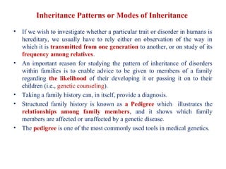
  • 1. Inheritance Patterns or Modes of Inheritance • If we wish to investigate whether a particular trait or disorder in humans is hereditary, we usually have to rely either on observation of the way in which it is transmitted from one generation to another, or on study of its frequency among relatives. • An important reason for studying the pattern of inheritance of disorders within families is to enable advice to be given to members of a family regarding the likelihood of their developing it or passing it on to their children (i.e., genetic counseling). • Taking a family history can, in itself, provide a diagnosis. • Structured family history is known as a Pedigree which illustrates the relationships among family members, and it shows which family members are affected or unaffected by a genetic disease. • The pedigree is one of the most commonly used tools in medical genetics.
  • 2. Basic Pedigree Structure, Drawing & Terminology • Pedigree usually begins with the person (first person in whom the disease is diagnosed) through whom the family came to the attention of the investigator. This person is referred to as the index case, proband, or propositus; or, if female, the proposita • The position of the proband in the family tree is indicated by an arrow. • When discussing relatives in families, one often refers to degrees of relationship (consanguinity), which the marriage between relatives is called consanguineous marriage. • First-degree relatives are those who are related at the parent–offspring or sibling (brother and sister) level. • Second-degree relatives are those who are removed by one additional generational step (e.g., grandparents and their grandchildren, uncles or aunts and their nieces or nephews). • Information about the health of the rest of the family is obtained by asking direct questions about brothers, sisters, parents, and maternal and paternal relatives, with the relevant information about the sex of the individual, affection status, and relationship to other individuals being carefully recorded in the pedigree chart.
  • 3.
  • 4.
  • 5.
  • 6. Genes in Individuals • Most human characteristics and common diseases are polygenic, whereas many of the disordered phenotypes thought of as “genetic” are monogenic but still influenced by other loci in a person’s genome. • Phenotypes due to alterations at a single gene (monogenic) are frequently referred to as Mendelian, after Gregor Mendel, the monk/biologist and the father of modern genetics. • Mendel showed that some traits were dominant relative to other traits; he called the latter traits recessive. Dominant traits require only one copy of a “factor” to be expressed, regardless of what the other copy is, whereas recessive traits require two copies before expression occurs. • The mode of inheritance for a given phenotypic trait or disease is determined by pedigree analysis. All affected and unaffected individuals in the family are recorded in a pedigree using standard symbols.
  • 7. Mendelian Inheritance • More than 16,000 traits or disorders in humans exhibit monogenic (single gene) or unifactorial. • A trait or disorder that is determined by a gene on an autosome is said to show autosomal inheritance, whereas a trait or disorder determined by a gene on one of the sex chromosomes is said to show sex-linked inheritance. • Based on the principles of allelic segregation, and the transmission of alleles from parents to children, one dominant (A) allele and one recessive (a) allele can display any of three Mendelian modes of inheritance: autosomal dominant, autosomal recessive, or chromosome sex-linked. • Approximately 65% of human monogenic disorders are autosomal dominant, 25% are autosomal recessive, and 5% are sex -linked. • Genetic testing is now available for many of these disorders and plays an increasingly important role in clinical medicine • .
  • 8. Autosomal Dominant Inheritance • An autosomal dominant trait is one that manifests or expresses in the heterozygous state, that is, in a person possessing both an abnormal or mutant allele and the normal allele. • It is often possible to trace a dominantly inherited trait or disorder through many generations of a family. • This pattern of inheritance is sometimes referred to as ‘vertical’ transmission and is confirmed when male–male (i.e., father to son) transmission is observed. • Autosomal dominant disorders are significant because mutations in a single allele are sufficient to cause the disease, in other word no carriers for the disease. • Genetic Risk: Each gamete from a heterozygous individual with a dominant trait or disorder will contain either the normal allele or the mutant allele. • Any child born to a heterozygous person affected with a dominant trait or disorder has a 1 in 2 (50%) chance of inheriting it and being similarly affected.
  • 9. Segregation of alleles in autosomal dominant inheritance D represents the mutated allele, whereas d represents the normal allele.
  • 10. Pedigree illustrating the inheritance pattern of postaxial polydactyly, an autosomal dominant disorder. Note the presence of male-to-male transmission
  • 11. A pedigree illustrating autosomal dominant inheritance
  • 12.
  • 13. Characteristics of Autosomal Dominant Inheritance • A vertical pattern is observed in the pedigree, multiple generations being affected. • Heterozygotes for the mutant allele show an abnormal phenotype (affected). • Males and females are affected with equal frequency and severity. • Only one parent must be affected for an offspring to be at risk for developing the phenotype. • When an affected heterozygous person mates with an unaffected one, each offspring has a 50% chance of inheriting the affected phenotype. This is true regardless of the sex of the affected parent—specifically, male-to-male transmission occurs. • Autosomal dominant phenotypes are often age dependent, less severe than autosomal recessive phenotypes, and associated with malformations or other physical features.
  • 14. Autosomal Recessive Inheritance • In the case of autosomal recessive disorders, mutated alleles result in a complete or partial loss of function. • Recessive disorders frequently involve enzymes in metabolic pathways, receptors, or proteins in signaling cascades. • The affected individual can be of either sex and either a homozygote or compound heterozygote for a single-gene defect. • Fortunately, autosomal recessive diseases are, for the most part, rare and often occur in the context of parental consanguinity. • The relatively high frequency of certain recessive disorders, such as sickle cell anemia, cystic fibrosis, and thalassemia, is partially explained by a selective biologic advantage for the heterozygous state. • Heterozygous carriers of a defective allele are usually clinically normal, but they may display indirect differences in phenotype that become apparent only with more precise testing or in the context of certain environmental influences
  • 15. Autosomal Recessive Inheritance • Recessive traits and disorders are manifest or express only when the mutant allele is present in a double dose (i.e., homozygosity or compound heterzygous). • Individuals heterozygous for such mutant alleles show no features of the disorder and are perfectly healthy; they are described as carriers. • Autosomal recessive phenotypes are often associated with deficient activity of enzymes and are thus termed inborn errors of metabolism. Such disorders include phenylketonuria, Tay-Sachs disease, and the various glycogen storage diseases. • Autosomal recessive phenotypes tend to be more severe, less variable, and less age dependent than dominant conditions. • When an autosomal recessive condition is quite rare, the chance that the parents of affected offspring are consanguineous for the phenotype is increased. As a result, the prevalence of rare recessive conditions is high among inbred groups. •
  • 16. Autosomal Recessive Inheritance • The pedigree for recessive traits differs markedly from that seen in autosomal dominant traits. • It is not possible to trace an autosomal recessive trait or disorder through the family, as all the affected individuals in a family are usually in a single sibship (i.e., brothers and sisters). • This is sometimes referred to as ‘horizontal’ transmission, but this is an inappropriate and misleading term.
  • 17. Segregation of alleles in autosomal recessive inheritance. Genetic Risks of Autosomal Recessive Inheritance • If we represent the normal dominant allele as ‘R’ and the recessive mutant allele as ‘r’, then each parental gamete carries either the mutant or the normal allele. • The various possible combinations of gametes mean that the offspring of two heterozygote parents have: • 1 in 4 (25%) chance of being homozygous affected • 1 in 2 (50%) chance of being heterozygous unaffected • 1 in 4 (25%) chance of being homozygous unaffected.
  • 18. Pedigree of an autosomal recessive trait
  • 19. • Is the situation in which the inheritance of a recessive trait mimics (imitate) a dominant pattern • If an individual who is affected (homozygous) by an autosomal recessive disorder marry a carrier of the same disorder, their offspring have a 1 in 2 (50%) chance of being affected. Such a pedigree is said to exhibit pseudodominance Pseudodominance A pedigree with a woman (I2) homozygous for an autosomal recessive disorder whose husband is heterozygous for the same disorder. They have a homozygous affected daughter so that the pedigree shows pseudodominant inheritance.
  • 20. Locus Heterogeneity • A disorder inherited in the same manner can be due to mutations in more than one gene, or what is known as locus heterogeneity. • For example, it is recognized that sensorineural hearing impairment/deafness most commonly shows autosomal recessive inheritance. • It would be expected that, if two deaf persons were homozygous for the same recessive gene, all of their children would be similarly affected. • Families have been described in which all the children born to parents who are deaf due to autosomal recessive genes have had perfectly normal hearing because they are double heterozygotes. The explanation is that the parents were homozygous for mutant alleles at different loci (i.e., different genes can cause autosomal recessive sensorineural deafness). • A very similar story applies to autosomal recessive retinitis pigmentosa. • Disorders with the same phenotype from different genetic loci are known as genocopies.
  • 21. Mutational Heterogeneity • Heterogeneity can also occur at the allelic level. In the majority of single-gene disorders (e.g., β-thalassemia) a large number of different mutations have been identified as being responsible. • There are individuals who have two different mutations at the same locus and are known as compound heterozygotes, constituting what is known as allelic or mutational heterogeneity. • Most individuals affected with an autosomal recessive disorder are probably compound heterozygotes rather than true homozygotes, unless their parents are related, when they are most likely to be homozygous for the same mutation by descent, having inherited the same mutation from a common ancestor.
  • 22. Characteristics of Autosomal Recessive Inheritance • A horizontal pattern is noted in the pedigree, with a single generation being affected. • Males and females are affected with equal frequency and severity. • Inheritance is from both parents, each of whom is a heterozygote (carrier) and each of whom is usually clinically unaffected by his or her carrier status. • Each offspring of two carriers has a 25% chance of being affected, a 50% chance of being a carrier, and a 25% chance of inheriting neither mutant allele. • Thus two-thirds of all clinically unaffected offspring are carriers of the autosomal recessive phenotype. • In matings between affected individuals, each with the same recessive phenotype, all offspring will be affected. • Affected individuals who mate with unaffected individuals who are not carriers have only unaffected offspring. • The rarer the recessive phenotype, the more likely it is that the parents are consanguineous.
  • 23. Sex-Linked Inheritance • Sex-linked inheritance refers to the pattern of inheritance shown by genes that are located on either of the sex chromosomes (X or Y). • Genes carried on the X chromosome are referred to as being X-linked, and those carried on the Y chromosome are referred to as exhibiting Y-linked or holandric inheritance.
  • 24. X-Linked Recessive Inheritance • An X-linked recessive trait is one determined by a gene carried on the X chromosome and usually manifests only in males. • A male with a mutant allele on his single X chromosome is said to be hemizygous for that allele. • Diseases inherited in an X-linked manner are transmitted by healthy heterozygous female carriers to affected males, as well as by affected males to their obligate carrier daughters, with a consequent risk to male grandchildren through these daughters. • This type of pedigree is sometimes said to show ‘diagonal’ or a ‘knight’s move’ pattern of transmission or criss-cross pattern. •
  • 25. Genetic Risks • A male transmits his X chromosome to each of his daughters and his Y chromosome to each of his sons. • If a male affected with hemophilia (as x-lined recessive disorder) marry to a normal female, then all of their daughters will be obligate carriers but none of their sons will be affected. • A male cannot transmit an X-linked trait to his son, with the very rare exception of uniparental heterodisomy (occurs as a consequence of nondisjunction when a person receives two copies of a chromosome, or of part of a chromosome, from one parent and no copy from the other parent). • For a carrier female of an X-linked recessive disorder who marry to a normal male, the chance of the children: each son has a 1 in 2 (50%) chance of being affected and each daughter has a 1 in 2 (50%) chance of being a carrier.
  • 26. Segregation of alleles in X-linked recessive inheritance, relating to the offspring of an affected male. r represents the mutated allele.
  • 27. Segregation of alleles in X-linked recessive inheritance, relating to the offspring of a carrier female. r represents the mutated allele.
  • 28. X-linked disorders • Some X-linked disorders are not compatible with survival to reproductive age and therefore, are not transmitted by affected males. • Duchenne muscular dystrophy is the commonest muscular dystrophy and is a severe disease. • The first sign is delayed walking followed by a waddling gait, difficulty in climbing stairs unaided, and a tendency to fall easily. By about the age of 10 years affected boys usually need to use a wheelchair. • The muscle weakness progresses gradually and affected males ultimately become confined to bed and often die in their late teenage years or early 20s. • Because affected boys do not usually survive to reproduce, the disease is transmitted by healthy female carriers, or may arise as a new mutation. •
  • 29. X-Chromosome Inactivation (Lyonization) • The X chromosome contains many important protein-coding genes, and it has long been known that human females have two X chromosomes and males have only one. Thus, females have two copies of each X-linked gene, and males have only one copy. Yet males and females do not differ in terms of the amounts of protein products (e.g., enzyme levels) encoded by most of these genes. What could account for this?. • In the early 1960s Mary Lyon hypothesized that one X chromosome in each somatic cell of the female is inactivated. This would result in dosage compensation, an equalization of the amount of X-linked gene products in males and females. • The Lyon hypothesis stated that X inactivation (Lyonization) occurs early in female embryonic development and that the X chromosome contributed by the father is inactivated in some cells, whereas in other cells the X chromosome contributed by the mother is inactivated.
  • 30. X-Chromosome Inactivation (Lyonization) • In each cell, one of the two X chromosomes is chosen at random for inactivation, so the maternally and paternally transmitted X chromosomes are each inactivated in about half of the embryo’s cells. Thus, inactivation, like gamete transmission, is analogous to a coin-tossing experiment • Once an X chromosome is inactivated in a cell, it will remain inactive in all descendants of that cell. • X inactivation is therefore a randomly determined, but fixed (or permanent), process. As a result of X inactivation, all normal females have two distinct populations of cells: one population has an active paternally derived X chromosome, and the other has an active maternally derived X chromosome. • Because they have two populations of cells, females are mosaics for X chromosome activity. Males, having only one copy of the X chromosome, are not mosaics but are hemizygous for the X chromosome (hemi means “half ”).
  • 31. The X inactivation process. The maternal (m) and paternal (p) X chromosomes are both active in the zygote and in early embryonic cells. X inactivation then takes place, resulting in cells having either an active paternal X or an active maternal X chromosome. Females are thus X chromosome mosaics.
  • 32. X-Chromosome Inactivation (Lyonization) • The Lyon hypothesis relied on several pieces of evidence, most of which were derived from animal studies. • First, it was known that females are typically mosaics for some X-linked traits and males are not. • For example, female “calico” cats have alternating black and orange patches of fur that correspond to two populations of cells: one that contains X chromosomes in which an “orange” allele is active and one that contains X chromosomes in which a “black” allele is active. Male cats of this breed do not exhibit alternating colors and have either black or orange patches of fur. • Another example, seen in humans, is X-linked ocular albinism. This is an X- linked recessive condition characterized by a lack of melanin production in the retina and by ocular problems such as nystagmus (rapid involuntary eye movements) and decreased visual acuity. Males who inherit the mutation show a relatively uniform lack of melanin in their retinas, whereas female heterozygotes exhibit alternating patches of pigmented and nonpigmented tissue
  • 33. X-Chromosome Inactivation in female “calico” cats
  • 34. Lyonization • The Lyon hypothesis was also supported by biochemical evidence. The enzyme glucose-6-phosphate dehydrogenase (G6PD) is encoded by a gene on the X chromosome and is present in equal quantities in males and females (dosage compensation). • In females who are heterozygous for two common G6PD alleles (labeled A and B), some skin cells produce only the A variant of the enzyme and others produce only the B variant. This is further proof of X chromosome mosaicism in females. • Cytogenetic studies in the 1940s showed that interphase cells of female cats often contained a densely staining chromatin mass in their nuclei. These masses were not seen in males. They were termed Barr bodies, after Murray Barr, one of the scientists who described them. • Barr and his colleague Ewart Bertram hypothesized that the Barr body represented a highly condensed X chromosome (heterochromatin) . Its condensed state is correlated with reduced transcriptional activity, and its DNA is replicated later in the S phase than that of other chromosomes
  • 35.
  • 36. Skewed X-Inactivation • The process of X-inactivation usually occurs randomly, there being an equal chance of either of the two X chromosomes in a heterozygous female being inactivated in any one cell. • After X-inactivation in embryogenesis, therefore, in roughly half the cells one of the X chromosomes is active, whereas in the other half it is the other X chromosome that is active. • Sometimes this process is not random (Skewed) , allowing for the possibility that the active X chromosome in most of the cells of a heterozygous female carrier is the one bearing the mutant allele. If this happens, a carrier female would exhibit some of the symptoms and signs of the disease and be a so-called manifesting heterozygote or carrier.
  • 37. A pedigree illustrating X-linked recessive inheritance A pedigree showing the inheritance of an X-linked recessive trait. Solid symbols represent affected individuals, and dotted symbols represent heterozygous carriers.
  • 38. Characteristics of X-linked recessive Inheritance • There is no male-to-male transmission of the phenotype. • Unaffected males do not transmit the phenotype. • All daughters of an affected male are heterozygous carriers. • Males are usually more severely affected than females. • Some mothers of affected males will not themselves be heterozygotes (i.e., they will be homozygous normal) but will have a germinal mutation. • • Heterozygous women transmit the mutant gene to 50% of their sons, who are affected, and to 50% of their daughters, who are heterozygotes. • If an affected male mates with a heterozygous female, 50% of the male offspring will be affected, giving the false impression of male-to-male transmission. Among the female offspring of such matings, 50% will be affected as severely as the average hemizygous male; in small pedigrees, this pattern may simulate autosomal dominant inheritance.
  • 39. X-Linked Dominant Inheritance • Although uncommon, there are disorders that are manifest in the heterozygous female as well as in the male who has the mutant allele on his single X chromosome. This is known as X-linked dominant inheritance. • X-linked dominant inheritance superficially resembles that of an autosomal dominant trait because both the daughters and sons of an affected female have a 1 in 2 (50%) chance of being affected. There is, however, an important difference. With an X-linked dominant trait, an affected male transmits the trait to all his daughters but to none of his sons. • Therefore, in families with an X-linked dominant disorder there is an excess of affected females and direct male-to-male transmission cannot occur. • X-linked dominant diseases display characteristic patterns of inheritance. They are about twice as common in females as in males, skipped generations are uncommon, and father-to-son transmission is not seen
  • 40. A pedigree illustrating X-linked dominant inheritance Pedigree demonstrating the inheritance of an X-linked dominant trait. X1, Chromosome with normal allele; X2, chromosome with disease allele
  • 41. Y-linked or holandric inheritance • Although it consists of approximately 60 Mb of DNA, the Y chromosome contains relatively few Y-linked, or holandric genes. • Y-linked, or holandric genes include the gene that initiates differentiation of the embryo into a male, several genes that encode testis specific spermatogenesis factors, a minor histocompatibility antigen (termed HY), and a gene in which mutations can cause hearing loss (DFNY1). • Y-linked or holandric inheritance implies that only males are affected. • Male-to-male transmission. An affected male transmits Y-linked traits to all of his sons but to none of his daughters.
  • 42. Pedigree demonstrating the inheritance of a Y-linked trait. Transmission is exclusively male to male. A pedigree illustrating Y-linked or holandric inheritance
  • 43. Sex-limited and sex-influenced inheritance • Confusion sometimes exists regarding traits that are sex-linked and those that are sex-limited or sex-influenced. • Sex limitation refers to the appearance of certain features only in individuals of a particular sex (limited to one sex). Examples include virilization (the development of male physical characteristics such as muscle bulk, body hair, and deep voice in a female) of female infants affected with the autosomal recessive endocrine disorder known as congenital adrenal hyperplasia. • While in sex influenced traits or inheritance, the phenotype of some autosomal traits could be encountered in both sexes however, expressed more frequently in one sex than in another—so-called sex influence. • Gout and baldness are examples of sex-influenced autosomal dominant traits, males being predominantly affected in both cases.
  • 44. Sex-limited and sex-influenced inheritance • The influence of sex in these two examples (Gout and baldness) is probably through the effect of male hormones. Gout, for example, is very rare in women before the menopause but the frequency increases in later life. Baldness does not occur in males who have been castrated (Castration also known as gonadectomy). • In hemochromatosis, the most common autosomal recessive disorder in Western society, homozygous females are much less likely than homozygous males to develop iron overload and associated symptoms; the explanation usually given is that women have a form of natural blood loss through menstruation. • .
  • 45. Mitochondrial Inheritance • The great majority of genetic diseases are caused by alterations in the nuclear genome. However, a small but significant number of diseases can be caused by mutations in mitochondrial DNA (mtDNA). • The mitochondria have their own DNA molecules, which occur in several copies per mitochondrial body and consist of 16,569 base pairs arranged on a double- stranded circular DNA molecule. • The mitochondrial genome encodes two ribosomal RNAs (rRNAs), 22 transfer RNAs (tRNAs), and 13 polypeptides involved in oxidative phosphorylation. • Transcription of mtDNA takes place in the mitochondrion, independently of the nucleus. Unlike nuclear genes, mtDNA genes contain no introns. • The mitochondrial genome is located in the cytoplasm and does not recombine from parents. • mtDNA is inherited exclusively through the maternal line. Males do not transmit mtDNA to their offspring because sperm, which contains only a small number of mtDNA molecules, does not contribute significant cytoplasmic components to the zygote, which are not incorporated into the developing embryo. • One isolated case of paternal transmission of mtDNA mutation has been reported, but such events appear to be extremely rare.
  • 46. Mutations in mtDNA • Mutations in the genes encoded by the mitochondrial chromosome cause a variety of diseases that affect (in particular) organs highly dependent on oxidative metabolism, such as the retina, brain, kidneys, and heart. • An affected woman can pass the defective mitochondrial chromosome to all of her offspring, whereas an affected man has little risk of passing his mutation to a child. • The mutation rate of mtDNA is about 10 times higher than that of nuclear DNA. This is caused by a relative lack of DNA repair mechanisms in the mtDNA and also by damage from free oxygen radicals released during the oxidative phosphorylation process. • Because each cell contains a population of mtDNA molecules, a single cell can harbor some molecules that have an mtDNA mutation and other molecules that do not. This heterogeneity in DNA composition, termed heteroplasmy, is an important cause of variable expression in mitochondrial diseases. • The larger the percentage of mutant mtDNA molecules, the more severe the expression of the disease.
  • 47. Locations of protein-encoding genes for: NADH dehydrogenase, cytochrome c oxidase, cytochrome c oxidoreductase, and ATP] synthase are shown, as are the locations of the two ribosomal RNA genes and 22 transfer RNA genes (designated by single letters). The circular mitochondrial DNA genome.
  • 48. Mitochondrial (maternal) Inheritance A mitochondrial genetic mutation, indicated by darkened symbols, is passed by the female to all of her offspring, including males. Among the subsequent offspring, the males do not transmit the mutation, but the females continue to transmit the mutation to all of their offspring because mitochondria are passed through ova, not sperm