SlideShare a Scribd company logo
1 of 43
Presented by
Loknath Goswami
B. Optom 3rd year
2015 - 2019
Ridley college of optometry
Introduction
• Bandage contact lenses are widely used in refractive
surgery
• The main purpose of this practice is to reduce
inflammatory cell infiltration into the corneal stroma
therefore decreasing the threat of corneal scarring
• Also, bandage lenses assist in the regeneration of
basement membrane and restoration of tight epithelial
stromal adhesion
• The bandage lens protects the loosely adherent and
regenerating epithelium from the relentless action of the
eyelids
• In addition, the lens provides comfort without affecting
the patient’s vision
Aim
• The therapeutic lenses principle aim is to
bandage the eye as a therapy which
– Provides relief from pain
– Serves mechanical protection by separating the
epithelial surface from the external agents such as lid
surfaces, thus protecting the epithelium
– Seal corneal perforations by acting as a splint for the
underlying weaker tissues and supports the area while
healing takes place
– Corrects the surface irregularities in irregular corneas
and improves visual acuity
Ideal contact lens
Conditions of eye which are suitable
for therapeutic lenses
• Eyelids abnormalities –
– Entropion
– Trichiasis
– Ectropion
– Lagophthalmos
Conditions of eye which are suitable
for therapeutic lenses
• Ocular surface disorders – chemical injuries,
dry eye, Stevens-Johnson syndrome
Conditions of eye which are suitable
for therapeutic lenses
• Corneal surface disorder
– Recurrent erosion syndrome
– Keratitis
– Traumatic epithelium abnormalities
– Filamentary keratitis
– Bullous keratopathy
Lens materials for extended wear use
Hydrogels
Silicone
hydrogels
High Dk RGP
lenses
Collagen
Sheilds
Scleral
lenses
HYDROGELS—SOFT LENSES
• HEMA lenses used for extended period are a
choice dependent on the corneal pathology.
i. High water content soft lenses:
• Lenses with water content 80% and Plano power are
available as bandage lens.
• They are suitable for epithelial defect patients.
• These lenses act as bandage which necessitate minimal
epithelial disturbance and help in relieving pain
HYDROGELS—SOFT LENSES
ii. Mid water content lenses:
– Lenses with 45 to 60% water content may be the
choice for small perforations or leaking wounds.
– They act as a splint.
iii. Low water content lenses:
– Low water (below 45%) thin lenses as bandage
lenses
– Used in disorders of lids such as trichiasis causing
trauma to cornea.
SILICONE HYDROGELS
• Silicon hydrogels are new generation lenses.
• They cause significantly lower level of hypoxia
related effects compared to the leading EW
hydrogel lenses.
• They also have lower level of bacterial binding
with them.
COLLAGEN SHIELDS
• Their main function is drug delivery.
• Shields soaked in the drug.
• Antibiotics are applied to the eye in case like
bacterial ulcers, post PK, etc. where the drug
is released in high concentration.
High Dk RGP
• Rigid lenses are frequently used for a combination
of optical and therapeutic indications.
• All corneal abnormalities leading to irregular
astigmatism or high amounts of astigmatism will
benefit visually only with rigid lenses.
• Conditions like post keratitis cornea, post PK,
traumatic cornea or keratoconus, the cornea is
already compromised so lenses with maximum Dk
should be fitted to these patients to prevent
further insult to the cornea
SCLERAL LENSES
• Scleral lenses have a host of therapeutic roles
• Their advantages include the following:
– There need be no corneal contact whatsoever.
– Any eye shape can be fitted.
– Complete protection of the cornea and bulbar
conjunctiva is provided.
– Sealed fits are possible, using gas-permeable
materials, which simplifes the ftting process and
minimizes ‘settling back’.
– Using gas-permeable materials, overnight wear is
possible.
SELECTION OF THE LENS
1. Oxygen Transmissibility
– High water content, thin mid water content lenses,
silicone hydrogels or high Dk RGP lenses give the best
transmissibility.
– Lenses which have to be worn for extended periods
should be selected from either of these materials.
– Select high water contact lens in eye conditions where
the lens has to act as a splint.
– RGP lenses are for irregular compromised corneas to
achieve better vision .
SELECTION OF THE LENS
2. Diameter
– Soft bandage lenses are usually larger in diameter
usually 14.0 to 15.0 mm.
– Larger diameter lenses (15mm to 20mm) may be
required where the specific function is to protect the
limbus or prevent wound leakage at suture or incision
sites
– Larger diameter lenses require flatter back optic zone
radii to achieve the desired fit.
3. Power
– Bandage lenses are usually plano in power.
SELECTION OF THE LENS
4. Disposable Lenses or FRP Lenses
– Disposable lenses or FRP lenses are selected as
bandage lenses.
– Therapeutic lenses should be preferably discarded
after every use.
– They are nowadays rarely cleaned and reinserted.
– Deposits formation is very likely and heavy in such
eye conditions.
Use after Refractive Surgery
• Surface Ablation
• Immediately after surface ablation procedures
such as LASEK or PRK, bandage contact lenses
are routinely applied to patients’ eyes to
encourage re-epithelialization and healing,
and to reduce discomfort and pain
LASIK
• Bandage contact lenses can be applied after, to
reduce discomfort and prevent epithelial in-growth
• However, some believe this actually increases the risk
for striae, or may not have any beneficial effects
• Some patients are at risk of developing an epithelial
slide, especially those with a history of anterior
basement membrane dystrophy. If an epithelial slide
occurs during the operation, a bandage contact lens
may be applied to the eye to improve healing and
protect the eye until the epithelium has regrown
Striae and epithelial in-growth
Striae
• Striae, or flap folds, are a complication of LASIK in up
to 3.5% of cases.
• The folds can disturb visual acuity, though often they
resolve on their own.
• Causes of striae include flap dessication, flap
misalignment and flap tenting.
• Striae can be classified as
– macrostriae or
– microstriae
• The treatment for both groups can involve the use of
bandage contact lenses.
Macrostriae
• These are caused by flap dislocation and often
involve the entire thickness of the flap. If the
macrostriae are not detected early, the flap
must be refloated and stretched again.
• Different methods have been proposed for
this procedure, but many involve the use of
bandage contact lenses after completion to
help the epithelium re-grow correctly.
Microstriae
• These are smaller flap folds that are caused by
problems in flap settling.
• These striae more often resolve on their own
with the help of artificial tears and bandage
contact lenses.
• However, if the striae persist, stretching or
refloating of the flap may be necessary, and a
bandage contact lens is used.
Epithelial In-growth
• Epithelial in-growth is an infrequent complication
of LASIK, that is caused either by implantation of
epithelial cells during surgery or from epithelial
cells growing underneath the flap.
• Removal of the in-growth involves lifting the flap,
irrigating the interface and subsequently placing
a bandage contact lens.
• This prevents epithelium from re-entering the
flap interface
Overcorrection
• In cases of consecutive hyperopia, also known as
overcorrection, bandage contact lenses may be
used in conjunction with non-steroidal anti-
inflammatory drugs (NSAIDs) to reduce the need
for a second surgery.
• The contact lens helps increase the NSAIDs
penetration into the cornea to stimulate stromal
re-growth.
• The tight fit of the contact lens also creates a
contour that helps correctly shape the growth.
Complications
• In some cases, bandage contact lenses can
lead to infectious keratitis.
• Other complications can include
– dry eye,
– corneal hypoxia and
– corneal edema
• Patients should be aware of proper lens
hygiene.
Bandage contact lens after photorefractive
keratotomy
• Although there are many conventional
therapeutic soft lenses on the market, disposable
contact lenses have become very popular
bandage lenses.
• The advantages of disposable lenses include less
expense, less risk of corneal infection, and less
risk of toxic complications with concurrent use of
topical medications.
• They are also easy to replace if a contact lens is
accidently dislodged or lost.
Bandage contact lens after photorefractive
keratotomy
• Standard hydrophillic disposable lenses (e.g.
Acuvue TM, Vistakon) approved for extended
wear have shown good clinical efficacy, but
silicone hydrogel disposable lenses
(PureVision TM, B&L; or focus Night & Day
TM, CIBAVision) also offer the advantage of
decreasing the risk of hypoxic complications
with continuous wear
Bandage contact lens after photorefractive
keratotomy
• Although the risk of infectious keratitis with the
use of therapeutic contact lenses after PRK is low,
bacterial contamination has been found on the
bandage contact lenses after removal.
• Microorganisms found on the lenses typically
represent the bacteria found in the normal ocular
flora, indicating the need for careful monitoring
and follow-up.
• Topical antibiotics combined with no handling of
the lenses by the patient reduce the risk of
bacterial infection
Fitting a therapeutic hydrophilic lens after
photorefractive keratotomy
• At the conclusion of the PRK procedure, the
surgeon generally places a disposable contact
lens on the eye. The contact lens should be
chosen based on the preoperative
measurements. The patient should be examined
at the slit lamp approximately 30 minutes later to
determine the contact lens-cornea relationship
• There may be both patient discomfort as well as
interference with reepithelialization. Check to be
sure the contact lens is not inverted before
changing base curves.
Fitting a therapeutic hydrophilic lens after
photorefractive keratotomy
• If the contact lenses does not have
movement, the causes may be:
– A steep base curve, which may produce corneal
edema. In this case, try a flatter curve.
– Edema of the conjunctiva produced by the use of
a device such as a suction ring. In this case, the
contact lens should not be disturbed.
Fitting a therapeutic hydrophilic lens after
photorefractive keratotomy
• If there is foreign material, rolled epithelial
edges, and/or significant debris under the
contact lens, it should be exchanged.
• On the other hand, if the quantity of debris is
very small, one should avoid disturbing the
lens. Its removal, aside from producing
discomfort, may actually increase the size of
the epithelial defect.
Advice to the patient
• Avoid removing the lens at home specially if the
patient have no experience with contact lenses.
Removal will typically be done at an office visit, once
the epithelium is intact. Manipulation of the contact
lenses by the patient can increase the risk of infection
• Avoid environmental pollutants and activities that
significantly reduce the blink rate, which may cause
dehydration of the contact lens. A dry contact lens
produces discomfort, facilitates deposit formation,
diminishes oxygenation of the cornea and increases
the risk of infection.
Advice to the patient
• Avoid cosmetic around the eyes until the
contact lens has been removed
• Use lubricants, preferably non-preserved,
between administration other topical
medications in order to lubricate and clean
the contact lens
• Discard the contact lens if it comes out of the
eye
Removing the lens
• The following steps are recommended
– Wet the contact lens and wait 2-5 mins before
removing it
– Use a drop of anaesthetic if necessary to reduce
trauma
– Ask the patient to look up
– Pull the contact lens inferiorly and remove it with
fingers or with a fine, non toothed forceps at the
slit lamp
FITTING GUIDELINES
• KERATOMETRY
– It is usually not possible to determine the corneal
curvature in such eye conditions.
– The mires are heavily distorted.
– Corneal topography or Keratoscopy can give some
useful information in selecting curvatures.
– In case of traumatic corneas, the good eye K reading
can form a base line to start with, on the assumption
that the corneal curvature may have been same
before trauma in the eye to be fitted with contact
lens.
FITTING GUIDELINES
• ANTERIOR SEGMENT ASSESSMENT
– Anterior segment assessment is important.
– Staining if possible should be done with rose Bengal
dye and recorded and graded.
– Also the eye should not be in acute infective state
during fitting.
– Tear film stability should also be measured.
• TYPE OF LENS
– Select the type of lens according to the eye condition
For Soft Lenses
• Allow the lens to settle on the eye may be for 15
to 20 minutes - Optimal fit—reasonably well
centered - Complete coverage of the cornea.
• Movement slightly restricted at the same time
does not allow the debris to accumulate
behind.About 0.3 to 0.5 mm with each blink.
• Observe the eye condition after 4 hours, then 24
hours of wear. Ensure there is no complication
developing because of the lens and the wound
has started healing.
For Soft Lenses
• In most cases the bandage lens is worn for short
periods of extended wear, regular follow-ups are
important.
• It is the practitioner who inserts and removes
these lenses whenever needed. However, the
patient should also be explained the emergency
removal technique and contact lens care and
maintenance. He should have a container with
the soaking solution at hand with him.
For RGP
• Measure the good eyes keratometry in case of
uniocular disorders as base line.
• The fitting is done on hit and trial basis.
• There should not be excessive bearing or
clearing areas.
• The tears should exchange properly and debris
should also not collect behind the lens
• Achieve a stable centered lens.
References
• Mark J. Mannis, contact lenses in Ophthalmic
Practice, 1st edition, page no. 148 – 187
• Monica Chaudhry, Contact Lens Primer, 1st
edition, page no. 162 – 173
• Therapeutic Contact lens, Manoj Aryal, B .
Optometry, Institute Of Medicine,
Maharajgunj Medical campus
• http://eyewiki.aao.org/Bandage_Contact_Len
ses_After_Refractive_Surgery
Thank you

More Related Content

What's hot

FUNDUS FLUORESCEIN ANGIOGRAPHY
FUNDUS FLUORESCEIN ANGIOGRAPHYFUNDUS FLUORESCEIN ANGIOGRAPHY
FUNDUS FLUORESCEIN ANGIOGRAPHYAnuraag Singh
 
Accommodation and convergence
Accommodation and convergenceAccommodation and convergence
Accommodation and convergenceSAMEEKSHA AGRAWAL
 
RGP Complications
RGP ComplicationsRGP Complications
RGP ComplicationsHira Dahal
 
Anomalies of accomodation ‫‬
Anomalies of accomodation ‫‬Anomalies of accomodation ‫‬
Anomalies of accomodation ‫‬Ayat AbuJazar
 
Diplopia charting
Diplopia charting Diplopia charting
Diplopia charting ANUJA DHAKAL
 
Diagnosis evaluation in strabismus
Diagnosis evaluation in strabismusDiagnosis evaluation in strabismus
Diagnosis evaluation in strabismusJayarajini
 
Anterior segment OCT & UBM
Anterior segment OCT & UBMAnterior segment OCT & UBM
Anterior segment OCT & UBMDinesh Madduri
 
Hess chart, diplopia chart, cover tests
Hess chart, diplopia chart, cover testsHess chart, diplopia chart, cover tests
Hess chart, diplopia chart, cover testsNikhil Rp
 
Amblyopia Management
Amblyopia ManagementAmblyopia Management
Amblyopia Managementsiraj safi
 
Measurements of the optical constants of the eye
Measurements  of  the optical constants of the eyeMeasurements  of  the optical constants of the eye
Measurements of the optical constants of the eyeDIVYAMURUGESAN8
 
Surgical induced astigmatism
Surgical induced astigmatismSurgical induced astigmatism
Surgical induced astigmatismNamrata Gupta
 
Multifocal iols
Multifocal iolsMultifocal iols
Multifocal iolsSSSIHMS-PG
 

What's hot (20)

FUNDUS FLUORESCEIN ANGIOGRAPHY
FUNDUS FLUORESCEIN ANGIOGRAPHYFUNDUS FLUORESCEIN ANGIOGRAPHY
FUNDUS FLUORESCEIN ANGIOGRAPHY
 
Accommodation and convergence
Accommodation and convergenceAccommodation and convergence
Accommodation and convergence
 
RGP Complications
RGP ComplicationsRGP Complications
RGP Complications
 
Anomalies of accomodation ‫‬
Anomalies of accomodation ‫‬Anomalies of accomodation ‫‬
Anomalies of accomodation ‫‬
 
Diplopia charting
Diplopia charting Diplopia charting
Diplopia charting
 
JCC -Jackson Cross Cylinder
JCC -Jackson Cross CylinderJCC -Jackson Cross Cylinder
JCC -Jackson Cross Cylinder
 
Diagnosis evaluation in strabismus
Diagnosis evaluation in strabismusDiagnosis evaluation in strabismus
Diagnosis evaluation in strabismus
 
Anisometropia
AnisometropiaAnisometropia
Anisometropia
 
Anterior segment OCT & UBM
Anterior segment OCT & UBMAnterior segment OCT & UBM
Anterior segment OCT & UBM
 
Hess chart, diplopia chart, cover tests
Hess chart, diplopia chart, cover testsHess chart, diplopia chart, cover tests
Hess chart, diplopia chart, cover tests
 
Esotropia
EsotropiaEsotropia
Esotropia
 
Amblyopia Management
Amblyopia ManagementAmblyopia Management
Amblyopia Management
 
Corneal topography
Corneal topographyCorneal topography
Corneal topography
 
Amsler grid
Amsler gridAmsler grid
Amsler grid
 
Measurements of the optical constants of the eye
Measurements  of  the optical constants of the eyeMeasurements  of  the optical constants of the eye
Measurements of the optical constants of the eye
 
Pentacam
Pentacam Pentacam
Pentacam
 
Scleral lenses
Scleral lensesScleral lenses
Scleral lenses
 
Paralytic strabismus
Paralytic strabismusParalytic strabismus
Paralytic strabismus
 
Surgical induced astigmatism
Surgical induced astigmatismSurgical induced astigmatism
Surgical induced astigmatism
 
Multifocal iols
Multifocal iolsMultifocal iols
Multifocal iols
 

Similar to Bandage Contact Lens

Indications and fitting philosophies of therapeutic contact lenses
Indications and fitting philosophies of therapeutic contact lensesIndications and fitting philosophies of therapeutic contact lenses
Indications and fitting philosophies of therapeutic contact lensesKrati Gupta
 
Therapeutic Contact lenses
Therapeutic Contact lensesTherapeutic Contact lenses
Therapeutic Contact lensesManoj Aryal
 
Therapeutic contact lens
Therapeutic contact lensTherapeutic contact lens
Therapeutic contact lensHira Dahal
 
Retinal detachment new
Retinal detachment newRetinal detachment new
Retinal detachment newyogesh tiwari
 
Recent Advances in treatment of Refractive Error.pptx
Recent Advances in treatment of Refractive Error.pptxRecent Advances in treatment of Refractive Error.pptx
Recent Advances in treatment of Refractive Error.pptxAlmaazAhmed
 
Ocular viscoelastic devices(OVD)
Ocular viscoelastic devices(OVD)Ocular viscoelastic devices(OVD)
Ocular viscoelastic devices(OVD)Sivateja Challa
 
Journal club contact lens.pptx
Journal club contact lens.pptxJournal club contact lens.pptx
Journal club contact lens.pptxAnisha Heka
 
Ocular Drug Delivery Systems - By Sujay Sawant
Ocular Drug Delivery Systems - By Sujay SawantOcular Drug Delivery Systems - By Sujay Sawant
Ocular Drug Delivery Systems - By Sujay Sawantsujay sawant
 
Laser in situ keratomileusis.pptx
Laser in situ keratomileusis.pptxLaser in situ keratomileusis.pptx
Laser in situ keratomileusis.pptxAdeelBaig22
 
Laser Eye Surgery Guide for Canadians
Laser Eye Surgery Guide for CanadiansLaser Eye Surgery Guide for Canadians
Laser Eye Surgery Guide for CanadiansOlympia Benefits Inc.
 

Similar to Bandage Contact Lens (20)

Indications and fitting philosophies of therapeutic contact lenses
Indications and fitting philosophies of therapeutic contact lensesIndications and fitting philosophies of therapeutic contact lenses
Indications and fitting philosophies of therapeutic contact lenses
 
Therapeutic Contact lenses
Therapeutic Contact lensesTherapeutic Contact lenses
Therapeutic Contact lenses
 
Therapeutic contact lens
Therapeutic contact lensTherapeutic contact lens
Therapeutic contact lens
 
Contact lens
Contact lensContact lens
Contact lens
 
Retinal detachment new
Retinal detachment newRetinal detachment new
Retinal detachment new
 
Ocular Prosthesis
Ocular ProsthesisOcular Prosthesis
Ocular Prosthesis
 
Corneal surgery
Corneal surgeryCorneal surgery
Corneal surgery
 
Corneal surgery
Corneal surgeryCorneal surgery
Corneal surgery
 
Contact Lenses
Contact LensesContact Lenses
Contact Lenses
 
Ocular emergencies
Ocular emergenciesOcular emergencies
Ocular emergencies
 
Cataract
CataractCataract
Cataract
 
Recent Advances in treatment of Refractive Error.pptx
Recent Advances in treatment of Refractive Error.pptxRecent Advances in treatment of Refractive Error.pptx
Recent Advances in treatment of Refractive Error.pptx
 
Ocular viscoelastic devices(OVD)
Ocular viscoelastic devices(OVD)Ocular viscoelastic devices(OVD)
Ocular viscoelastic devices(OVD)
 
Cataract surgery
Cataract surgeryCataract surgery
Cataract surgery
 
Journal club contact lens.pptx
Journal club contact lens.pptxJournal club contact lens.pptx
Journal club contact lens.pptx
 
Ocular Drug Delivery Systems - By Sujay Sawant
Ocular Drug Delivery Systems - By Sujay SawantOcular Drug Delivery Systems - By Sujay Sawant
Ocular Drug Delivery Systems - By Sujay Sawant
 
Cataract
CataractCataract
Cataract
 
Laser in situ keratomileusis.pptx
Laser in situ keratomileusis.pptxLaser in situ keratomileusis.pptx
Laser in situ keratomileusis.pptx
 
Retinal detachment
Retinal detachmentRetinal detachment
Retinal detachment
 
Laser Eye Surgery Guide for Canadians
Laser Eye Surgery Guide for CanadiansLaser Eye Surgery Guide for Canadians
Laser Eye Surgery Guide for Canadians
 

More from Loknath Goswami

Pigment epithelial defect and intraretinal fluid
Pigment epithelial defect and intraretinal fluidPigment epithelial defect and intraretinal fluid
Pigment epithelial defect and intraretinal fluidLoknath Goswami
 
Synoptophore and its parts
Synoptophore and its partsSynoptophore and its parts
Synoptophore and its partsLoknath Goswami
 
Slit lamp biomicroscopy and illumination techniques
Slit lamp biomicroscopy and illumination techniquesSlit lamp biomicroscopy and illumination techniques
Slit lamp biomicroscopy and illumination techniquesLoknath Goswami
 
Retinoscope and retinoscopy
Retinoscope and retinoscopyRetinoscope and retinoscopy
Retinoscope and retinoscopyLoknath Goswami
 
Tonometry and tonography
Tonometry and tonographyTonometry and tonography
Tonometry and tonographyLoknath Goswami
 
Keratometer and keratometry
Keratometer and keratometryKeratometer and keratometry
Keratometer and keratometryLoknath Goswami
 
Recumbent prisms and fresnel prisms
Recumbent prisms and fresnel prismsRecumbent prisms and fresnel prisms
Recumbent prisms and fresnel prismsLoknath Goswami
 

More from Loknath Goswami (8)

Pigment epithelial defect and intraretinal fluid
Pigment epithelial defect and intraretinal fluidPigment epithelial defect and intraretinal fluid
Pigment epithelial defect and intraretinal fluid
 
Synoptophore and its parts
Synoptophore and its partsSynoptophore and its parts
Synoptophore and its parts
 
Slit lamp biomicroscopy and illumination techniques
Slit lamp biomicroscopy and illumination techniquesSlit lamp biomicroscopy and illumination techniques
Slit lamp biomicroscopy and illumination techniques
 
Retinoscope and retinoscopy
Retinoscope and retinoscopyRetinoscope and retinoscopy
Retinoscope and retinoscopy
 
Tonometry and tonography
Tonometry and tonographyTonometry and tonography
Tonometry and tonography
 
Keratometer and keratometry
Keratometer and keratometryKeratometer and keratometry
Keratometer and keratometry
 
Recumbent prisms and fresnel prisms
Recumbent prisms and fresnel prismsRecumbent prisms and fresnel prisms
Recumbent prisms and fresnel prisms
 
Cardinal points
Cardinal pointsCardinal points
Cardinal points
 

Recently uploaded

Advanced Views - Calendar View in Odoo 17
Advanced Views - Calendar View in Odoo 17Advanced Views - Calendar View in Odoo 17
Advanced Views - Calendar View in Odoo 17Celine George
 
Basic Civil Engineering first year Notes- Chapter 4 Building.pptx
Basic Civil Engineering first year Notes- Chapter 4 Building.pptxBasic Civil Engineering first year Notes- Chapter 4 Building.pptx
Basic Civil Engineering first year Notes- Chapter 4 Building.pptxDenish Jangid
 
fourth grading exam for kindergarten in writing
fourth grading exam for kindergarten in writingfourth grading exam for kindergarten in writing
fourth grading exam for kindergarten in writingTeacherCyreneCayanan
 
Grant Readiness 101 TechSoup and Remy Consulting
Grant Readiness 101 TechSoup and Remy ConsultingGrant Readiness 101 TechSoup and Remy Consulting
Grant Readiness 101 TechSoup and Remy ConsultingTechSoup
 
Z Score,T Score, Percential Rank and Box Plot Graph
Z Score,T Score, Percential Rank and Box Plot GraphZ Score,T Score, Percential Rank and Box Plot Graph
Z Score,T Score, Percential Rank and Box Plot GraphThiyagu K
 
ICT Role in 21st Century Education & its Challenges.pptx
ICT Role in 21st Century Education & its Challenges.pptxICT Role in 21st Century Education & its Challenges.pptx
ICT Role in 21st Century Education & its Challenges.pptxAreebaZafar22
 
Unit-IV- Pharma. Marketing Channels.pptx
Unit-IV- Pharma. Marketing Channels.pptxUnit-IV- Pharma. Marketing Channels.pptx
Unit-IV- Pharma. Marketing Channels.pptxVishalSingh1417
 
Paris 2024 Olympic Geographies - an activity
Paris 2024 Olympic Geographies - an activityParis 2024 Olympic Geographies - an activity
Paris 2024 Olympic Geographies - an activityGeoBlogs
 
Application orientated numerical on hev.ppt
Application orientated numerical on hev.pptApplication orientated numerical on hev.ppt
Application orientated numerical on hev.pptRamjanShidvankar
 
Introduction to Nonprofit Accounting: The Basics
Introduction to Nonprofit Accounting: The BasicsIntroduction to Nonprofit Accounting: The Basics
Introduction to Nonprofit Accounting: The BasicsTechSoup
 
microwave assisted reaction. General introduction
microwave assisted reaction. General introductionmicrowave assisted reaction. General introduction
microwave assisted reaction. General introductionMaksud Ahmed
 
Presentation by Andreas Schleicher Tackling the School Absenteeism Crisis 30 ...
Presentation by Andreas Schleicher Tackling the School Absenteeism Crisis 30 ...Presentation by Andreas Schleicher Tackling the School Absenteeism Crisis 30 ...
Presentation by Andreas Schleicher Tackling the School Absenteeism Crisis 30 ...EduSkills OECD
 
PROCESS RECORDING FORMAT.docx
PROCESS      RECORDING        FORMAT.docxPROCESS      RECORDING        FORMAT.docx
PROCESS RECORDING FORMAT.docxPoojaSen20
 
Gardella_Mateo_IntellectualProperty.pdf.
Gardella_Mateo_IntellectualProperty.pdf.Gardella_Mateo_IntellectualProperty.pdf.
Gardella_Mateo_IntellectualProperty.pdf.MateoGardella
 
1029-Danh muc Sach Giao Khoa khoi 6.pdf
1029-Danh muc Sach Giao Khoa khoi  6.pdf1029-Danh muc Sach Giao Khoa khoi  6.pdf
1029-Danh muc Sach Giao Khoa khoi 6.pdfQucHHunhnh
 
Russian Escort Service in Delhi 11k Hotel Foreigner Russian Call Girls in Delhi
Russian Escort Service in Delhi 11k Hotel Foreigner Russian Call Girls in DelhiRussian Escort Service in Delhi 11k Hotel Foreigner Russian Call Girls in Delhi
Russian Escort Service in Delhi 11k Hotel Foreigner Russian Call Girls in Delhikauryashika82
 

Recently uploaded (20)

Advanced Views - Calendar View in Odoo 17
Advanced Views - Calendar View in Odoo 17Advanced Views - Calendar View in Odoo 17
Advanced Views - Calendar View in Odoo 17
 
Basic Civil Engineering first year Notes- Chapter 4 Building.pptx
Basic Civil Engineering first year Notes- Chapter 4 Building.pptxBasic Civil Engineering first year Notes- Chapter 4 Building.pptx
Basic Civil Engineering first year Notes- Chapter 4 Building.pptx
 
Mattingly "AI & Prompt Design: Structured Data, Assistants, & RAG"
Mattingly "AI & Prompt Design: Structured Data, Assistants, & RAG"Mattingly "AI & Prompt Design: Structured Data, Assistants, & RAG"
Mattingly "AI & Prompt Design: Structured Data, Assistants, & RAG"
 
fourth grading exam for kindergarten in writing
fourth grading exam for kindergarten in writingfourth grading exam for kindergarten in writing
fourth grading exam for kindergarten in writing
 
Grant Readiness 101 TechSoup and Remy Consulting
Grant Readiness 101 TechSoup and Remy ConsultingGrant Readiness 101 TechSoup and Remy Consulting
Grant Readiness 101 TechSoup and Remy Consulting
 
Z Score,T Score, Percential Rank and Box Plot Graph
Z Score,T Score, Percential Rank and Box Plot GraphZ Score,T Score, Percential Rank and Box Plot Graph
Z Score,T Score, Percential Rank and Box Plot Graph
 
Mehran University Newsletter Vol-X, Issue-I, 2024
Mehran University Newsletter Vol-X, Issue-I, 2024Mehran University Newsletter Vol-X, Issue-I, 2024
Mehran University Newsletter Vol-X, Issue-I, 2024
 
INDIA QUIZ 2024 RLAC DELHI UNIVERSITY.pptx
INDIA QUIZ 2024 RLAC DELHI UNIVERSITY.pptxINDIA QUIZ 2024 RLAC DELHI UNIVERSITY.pptx
INDIA QUIZ 2024 RLAC DELHI UNIVERSITY.pptx
 
ICT Role in 21st Century Education & its Challenges.pptx
ICT Role in 21st Century Education & its Challenges.pptxICT Role in 21st Century Education & its Challenges.pptx
ICT Role in 21st Century Education & its Challenges.pptx
 
Unit-IV- Pharma. Marketing Channels.pptx
Unit-IV- Pharma. Marketing Channels.pptxUnit-IV- Pharma. Marketing Channels.pptx
Unit-IV- Pharma. Marketing Channels.pptx
 
Paris 2024 Olympic Geographies - an activity
Paris 2024 Olympic Geographies - an activityParis 2024 Olympic Geographies - an activity
Paris 2024 Olympic Geographies - an activity
 
Application orientated numerical on hev.ppt
Application orientated numerical on hev.pptApplication orientated numerical on hev.ppt
Application orientated numerical on hev.ppt
 
Introduction to Nonprofit Accounting: The Basics
Introduction to Nonprofit Accounting: The BasicsIntroduction to Nonprofit Accounting: The Basics
Introduction to Nonprofit Accounting: The Basics
 
Advance Mobile Application Development class 07
Advance Mobile Application Development class 07Advance Mobile Application Development class 07
Advance Mobile Application Development class 07
 
microwave assisted reaction. General introduction
microwave assisted reaction. General introductionmicrowave assisted reaction. General introduction
microwave assisted reaction. General introduction
 
Presentation by Andreas Schleicher Tackling the School Absenteeism Crisis 30 ...
Presentation by Andreas Schleicher Tackling the School Absenteeism Crisis 30 ...Presentation by Andreas Schleicher Tackling the School Absenteeism Crisis 30 ...
Presentation by Andreas Schleicher Tackling the School Absenteeism Crisis 30 ...
 
PROCESS RECORDING FORMAT.docx
PROCESS      RECORDING        FORMAT.docxPROCESS      RECORDING        FORMAT.docx
PROCESS RECORDING FORMAT.docx
 
Gardella_Mateo_IntellectualProperty.pdf.
Gardella_Mateo_IntellectualProperty.pdf.Gardella_Mateo_IntellectualProperty.pdf.
Gardella_Mateo_IntellectualProperty.pdf.
 
1029-Danh muc Sach Giao Khoa khoi 6.pdf
1029-Danh muc Sach Giao Khoa khoi  6.pdf1029-Danh muc Sach Giao Khoa khoi  6.pdf
1029-Danh muc Sach Giao Khoa khoi 6.pdf
 
Russian Escort Service in Delhi 11k Hotel Foreigner Russian Call Girls in Delhi
Russian Escort Service in Delhi 11k Hotel Foreigner Russian Call Girls in DelhiRussian Escort Service in Delhi 11k Hotel Foreigner Russian Call Girls in Delhi
Russian Escort Service in Delhi 11k Hotel Foreigner Russian Call Girls in Delhi
 

Bandage Contact Lens

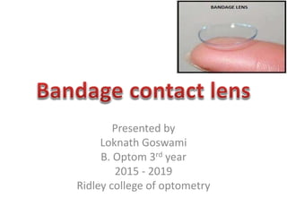
  • 1. Presented by Loknath Goswami B. Optom 3rd year 2015 - 2019 Ridley college of optometry
  • 2. Introduction • Bandage contact lenses are widely used in refractive surgery • The main purpose of this practice is to reduce inflammatory cell infiltration into the corneal stroma therefore decreasing the threat of corneal scarring • Also, bandage lenses assist in the regeneration of basement membrane and restoration of tight epithelial stromal adhesion • The bandage lens protects the loosely adherent and regenerating epithelium from the relentless action of the eyelids • In addition, the lens provides comfort without affecting the patient’s vision
  • 3. Aim • The therapeutic lenses principle aim is to bandage the eye as a therapy which – Provides relief from pain – Serves mechanical protection by separating the epithelial surface from the external agents such as lid surfaces, thus protecting the epithelium – Seal corneal perforations by acting as a splint for the underlying weaker tissues and supports the area while healing takes place – Corrects the surface irregularities in irregular corneas and improves visual acuity
  • 5. Conditions of eye which are suitable for therapeutic lenses • Eyelids abnormalities – – Entropion – Trichiasis – Ectropion – Lagophthalmos
  • 6. Conditions of eye which are suitable for therapeutic lenses • Ocular surface disorders – chemical injuries, dry eye, Stevens-Johnson syndrome
  • 7. Conditions of eye which are suitable for therapeutic lenses • Corneal surface disorder – Recurrent erosion syndrome – Keratitis – Traumatic epithelium abnormalities – Filamentary keratitis – Bullous keratopathy
  • 8. Lens materials for extended wear use Hydrogels Silicone hydrogels High Dk RGP lenses Collagen Sheilds Scleral lenses
  • 9. HYDROGELS—SOFT LENSES • HEMA lenses used for extended period are a choice dependent on the corneal pathology. i. High water content soft lenses: • Lenses with water content 80% and Plano power are available as bandage lens. • They are suitable for epithelial defect patients. • These lenses act as bandage which necessitate minimal epithelial disturbance and help in relieving pain
  • 10. HYDROGELS—SOFT LENSES ii. Mid water content lenses: – Lenses with 45 to 60% water content may be the choice for small perforations or leaking wounds. – They act as a splint. iii. Low water content lenses: – Low water (below 45%) thin lenses as bandage lenses – Used in disorders of lids such as trichiasis causing trauma to cornea.
  • 11. SILICONE HYDROGELS • Silicon hydrogels are new generation lenses. • They cause significantly lower level of hypoxia related effects compared to the leading EW hydrogel lenses. • They also have lower level of bacterial binding with them.
  • 12. COLLAGEN SHIELDS • Their main function is drug delivery. • Shields soaked in the drug. • Antibiotics are applied to the eye in case like bacterial ulcers, post PK, etc. where the drug is released in high concentration.
  • 13. High Dk RGP • Rigid lenses are frequently used for a combination of optical and therapeutic indications. • All corneal abnormalities leading to irregular astigmatism or high amounts of astigmatism will benefit visually only with rigid lenses. • Conditions like post keratitis cornea, post PK, traumatic cornea or keratoconus, the cornea is already compromised so lenses with maximum Dk should be fitted to these patients to prevent further insult to the cornea
  • 14. SCLERAL LENSES • Scleral lenses have a host of therapeutic roles • Their advantages include the following: – There need be no corneal contact whatsoever. – Any eye shape can be fitted. – Complete protection of the cornea and bulbar conjunctiva is provided. – Sealed fits are possible, using gas-permeable materials, which simplifes the ftting process and minimizes ‘settling back’. – Using gas-permeable materials, overnight wear is possible.
  • 15. SELECTION OF THE LENS 1. Oxygen Transmissibility – High water content, thin mid water content lenses, silicone hydrogels or high Dk RGP lenses give the best transmissibility. – Lenses which have to be worn for extended periods should be selected from either of these materials. – Select high water contact lens in eye conditions where the lens has to act as a splint. – RGP lenses are for irregular compromised corneas to achieve better vision .
  • 16. SELECTION OF THE LENS 2. Diameter – Soft bandage lenses are usually larger in diameter usually 14.0 to 15.0 mm. – Larger diameter lenses (15mm to 20mm) may be required where the specific function is to protect the limbus or prevent wound leakage at suture or incision sites – Larger diameter lenses require flatter back optic zone radii to achieve the desired fit. 3. Power – Bandage lenses are usually plano in power.
  • 17. SELECTION OF THE LENS 4. Disposable Lenses or FRP Lenses – Disposable lenses or FRP lenses are selected as bandage lenses. – Therapeutic lenses should be preferably discarded after every use. – They are nowadays rarely cleaned and reinserted. – Deposits formation is very likely and heavy in such eye conditions.
  • 18. Use after Refractive Surgery • Surface Ablation • Immediately after surface ablation procedures such as LASEK or PRK, bandage contact lenses are routinely applied to patients’ eyes to encourage re-epithelialization and healing, and to reduce discomfort and pain
  • 19. LASIK • Bandage contact lenses can be applied after, to reduce discomfort and prevent epithelial in-growth • However, some believe this actually increases the risk for striae, or may not have any beneficial effects • Some patients are at risk of developing an epithelial slide, especially those with a history of anterior basement membrane dystrophy. If an epithelial slide occurs during the operation, a bandage contact lens may be applied to the eye to improve healing and protect the eye until the epithelium has regrown
  • 21. Striae • Striae, or flap folds, are a complication of LASIK in up to 3.5% of cases. • The folds can disturb visual acuity, though often they resolve on their own. • Causes of striae include flap dessication, flap misalignment and flap tenting. • Striae can be classified as – macrostriae or – microstriae • The treatment for both groups can involve the use of bandage contact lenses.
  • 22. Macrostriae • These are caused by flap dislocation and often involve the entire thickness of the flap. If the macrostriae are not detected early, the flap must be refloated and stretched again. • Different methods have been proposed for this procedure, but many involve the use of bandage contact lenses after completion to help the epithelium re-grow correctly.
  • 23. Microstriae • These are smaller flap folds that are caused by problems in flap settling. • These striae more often resolve on their own with the help of artificial tears and bandage contact lenses. • However, if the striae persist, stretching or refloating of the flap may be necessary, and a bandage contact lens is used.
  • 24. Epithelial In-growth • Epithelial in-growth is an infrequent complication of LASIK, that is caused either by implantation of epithelial cells during surgery or from epithelial cells growing underneath the flap. • Removal of the in-growth involves lifting the flap, irrigating the interface and subsequently placing a bandage contact lens. • This prevents epithelium from re-entering the flap interface
  • 25. Overcorrection • In cases of consecutive hyperopia, also known as overcorrection, bandage contact lenses may be used in conjunction with non-steroidal anti- inflammatory drugs (NSAIDs) to reduce the need for a second surgery. • The contact lens helps increase the NSAIDs penetration into the cornea to stimulate stromal re-growth. • The tight fit of the contact lens also creates a contour that helps correctly shape the growth.
  • 26. Complications • In some cases, bandage contact lenses can lead to infectious keratitis. • Other complications can include – dry eye, – corneal hypoxia and – corneal edema • Patients should be aware of proper lens hygiene.
  • 27. Bandage contact lens after photorefractive keratotomy • Although there are many conventional therapeutic soft lenses on the market, disposable contact lenses have become very popular bandage lenses. • The advantages of disposable lenses include less expense, less risk of corneal infection, and less risk of toxic complications with concurrent use of topical medications. • They are also easy to replace if a contact lens is accidently dislodged or lost.
  • 28. Bandage contact lens after photorefractive keratotomy • Standard hydrophillic disposable lenses (e.g. Acuvue TM, Vistakon) approved for extended wear have shown good clinical efficacy, but silicone hydrogel disposable lenses (PureVision TM, B&L; or focus Night & Day TM, CIBAVision) also offer the advantage of decreasing the risk of hypoxic complications with continuous wear
  • 29. Bandage contact lens after photorefractive keratotomy • Although the risk of infectious keratitis with the use of therapeutic contact lenses after PRK is low, bacterial contamination has been found on the bandage contact lenses after removal. • Microorganisms found on the lenses typically represent the bacteria found in the normal ocular flora, indicating the need for careful monitoring and follow-up. • Topical antibiotics combined with no handling of the lenses by the patient reduce the risk of bacterial infection
  • 30. Fitting a therapeutic hydrophilic lens after photorefractive keratotomy • At the conclusion of the PRK procedure, the surgeon generally places a disposable contact lens on the eye. The contact lens should be chosen based on the preoperative measurements. The patient should be examined at the slit lamp approximately 30 minutes later to determine the contact lens-cornea relationship • There may be both patient discomfort as well as interference with reepithelialization. Check to be sure the contact lens is not inverted before changing base curves.
  • 31. Fitting a therapeutic hydrophilic lens after photorefractive keratotomy • If the contact lenses does not have movement, the causes may be: – A steep base curve, which may produce corneal edema. In this case, try a flatter curve. – Edema of the conjunctiva produced by the use of a device such as a suction ring. In this case, the contact lens should not be disturbed.
  • 32. Fitting a therapeutic hydrophilic lens after photorefractive keratotomy • If there is foreign material, rolled epithelial edges, and/or significant debris under the contact lens, it should be exchanged. • On the other hand, if the quantity of debris is very small, one should avoid disturbing the lens. Its removal, aside from producing discomfort, may actually increase the size of the epithelial defect.
  • 33. Advice to the patient • Avoid removing the lens at home specially if the patient have no experience with contact lenses. Removal will typically be done at an office visit, once the epithelium is intact. Manipulation of the contact lenses by the patient can increase the risk of infection • Avoid environmental pollutants and activities that significantly reduce the blink rate, which may cause dehydration of the contact lens. A dry contact lens produces discomfort, facilitates deposit formation, diminishes oxygenation of the cornea and increases the risk of infection.
  • 34. Advice to the patient • Avoid cosmetic around the eyes until the contact lens has been removed • Use lubricants, preferably non-preserved, between administration other topical medications in order to lubricate and clean the contact lens • Discard the contact lens if it comes out of the eye
  • 35. Removing the lens • The following steps are recommended – Wet the contact lens and wait 2-5 mins before removing it – Use a drop of anaesthetic if necessary to reduce trauma – Ask the patient to look up – Pull the contact lens inferiorly and remove it with fingers or with a fine, non toothed forceps at the slit lamp
  • 36. FITTING GUIDELINES • KERATOMETRY – It is usually not possible to determine the corneal curvature in such eye conditions. – The mires are heavily distorted. – Corneal topography or Keratoscopy can give some useful information in selecting curvatures. – In case of traumatic corneas, the good eye K reading can form a base line to start with, on the assumption that the corneal curvature may have been same before trauma in the eye to be fitted with contact lens.
  • 37. FITTING GUIDELINES • ANTERIOR SEGMENT ASSESSMENT – Anterior segment assessment is important. – Staining if possible should be done with rose Bengal dye and recorded and graded. – Also the eye should not be in acute infective state during fitting. – Tear film stability should also be measured. • TYPE OF LENS – Select the type of lens according to the eye condition
  • 38. For Soft Lenses • Allow the lens to settle on the eye may be for 15 to 20 minutes - Optimal fit—reasonably well centered - Complete coverage of the cornea. • Movement slightly restricted at the same time does not allow the debris to accumulate behind.About 0.3 to 0.5 mm with each blink. • Observe the eye condition after 4 hours, then 24 hours of wear. Ensure there is no complication developing because of the lens and the wound has started healing.
  • 39. For Soft Lenses • In most cases the bandage lens is worn for short periods of extended wear, regular follow-ups are important. • It is the practitioner who inserts and removes these lenses whenever needed. However, the patient should also be explained the emergency removal technique and contact lens care and maintenance. He should have a container with the soaking solution at hand with him.
  • 40. For RGP • Measure the good eyes keratometry in case of uniocular disorders as base line. • The fitting is done on hit and trial basis. • There should not be excessive bearing or clearing areas. • The tears should exchange properly and debris should also not collect behind the lens • Achieve a stable centered lens.
  • 41.
  • 42. References • Mark J. Mannis, contact lenses in Ophthalmic Practice, 1st edition, page no. 148 – 187 • Monica Chaudhry, Contact Lens Primer, 1st edition, page no. 162 – 173 • Therapeutic Contact lens, Manoj Aryal, B . Optometry, Institute Of Medicine, Maharajgunj Medical campus • http://eyewiki.aao.org/Bandage_Contact_Len ses_After_Refractive_Surgery

Editor's Notes

  1. Unlike normal corneas the eye to be fitted with therapeutic lens is a compromised eye and is at higher risk of hypoxia and infections. The balance between benefits and potential risk must be carefully considered
  2. (Tan et al., 1995; Romero-Rangel et al., 2000; Pullum and Buckley, 2007).
  3. The lens type is selected on the following criterias: