CLABSI
•Download as PPTX, PDF•
23 likes•29,476 views

Central-Line-Associated Bloodstream Infections (CLABSI) pause a major health problem in hospitalized patients. This disease is associated with people with a central line/tube inserted through the skin into the large vein, which can be used to give medicines, fluids, nutrients, or blood products to patients in critical conditions. The disease occurs when microbes enter through the central line invading the bloodstream.

Recommended
Recommended
More Related Content
What's hot
What's hot (20)
Similar to CLABSI
Similar to CLABSI (20)
More from Mary Mwinga
Recently uploaded
Recently uploaded (20)
CLABSI
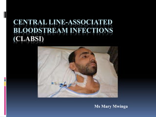
- 1. CENTRAL LINE-ASSOCIATED BLOODSTREAM INFECTIONS (CLABSI) Ms Mary Mwinga
- 2. INTRODUCTION A CLABSI is a serious infection that occurs when microbes enter the bloodstream through a central line. Central line: also called central venous access device -Is a long, thin, flexible tube used to give medicines, fluids, nutrients, or blood products over a long period of time, usually several weeks or more.
- 4. INTRODUCTION A catheter is inserted in the arm, neck or chest through the skin into a large vein. Is threaded through this vein until it reaches a large vein near the heart. Examples of central venous access device Multi-Lumen Catheter PICC Line Implanted Port Quinton Catheter Tunnel Catheter
- 5. EPIDERMIOLOGY CLABSI results in thousands of deaths each year The infections are preventable. Risk factors Age: 1 year or younger or 60 years or older. Malnutrition. Immunosuppressive chemotherapy. Loss of skin integrity (e.g. burn) Severe underlying illness. Indwelling device (e.g. Catheter). Intensive care unit stay. Prolonged hospital stay.
- 6. CLABSI Can lead to serious consequences such as : - Shock - Multiple organ failure - DIC (disseminated intravascular coagulopathies) - BSI can be cause by all microbes bacteria, viruses, fungi and parasites. - Bacteria cause majority BSI.
- 7. Bacteria and BSI Bacteraemia: presence of bacteria in blood without any multiplication. Septicaemia: circulation and multiplication of bacteria in the bloodstream May produce products e.g. Toxins, that cause harm to host.
- 8. Bacteraemia Three types 1.Transient bacteraemia. 2. Continuous bacteraemia. 3. Intermittent bacteraemia.
- 9. bacteraemia 1. Transient bacteraemia: occurs spontaneously or with minor events (such as brushing teeth or chewing food, instrumentation of contaminated mucosal site & surgery involving non-sterile site. These may lead to septicaemia.
- 10. Bacteraemia 2. Continuous bacteraemia Organisms released into bloodstream at a constant rate. Occurs in: - Septic shock, endocarditis & other endovascular infection. - Early stages involved in enteric fever, brucellosis & leptospirosis.
- 11. Bacteraemia 3. Intermittent bacteraemia Release of blood intermittently (unsteady) E.g. - Undrained abscess (bacteria released approx. 45 minutes before a febrile episode). - Early course of meningitis, pneumonia, pyogenic arthritis & osteomyelitis.
- 12. Etiologic agents of BSI Bacteria S.aureus CoNS Β-haemo. streptococci Enterococcus spp. S. pneumoniae Viridans streptococci Salmonella spp E.coli K. pneumoniae Enterobacter cloacae Proteus spp Pseudomonas spp Brucella spp Viruses HIV Epstein-Barr V Cytomegalovirus Fungi Candida Cryptococcus Coccidioides immitis Histoplasma capsulatum Blastomyces dermatitidis Mucor spp Aspergillus spp Parasites Plasmodium Trypanosoma Babesia Wuchereria Loa Loa
- 13. Types of bloodstream infections (BSI) A. Intravascular B. Extra vascular Factors contributing to initiation of BSI Immunosupression Use of broad spectrum antimicrobial agents – suppresses normal flora allows emergence of resistant strains of bacteria. Invasive procedures or extensive surgeries- allowing bacteria to access the blood. Prolonged survival of debilitated patients.
- 14. Intravascular BSI Def: originates within the cardiovascular system. Includes - infective endocarditis - mycotic aneurysm - suppurative thrombophlebitis - Intravenous catheter associated bacteraemia Complications: lead to continuous bacteraemia resulting into serious and life threatening events.
- 15. Intravascular BSI INFECTIVE ENDOCARDITIS DEF: Infection of the endocardium, characterized by presence of ‘vegetation’ Composed of mass of platelets, fibrin, micro colonies of organisms and scanty inflammatory cells. Vegetative- most common in heart valves - Followed by low pressure side of a ventricular septal defect, & on the mural endocardium.
- 16. Intravascular BSI- Infective Endocarditis Classification of infective endocardium I. Onset-acute (rapid damage of cardiac structures- spreads to extracardiac site, rapidly fatal). II. Sub-acute: slow evolution Type of valve affected- occur in native or prosthetic valve. Associated with intravenous drug abuse
- 17. Agents of Endocarditis Streptococci viridans and others Pneumococci Enterococci S. aureus CoNS – S. epidermidis Fastidious GNCB (HACEK group) GNB Candida spp Culture- negative endocarditis- Bartonella, Coxiella
- 18. Agents of Endocarditis Types Native valve endocarditis: S.aureus Prosthetic valve endocarditis: occurs after valve repacement. Early prosthetic: occurs within 12 months of valve replacement - S.epidermidis Late prosthetic valve endocarditis: occurs after 12 months of valve replacement -due to Viridans streptococci.
- 19. Agents of Endocarditis in IV drug abusers Common in young males Skin- common source of infection - Right sided: S.aureus - Left sided: Enterococcus, S. aureus - Subacute endocarditis: Viridans streptococcus.
- 20. Extra vascular bloodstream infection Organisms multiply at primary site e.g. Lungs - Drained by lymphatics and reach bloodstream. - Cells are removed by reticuloendothelial cells or multiply more cause septicaemia. - portal of entry: Genitourinary tract (25%) respiratory tract (20%) abscesses (10%) surgical site wound infections (5%) biliary tract (5%) 25% cases unknown.
- 21. Extra vascular bloodstream infectionAGENTS Organism Portal of entry E. coli & other GNB – kleb, proteus Common- UTI Enterobacter, pseudomonas rare- intestinal Haemophilus influenzae-b meninges, epiglottis, lungs Pneumococcus Meninges, lungs Brucella Reticuloendothelial system S. Typhi Small intestines, lymph nodes, reticuloendothelial system
- 22. Clinical manifestations Infections are evident during septicaemic stage- during multiplication and production of toxins. Signs & symptoms - Fever or hypothermia with/with no chills & rigors. - Hyperventilation – leads to excess loss of CO2 & subsequent respiratory alkalosis. - Skin lesions, change of mental state & diarrhoea.
- 23. Signs & symptoms Septic shock - Complication of septicaemia. - Mediated by activated mononuclear cells producing cytokines (e.g. TNF & interleukins) - GNB release endotoxins - LPS : has direct effect on pathogenesis of septic (endotoxic) shock. Effect mediates responses e.g. Fever, activation of complement and clotting factors. - Manifestations: hypotension, acute respiratory distress, renal failure, tissue destruction.
- 24. Laboratory diagnosis Depends on isolation and causative agents from blood. Preparation of site: 70% isopropyl alcohol is used to clean skin Site: antecubital vein, at a point below the existing IV line -blood above not collected- diluted by infused fluid. Time of collection: before start of antimicrobial therapy.
- 25. Laboratory diagnosis Specimen volume: - Infants & small children: 1-5ml blood for culture. - Adults: 10-20ml blood. No of samples: -continuous bacteraemia: single blood culture collected before therapy. - Other situations: 2-3 blood cultures-for good isolation.
- 26. Culture Blood culture broths reccommended. Media - Monophasic medium: contains 50-100 ml BHI broth, trypticase soy broth or thyoglycolate broth. Incubated for two weeks at 37 °C, Subculture on MA, BA & CA after 24 hrs, 48 hrs, 5th day, 7th and 14th day of incubation. - Castaneda’s biphasic medium: contains BHI broth (50-100ml) and BHI agar slope. Dilution: 1:5 SPS (sodium polyanethol sulphonate) added to medium as anticoagulant & counteracts the bactericidal action in blood.
- 27. Anaerobic adult aerobic Paediatric aerobic
- 28. Identification Isolated organisms further identified by colony morphology, Gram staining, biochem. Reactions and serological tests (for Coxiella burnetti & Chlamydia) Antibiotic Susceptibility Testing Done by Kirby-Bauer disc diffusion method and MIC and MBC (minimum bactericidal concentration) -serves a s guide for treatment. Other tests Total leukocyte count, ESR, CRP, urine culture, chest X-ray, endocardiography, endotoxin-limulus lysate test (detects endotoxin).
- 29. prevention