2. Mini-Reviews and Perspectives continued
tropin (ACTH) insensitivity. Achalasia can be caused by include slow eating, stereotactic movements with eating,
infiltration of the esophagus and LES in amyloidosis or and avoidance of social functions that involve meals.
owing to extrinsic compression of the gastroesophageal With these self-taught accommodating techniques, the
junction, as may occur with tight fundoplication during onset of symptoms in patients can be slow in progression
antireflux procedures19 or laparoscopic adjustable gastric and many patients experience symptoms for years before
banding.20 Achalasia is known to occur after infection by coming to medical attention.21 Patients with achalasia
Trypanosoma cruzi, also known as Chagas disease. Pa- may also present with heartburn owing to bacterial fer-
tients with this infection often have other features of mentation, and thus acidification, of food products
diffuse enteric myenteric destruction, including megaco- within the esophagus. As a result, reflux symptoms un-
lon, heart disease, and neurologic disorders, although the responsive to reflux therapy may suggest achalasia.
phenotypic manifestations are linked to geographic vari-
ation in endemic countries. Diagnosis
The most common cause of secondary achalasia is Patients who have a history that suggests achala-
malignancy through one of several mechanisms. Tumors sia commonly require at least 2, and sometimes 3, mo-
that infiltrate the gastroesophageal junction may cause dalities for diagnosis.22 Barium esophagram is often the
an achalasia-like picture from extrinsic pressure or direct first study performed for patients with dysphagia. In
tumor invasion of the myenteric plexus. This is most patients with achalasia with severe symptoms, there is
commonly described with adenocarcinoma of the gastro- typically a dilated esophagus, absence of peristalsis, and
esophageal junction or proximal stomach, but may occur narrowing of the distal esophagus in a typical “bird’s
with pancreatic, breast, lung, or hepatocellular cancers. beak” appearance but the radiographic appearance of
Cancer, most commonly small cell lung cancer, may pro- achalasia can be variable (Figure 1). The number and
duce achalasia through a paraneoplastic effect by secre- severity of findings on esophagram often do not correlate
tion of an antineuronal antibody. with the degree of symptoms a patient with achalasia has,
but at least 1 of these features is present in virtually all
Clinical Presentation cases of achalasia.23 This study alone may be diagnostic,
Most patients with achalasia present with esoph- particularly in advanced cases.
ageal dysphagia (present in up to 90% of patients), often Endoscopic evaluation of the esophagus and stomach
for both solids and liquids, a distinction from anatomic is recommended in every patient with achalasia to ensure
disorders of the esophagus. Other common symptoms that there is not a malignancy causing the disease or
include chest pain, heartburn, regurgitation, and weight esophageal squamous cell carcinoma complicating acha-
loss, all of which occur in up to 60% of patients. Patients lasia. At endoscopy, a dilated esophagus with a tight LES
with achalasia may also present with more subtle symp- that “pops” open with gentle pressure is often observed,
toms owing to accommodation. These symptoms might as is retained food and saliva. However, a normal esopha-
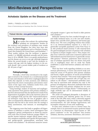
Figure 1. Barium esophagrams on achalasia patients
demonstrating a dilated esophagus and tapering of the
distal esophagus with the classic “bird’s beak” (A), dif-
fuse narrowing of the esophagus with a spastic appear-
ance often seen in “vigorous” achalasia and a large epi-
phrenic diverticulum (B), and a dilated and tortuous
esophagus with early “sigmoidization” of the esophagus
(because of its visual similarity to the sigmoid colon on
barium enema) observed in long standing achalasia ac-
companied by multiple pulsion diverticuli (C).
370
3. Mini-Reviews and Perspectives continued
Figure 2. High-resolution esophageal manometry with
impedance on an achalasia patient revealing aperistalsis
and incomplete bolus transit demonstrated by imped-
ance (A), isobaric simultaneous contractions (B), and a
hypertensive and poorly relaxing LES (C).
gogastroduodenoscopy should not dissuade a clinician with an esophageal pressurization 30 mm Hg, and type
from making the diagnosis because up to 40% of patients III (spastic achalasia) patients have 2 spastic contrac-
with achalasia will have a normal endoscopy.24 tions with or without a period of compartmentalized
Esophageal manometry is considered the gold stan- pressurization. These investigators used logistic regres-
dard diagnostic modality for achalasia. Manometrically, sion and found that type II achalasia patients were more
classic achalasia is defined by absence of peristalsis in the likely to have good symptom response and less likely to
esophageal body, a hypertensive LES (resting pressure require multiple treatments than the other 2 groups.
45 mm Hg) and a poorly relaxing LES, with a residual With reevaluation of the manometric criteria for acha-
pressure 8 mm Hg. However, it is well understood that lasia, some diseases may commonly be called achalasia
as many as 50% of patients that are given the clinical variants.28 For example, diffuse esophageal spasm, which
diagnosis of achalasia do not have a hypertensive LES,25 may be defined manometrically by a hypertensive LES
although the diagnosis does require aperistalsis and a and some simultaneous contractions, may progress to
poorly relaxing LES.26,27 achalasia. Indeed, some patients with insufficient mano-
High-resolution manometry and multichannel intralu- metric criteria undergo classic barium esophagograms
minal impedance with manometry have been applied to for achalasia. As a result, diagnosis of achalasia often
achalasia (Figure 2). Using high-resolution manometry, becomes a compilation of compatible clinical and objec-
achalasia has been divided into 3 subtypes based on the tive criteria rather than reliance on a single test.
function of the esophageal body with the notion that
different subtypes may respond to treatment in a variable
fashion. Type I (classic achalasia has no distal esophageal Treatment
pressurization 30 mm Hg); type II (achalasia with Therapy of achalasia focuses on relaxation or me-
esophageal compression) has 2 test swallows associated chanical disruption of the LES. Achalasia is rare so there
371
4. Mini-Reviews and Perspectives continued
are few randomized, controlled clinical trials that can Some investigators debate the role of injection of bot-
define the optimal strategy. The safety, effectiveness, and ulinum toxin injection versus pneumatic dilation. A re-
durability of current treatment options vary widely. cent Cochrane Review that included 6 randomized, con-
trolled trials with 178 patients evaluated symptom
Medications recurrence after esophageal dilation versus endoscopic
A number of medications have been used in acha- botulinum toxin injection at 1, 6, and 12 months after
lasia including nitrates,29 calcium channel blockers,30 and treatment. Thirty percent of patients undergoing dilation
nitric oxide donors (sildenafil)31 in an attempt to either experienced symptom recurrence and treatment failure at
facilitate LES relaxation and/or augment esophageal 12 months versus 74% of patients who received EBTI.43
peristalsis. Unfortunately, adverse side effects and a gen- Furthermore, a systematic review and meta-analysis of
eral lack of efficacy have precluded common use of these 105 articles that reported on 7855 patients with achalasia
medications for achalasia. who underwent endoscopic treatment with esophageal
dilation or EBTI showed that symptom relief was better
Endoscopic Treatments for dilation than for EBTI and the necessity of further
procedures was reduced.44
Endoscopic botulinum toxin injection (EBTI) into
The role of pneumatic dilation in comparison to sur-
the LES inhibits the release of acetylcholine from nerve
gery is less clear. There has been a single randomized
endings thereby relaxing muscles. The injection of botu-
linum toxin into the LES is an appealing strategy; it is prospective trial examining esophagomyotomy versus
safe, easy to perform, inexpensive, and effective.32,33 A pneumatic dilation.45 This study showed equivalent re-
variety of studies have looked at the efficacy and dura- sults regarding effectiveness at relieving symptoms ini-
bility of botulinum toxin injection and have found excel- tially, but at follow-up showed that patients who had
lent short-term symptomatic improvement, although re- esophagomyotomy had fewer recurrent symptoms than
peat injections are commonly required.34 The long-term those who had pneumatic dilation. Some studies suggest
safety and efficacy are less certain.34,35 There is some that pneumatic dilation may achieve long-term relief of
evidence that injection of botulinum toxin into the LES symptoms,46 particularly in patients 50 or 60 years old
is associated with increased difficulty of performing or when compared retrospectively with myotomy and,
esophagomyotomy at a later date.36 For this reason, many consequently, should be offered as a treatment to pa-
clinicians reserve the use of botulinum toxin for patients tients with achalasia.
who are of extreme advanced age or who have severe
comorbidities that preclude treatment with pneumatic Surgery
balloon dilation or esophagomyotomy because of their Esophagomyotomy or Heller myotomy divides
associated risks. LES from serosa to mucosa, thereby completely disrupt-
Dilation of the LES in patients with achalasia should ing the muscular layers. A longitudinal incision is initi-
be aimed at fracturing the muscularis propria. Bougien- ated on the gastric side approximately 2 cm distal to the
age or standard balloon dilation are typically ineffective gastroesophageal junction and extended proximally 7 cm
in achalasia, but pneumatic balloon dilation with a rigid above the junction. Over the past 20 years, this procedure
balloon across the LES has been shown to be effective has been performed safely and successfully laparoscopi-
and inexpensive.37,38 It is typically performed using a cally47,48 and more recently with the assistance of com-
guidewire and fluoroscopy to position the balloon across puter guidance and a robotic arm.49 Long-term studies
the LES. Recently, investigators have performed pneu- show that surgical myotomy may result in symptomatic
matic dilation using direct visualization rather than flu- relief in 80% to 85% of patients when followed for 10
oroscopy to limit radiation exposure and to improve years.50,51 There is mounting evidence, however, that the
clinical remission and complications. Although this en- learning curve, particularly when performed laparoscopi-
doscopically guided dilation is as effective as the tradi- cally, is steep, requiring 200 procedures. This has been
tional fluoroscopically guided technique, it did not have shown to lead to fewer complications and a shorter
fewer complications or better response rates.39 The draw- duration of hospitalization.52
backs of pneumatic dilation are that there is an esopha- Heller myotomy is not effective in every case, and some
geal perforation rate between 2% and 6% (depending on patients require reoperation or esophagectomy. The best
the series and technique)40 and lack of durability com- predictor of patients who will require additional inter-
pared with operative treatment in most studies.41 Al- vention after Heller myotomy is esophageal dilation of
though patients who sustain perforation may have effec- 6 cm in diameter before surgery, a condition known as
tive completion myotomy emergently, experience with megaesophagus. Patients who have severe dilation of the
emergent laparoscopic repair is preliminary42 and often esophagus have a higher rate of reoperation with esoph-
necessitates open laparotomy. agectomy, although the majority of these patients
372
5. Mini-Reviews and Perspectives continued
( 90%) do not require esophagectomy.53,54 Most experts the more invasive nature of the laparoscopic procedure,
advocate that patients with megaesophagus be treated but differences in baseline patient characteristics and
with Heller myotomy initially. severity of the disease may have an impact on the results
Nearly 50% of patients who have a modified Heller of each method reported.
myotomy progress to develop gastroesophageal reflux A decision analysis model has been pursued for the
disease,55 with some developing erosive esophagitis, stric- treatment of achalasia. They evaluated 4 strategies: (1)
ture, and Barrett’s esophagus. This has led to the com- Laparoscopic Heller myotomy and partial fundoplica-
mon practice of coupling the modified Heller myotomy tion, (2) pneumatic dilation, (3) botulinum toxin injec-
with a fundoplication in most centers. Initially, a tion, and (4) thoracoscopic Heller myotomy. Based on
“floppy” Nissen fundoplication was used, but more re- complications, need for repeated procedures, and overall
cently there has been widespread adoption of the Dor (or treatment cost, they found that laparoscopic Heller my-
anterior) fundoplication. Investigators have compared otomy with fundoplication was preferred treatment strat-
the 2 in reference to dysphagia and control of gastro- egy unless the patient’s risk of operative mortality was
esophageal reflux and have found that the Dor fundopli- 0.7%.62
cation is associated with good long-term control of gas- These authors recommend that those patients who
troesophageal reflux and less dysphagia than a Nissen meet criteria for achalasia (manometric, endoscopic, ra-
fundoplication.56 Furthermore, a prospective, random- diographic) and who are good surgical candidates should
ized clinical trial of myotomy with and without Dor be referred to an experienced center for minimally inva-
fundoplication showed that patients with the Dor pro- sive modified Heller myotomy. Patients who are not good
cedure had much less gastroesophageal reflux assessed by surgical candidates and cannot accept the risk of pneu-
24-hour esophageal pH testing than those without the matic dilation can be treated with endoscopically di-
fundoplication (9% vs 48%). Despite the theoretical con- rected botulinum toxin injection or medications.
cerns, dysphagia was not a long-term complication in
either group.57
Outcomes
New Treatments Patients who have had treatment for achalasia
The utility of self-expanding, 30-mm metallic typically have their response measured by amelioration of
stents for achalasia has been prospectively evaluated in their symptoms. Others suggest, however, that there
75 patients with achalasia at a single center over a 13-year should be more objective testing for response. Manomet-
period. The clinical success rate after 10 years of fol- rically, an LES pressure 10 mm Hg has been shown to
low-up after the stent was removed was high (83%). There be an accurate indicator of a complete myotomy.63 Rou-
were no perforations or mortality associated with the tine barium esophagram has been demonstrated to have
treatment, but stent migration occurred in 5% of pa- poor positive predictive value postmyotomy;64 however,
tients, reflux in 20% and chest pain in 38.7%.58 the use of timed barium study to assess esophageal emp-
Several centers are developing esophagomyotomy that tying correlates well with clinical outcome after myot-
is performed via the intestinal lumen. Pasricha et al omy.65 Some authors have voiced concern that some
reported a submucosal esophagomyotomy successfully patients may progress to advanced stages of achalasia
performed on porcine models endoscopically.59 Recently, with minimal symptoms until they present with a megae-
2 groups of investigators have performed peroral endo- sophagus. Once this stage is reached, complete esopha-
scopic myotomy. One group published its experience in 17 gectomy may be required, although myotomy is at-
patients, all of whom had good clinical response with no tempted initially. As a result, periodic timed barium
significant complications in short-term follow-up.60,61 swallows may be employed in patients after treatment.66
Several studies have reported long-term outcomes.
Overall, there is no difference in life expectancy or mor-
Treatment Recommendations tality in patients with treated achalasia versus the general
The appropriate treatment for any given patient population.67 Two decades after an esophagomyotomy
with achalasia depends on their willingness to undergo and anterior fundoplication is performed, there is even-
invasive procedures and on their physical ability to en- tual evidence of clinical deterioration after initial good
dure them. The perforation rate for endoscopic pneu- results owing primarily to an increase in acid reflux and
matic dilation with a rigid balloon is approximately 2%. A its associated complications.68 Symptoms may recur also
systematic review of the results of 3086 patients who owing to either initial incomplete myotomy, regrowth of
underwent a laparoscopic myotomy found that compli- muscle fibers, or stricturing. Finally, there is clear evi-
cations were reported in 6% and death in 0.1%.44 When dence that achalasia patients (treated or untreated) are at
comparing the overall complication rates of laparoscopic increased risk for esophageal squamous cell carcinoma.47
myotomy with dilation, differences are likely related to Although there is no consensus on screening we recom-
373
6. Mini-Reviews and Perspectives continued
mend an endoscopy at least once in the decade following References
the diagnosis of achalasia. 1. Birgisson S, Richter JE. Achalasia in Iceland, 1952–2002: an
epidemiologic study. Dig Dis Sci 2007;52:1855–1860.
Conclusion 2. Mayberry JF, Atkinson M. Variations in the prevalence of achalasia
in Great Britain and Ireland: an epidemiological study based on
Achalasia is a rare disorder with a possible viral- hospital admissions. Q J Med 1987;62:67–74.
mediated, autoimmune etiology. When it is diagnosed, it 3. Arber N, Grossman A, Lurie B, et al. Epidemiology of achalasia in
should be treated and the best treatments available are central Israel. Rarity of esophageal cancer. Dig Dis Sci 1993;38:
aimed at disrupting the LES either with endoscopic di- 1920 –1925.
lation or surgically. Further study is required to deter- 4. Mayberry JF, Atkinson M. Incidence of achalasia in New Zealand,
1980 –1984. An epidemiological study based on hospital dis-
mine the cause of achalasia with identifying mechanisms charges. J Gastroenterol Hepatol 1988;3:247–257.
that would facilitate medical intervention. In the near 5. Stein CM, Gelfand M, Taylor HG. Achalasia in Zimbabwean blacks.
future, we anticipate increased experience with and eval- S Afr Med J 1985;67:261–262.
uation of less invasive intraluminal myotomy.
Supplementary Material Reprint requests
The first 5 references associated with this article are Address requests for reprints to: David A. Katzka, MD, Division of
available below in print. The remaining references accom- Gastroenterology and Hepatology, Mayo Clinic, 200 First Avenue
SW, Rochester, Minnesota 55905. e-mail: katzka.david@mayo.edu;
panying this article are available online only with the elec-
fax: (507) 266-9081.
tronic version of the article. To access the remaining refer-
ences, visit the online version of Gastroenterology at www. Conflicts of interest
gastrojournal.org, and at doi:10.1053/j.gastro.2010.06.024. The authors disclose no conflicts.
374
7. August 2010 Mini-Reviews and Perspectives 374.e1
References (Online Only) 29. Gelfond M, Rozen P, Gilat T. Isosorbide dinitrate and nifedipine
treatment of achalasia: a clinical, manometric and radionuclide
6. Mayberry JF, Mayell MJ. Epidemiological study of achalasia in
evaluation. Gastroenterology 1982;83:963–969.
children. Gut 1988;29:90 –93.
30. Coccia G, Bortolotti M, Michetti P, et al. Return of esophageal
7. Sonnenberg A. Hospitalization for achalasia in the United States
peristalsis after nifedipine therapy in patients with idiopathic
1997–2006. Dig Dis Sci 2009;54:1680 –1685.
esophageal achalasia. Am J Gastroenterol 1992;87:1705–
8. Stein DT, Knauer CM. Achalasia in monozygotic twins. Dig Dis Sci
1708.
1982;27:636 – 640.
31. Bortolotti M, Mari C, Lopilato C, et al. Effects of sildenafil on
9. Clark SB, Rice TW, Tubbs RR, et al. The nature of the myenteric
esophageal motility of patients with idiopathic achalasia. Gastro-
infiltrate in achalasia: an immunohistochemical analysis. Am J
enterology 2000;118:253–257.
Surg Pathol 2000;24:1153–1158. 32. Pasricha PJ, Ravich WJ, Hendrix TR, et al. Treatment of achalasia
10. Park W, Vaezi MF. Etiology and pathogenesis of achalasia: the with intrasphincteric injection of botulinum toxin. A pilot trial. Ann
current understanding. Am J Gastroenterol 2005;100:1404 – Intern Med 1994;121:590 –591.
1414. 33. Pasricha PJ, Ravich WJ, Hendrix TR, et al. Intrasphincteric botu-
11. Wong RK, Maydonovitch CL, Metz SJ, et al. Significant DQw1 linum toxin for the treatment of achalasia. N Engl J Med 1995;
association in achalasia. Dig Dis Sci 1989;34:349 –352. 332:774 –778.
12. Ruiz-de-Leon A, Mendoza J, Sevilla-Mantilla C, et al. Myenteric 34. Annese V, D’Onofrio V, Andriulli A. Botulinum toxin in long-term
antiplexus antibodies and class II HLA in achalasia. Dig Dis Sci therapy for achalasia. Ann Intern Med 1998;128:696.
2002;47:15–19. 35. Pasricha PJ, Rai R, Ravich WJ, et al. Botulinum toxin for achala-
13. Santiago JL, Martinez A, Benito MS, et al. Gender-specific asso- sia: long-term outcome and predictors of response. Gastroenter-
ciation of the PTPN22 C1858T polymorphism with achalasia. ology 1996;110:1410 –1415.
Hum Immunol 2007;68:867– 870. 36. Smith CD, Stival A, Howell DL, et al. Endoscopic therapy for
14. Paladini F, Cocco E, Cascino I, et al. Age-dependent association achalasia before Heller myotomy results in worse outcomes than
of idiopathic achalasia with vasoactive intestinal peptide recep- Heller myotomy alone. Ann Surg 2006;243:579 –584.
tor 1 gene. Neurogastroenterol Motil 2009;21:597– 602. 37. Kadakia SC, Wong RK. Graded pneumatic dilation using Rigiflex
15. Facco M, Brun P, Baesso I, et al. T cells in the myenteric plexus achalasia dilators in patients with primary esophageal achalasia.
of achalasia patients show a skewed TCR repertoire and react to Am J Gastroenterol 1993;88:34 –38.
HSV-1 antigens. Am J Gastroenterol 2008;103:1598 –1609. 38. Hulselmans M, Vanuytsel T, Degreef T, et al. Long-term outcome
16. Kraichely RE, Farrugia G, Pittock SJ, et al. Neural autoantibody of pneumatic dilation in the treatment of achalasia. Clin Gastro-
profile of primary achalasia. Dig Dis Sci ;55:307–311. enterol Hepatol 2010;8:30 –35.
17. Gockel I, Bohl JR, Eckardt VF, et al. Reduction of interstitial cells 39. Chuah SK, Hu TH, Wu KL, et al. Clinical remission in endoscope-
of Cajal (ICC) associated with neuronal nitric oxide synthase guided pneumatic dilation for the treatment of esophageal acha-
(n-NOS) in patients with achalasia. Am J Gastroenterol 2008; lasia: 7-year follow-up results of a prospective investigation. J
103:856 – 864. Gastrointest Surg 2009;13:862– 867.
18. Goin JC, Sterin-Borda L, Bilder CR, et al. Functional implications 40. Reynolds JC, Parkman HP. Achalasia. Gastroenterol Clin North
of circulating muscarinic cholinergic receptor autoantibodies in Am 1989;18:223–255.
chagasic patients with achalasia. Gastroenterology 1999;117: 41. West RL, Hirsch DP, Bartelsman JF, et al. Long term results of
798 – 805. pneumatic dilation in achalasia followed for more than 5 years.
19. Stylopoulos N, Bunker CJ, Rattner DW. Development of achalasia Am J Gastroenterol 2002;97:1346 –1351.
secondary to laparoscopic Nissen fundoplication. J Gastrointest 42. Sanchez-Pernaute A, Aguirre EP, Talavera P, et al. Laparoscopic
Surg 2002;6:368 – 6376. approach to esophageal perforation secondary to pneumatic di-
20. Jomni T, Dray X, Merrouche M, et al. [Achalasia in an obese lation for achalasia. Surg Endosc 2009;23:1106 –1109.
43. Leyden JE, Moss AC, MacMathuna P. Endoscopic pneumatic dila-
woman treated by laparoscopic gastric banding]. Gastroenterol
tion versus botulinum toxin injection in the management of primary
Clin Biol 2008;32:973–975.
achalasia. Cochrane Database Syst Rev 2006:CD005046.
21. Eckardt VF, Kohne U, Junginger T, et al. Risk factors for diagnos-
44. Campos GM, Vittinghoff E, Rabl C, et al. Endoscopic and surgical
tic delay in achalasia. Dig Dis Sci 1997;42:580 –585.
treatments for achalasia: a systematic review and meta-analysis.
22. Richter JE. Oesophageal motility disorders. Lancet 2001;358:
Ann Surg 2009;249:45–57.
823– 828.
45. Csendes A, Braghetto I, Henriquez A, et al. Late results of a
23. Hart P, Francis D. Barium esophagram remains a highly sensitive
prospective randomised study comparing forceful dilatation and
screening examination for the diagnosis of achalasia. Am J Gas-
oesophagomyotomy in patients with achalasia. Gut 1989;30:
troenterol 2009;104(suppl 3):3. 299 –304.
24. Howard PJ, Maher L, Pryde A, et al. Five year prospective study of 46. Vela MF, Richter JE, Khandwala F, et al. The long-term efficacy of
the incidence, clinical features, and diagnosis of achalasia in pneumatic dilatation and Heller myotomy for the treatment of
Edinburgh. Gut 1992;33:1011–1015. achalasia. Clin Gastroenterol Hepatol 2006;4:580 –587.
25. Pandolfino JE, Kahrilas PJ. AGA technical review on the clinical 47. Zaninotto G, Rizzetto C, Zambon P, et al. Long-term outcome and
use of esophageal manometry. Gastroenterology 2005;128:209 – risk of oesophageal cancer after surgery for achalasia. Br J Surg
224. 2008;95:1488 –1494.
26. Fisichella PM, Raz D, Palazzo F, et al. Clinical, radiological, and 48. Rosen MJ, Novitsky YW, Cobb WS, et al. Laparoscopic Heller
manometric profile in 145 patients with untreated achalasia. myotomy for achalasia in 101 patients: can successful symptom-
World J Surg 2008;32:1974 –1979. atic outcomes be predicted? Surg Innov 2007;14:177–183.
27. Agrawal A, Hila A, Tutuian R, et al. Manometry and impedance 49. Melvin WS, Needleman BJ, Krause KR, et al. Computer-assisted
characteristics of achalasia. Facts and myths. J Clin Gastroen- robotic Heller myotomy: initial case report. J Laparoendosc Adv
terol 2008;42:266 –270. Surg Tech A 2001;11:251–253.
28. Hirano I, Tatum RP, Shi G, et al. Manometric heterogeneity in 50. Malthaner RA, Tood TR, Miller L, et al. Long-term results in
patients with idiopathic achalasia. Gastroenterology 2001;120: surgically managed esophageal achalasia. Ann Thorac Surg
789 –798. 1994;58:1343–1346.
8. 374.e2 Mini-Reviews and Perspectives GASTROENTEROLOGY Vol. 139, No. 2
51. Kilic A, Schuchert MJ, Pennathur A, et al. Long-term outcomes of 60. Savides T, Horgan S. Experience with peroral endoscopic myot-
laparoscopic Heller myotomy for achalasia. Surgery 2009;146: omy in two patients with achalasia.
826 – 831. 61. Inoue H, Minami H, Kobayashi Y, et al. Peroral endoscopic my-
52. Wang YR, Dempsey DT, Friedenberg FK, et al. Trends of Heller otomy (POEM) for esophageal achalasia. Endoscopy;42:265-
myotomy hospitalizations for achalasia in the United States, 271.
1993–2005: effect of surgery volume on perioperative out- 62. Urbach DR, Hansen PD, Khajanchee YS, et al. A decision analysis
comes. Am J Gastroenterol 2008;103:2454 –2464. of the optimal initial approach to achalasia: laparoscopic Heller
53. Eldaif SM, Mutrie CJ, Rutledge WC, et al. The risk of esophageal myotomy with partial fundoplication, thoracoscopic Heller myot-
resection after esophagomyotomy for achalasia. Ann Thorac Surg omy, pneumatic dilatation, or botulinum toxin injection. J Gastroi-
2009;87:1558 –1562. ntest Surg 2001;5:192–205.
54. Scott PD, Harold KL, Heniford BT, et al. Results of laparoscopic 63. Eckardt VF, Aignherr C, Bernhard G. Predictors of outcome in
Heller myotomy for extreme megaesophagus: an alternative to patients with achalasia treated by pneumatic dilation. Gastroen-
esophagectomy. Surg Laparosc Endosc Percutan Tech 2009;19: terology 1992;103:1732–1738.
198 –200. 64. Melman L, Quinlan JA, Hall BL, et al. Clinical utility of routine
55. Torquati A, Lutfi R, Khaitan L, et al. Heller myotomy vs Heller barium esophagram after laparoscopic anterior esophageal my-
myotomy plus Dor fundoplication: cost-utility analysis of a ran- otomy for achalasia. Surg Endosc 2009;23:606 – 610.
domized trial. Surg Endosc 2006;20:389 –393. 65. Oezcelik A, Hagen JA, Halls JM, et al. An improved method of
56. Rebecchi F, Giaccone C, Farinella E, et al. Randomized controlled assessing esophageal emptying using the timed barium study
trial of laparoscopic Heller myotomy plus Dor fundoplication ver- following surgical myotomy for achalasia. J Gastrointest Surg
sus Nissen fundoplication for achalasia: long-term results. Ann 2009;13:14 –18.
Surg 2008;248:1023–1030. 66. Vaezi MF, Baker ME, Achkar E, et al. Timed barium oesophagram:
57. Richards WO, Torquati A, Holzman MD, et al. Heller myotomy better predictor of long term success after pneumatic dilation in
versus Heller myotomy with Dor fundoplication for achalasia: a achalasia than symptom assessment. Gut 2002;50:765–770.
prospective randomized double-blind clinical trial. Ann Surg 67. Eckardt VF, Hoischen T, Bernhard G. Life expectancy, complica-
2004;240:405– 412. tions, and causes of death in patients with achalasia: results of
58. Zhao JG, Li YD, Cheng YS, et al. Long-term safety and outcome of a 33-year follow-up investigation. Eur J Gastroenterol Hepatol
a temporary self-expanding metallic stent for achalasia: a pro- 2008;20:956 –960.
spective study with a 13-year single-center experience. Eur Radiol 68. Csendes A, Braghetto I, Burdiles P, et al. Very late results of
2009;19:1973–1980. esophagomyotomy for patients with achalasia: clinical, endo-
59. Pasricha PJ, Hawari R, Ahmed I, et al. Submucosal endoscopic scopic, histologic, manometric and acid reflux studies in 67
esophageal myotomy: a novel experimental approach for the patients for a mean follow-up of 190 months. Ann Surg 2007;
treatment of achalasia. Endoscopy 2007;39:761–764. 245:334 –335.