SlideShare a Scribd company logo
1 of 59
Objectives
• Introduction
• General Features
• Development
• Classifications
• Examples
• Applied
Gland - Introduction
• Some epithelial cells converted in to specialized
cells to perform a secretory function - form glands
• Definition:
An organ of secretion made up of specialized
secretory cells; derived from surface epithelium
on which it opens
General Features of Glands
• Can be present as “Discrete Organ” or “ in the
layers of viscera”
• Epithelial in origin (Derived from surface
epithelium)
• Functional unit of gland is formed by specialized
secretory cells known as – Secretory End Piece
• Fluid secreted by glands contain enzymes, mucus,
hormones, protein, fat etc…
General Features of Glands
• Rate of secretion is modulated by Nervous &
Hormonal influence
• Secretory end piece of some exocrine glands is
surrounded by star shaped contractile cells that lies
between cells & basement membrane
–Called Myo-epithelial cells
–Share features of both
epithelium & muscle cell
–Help in expulsion of
secretions
Glandular Epithelium
Development of Glands
Developed as cords of
epithelial cells from the
surface of membrane
Invaginates in to underlying
Connective tissue &
Form 2 parts
Proximal
Part
Distal Part
Development of Glands
Form duct
Connect secretory end piece
with surface of epithelium
Proximal Part
Distal Part
Exocrine Glands
Differentiated in to
the secretory cells
Secretory End Piece
Pours the secretion through ducts / directly on
surface of the glands or epithelium
Development of Glands
DisappearProximal Part
Distal Part
Endocrine Glands
Form islands of the secretory
cells permeated / surrounded
by the blood capillaries
Pours their secretion in to directly in to blood
through the blood capillaries
Ductless Gland *
• Develop as invagination of the epithelium into
the underlying vascular connective tissue.
• Distal part forms glandular or Secretory end
Piece – functionally an active portion.
• Proximal part - Excretory Duct-opens on the
surface of the epithelium = Exocrine Gland
• Some cells get detached from the epithelial
surface- Ductless or endocrine glands
Development of Glands
Based on Site of Secretion
Exocrine
With Ducts
Endocrine
Ductless
Exocrine +
Endocrine
Paracrine
Classification of Glands
Unicellular /
Multicellular
Cord & Clump type/
Follicular Type
Pancreas &
Liver
APUD &
DNES
APUD – Amine Precursor Uptake & Decarboxylation
DNES - Diffuse NeuroEndocrine System
Classifications of Exocrine Glands
Based on numbers of cell
Unicellular Multicellular
Goblet Cell
Multicellular Exocrine Glands
1. Based on branching pattern of ducts
Simple
No Branching
Compound
Branched
Multicellular Exocrine Glands
2. Based on Shape of Secretory End Piece
Tubular Alveolar /
Acinar
Tubulo-
alveolar
Multicellular Exocrine Glands
3. Based on Nature of Secretion
Mucous Serous Mixed /
Sero-mucous
Multicellular Exocrine Glands
4. Based on Manner of Secretion
Merocrine Apocrine Holocrine Cytocrine
Multicellular Exocrine Glands
5. Based on Development of gland
Ectodermal Endodermal Mesodermal
Glands -Based on Site of Secretion
• Endocrine :- Ductless
• Exocrine :- With Ducts
• Exocrine + Endocrine
• Paracrine
• Ductless / Internally secretory gland
• Secrete hormones & useful chemical
substances in blood capillaries
• Cells & blood vessels are supported
by connective tissue & surrounded by
capsule
• 2 types:
– Cord & Clump type
– Follicular Type
Endocrine Glands
Cord & Clump Type Follicular Type
Endocrine Glands
Cells arranged in irregular
Cords / clumps permeated
by capillaries
Cells arranged in follicles
surrounded by capillaries
Secretions directly delivered
outward in capillaries
Secretions delivered
inward inside the follicles
Cord & Clump Type Follicular Type
Endocrine Glands
Secretions stored inside
the cells
“Intra-cellular method”
Secretions stored outside
the cells inside follicles
“Extra-cellular method”
E.g. Most endocrine glands
Pituitary, Adrenal,
Parathyroid, Pineal
E.g.
Thyroid Gland
• Act locally / Secretions do not
travel through blood
• Secrete its products(hormones) in
to local extracellular environment
• Exert effects only on neighboring
cells in vicinity of secreting cells
• E.G.
– Enteroendocrine cells of GIT
– Diffuse neuroendocrine system
(DNES)
Paracrine Glands
Entero-endocrine cells of GIT
• With Ducts / Externally secretory
gland
• Secrete its products on to the
surface through it’s duct
• Consist of 3 components
– Secretory end piece
– Duct System
– Supporting Connective
tissues
• E.G. All Salivary Glands
Exocrine Glands
Parenchyma
Stroma
Parenchyma
Exocrine Glands
Lobes
Lobules
Blood Vessels & Nerves
Parenchyma Stroma
Inter-lobar
Septa
Capsule
Ducts
Inter-lobular Septa
General Architecture of a Exocrine Gland
Parenchyma-
• Secretory end pieces- Acini / tubules / tubulo-acinar
• Ducts- Intralobular, interlobular, main excretory duct
Stroma-
• Capsule
• Septa (interlobular, interlobar)
• Loose inter-lobular connective tissue supporting the
parenchyma
• Divide the gland into lobes and lobules.
Based on Number of cells:
• Unicellular
• Multicellular
Exocrine Glands - Classification
• Has only single cell
• Interspersed among other non-secretory epithelial cells
• E.G.
– Goblet cells in respiratory & intestinal tract
• Secrete mucous by partial destruction of cell memb.
• In H&E staining – looks empty
• Mucous stained by PAS (Periodic acid schiff) & Alcian
Blue staining
Unicellular Gland
Unicellular Gland
PAS (Periodic acid schiff) Alcian Blue
Exocrine glands
Based on number of ducts
• Simple(single duct)
• Compound(minor & major ducts)
Based on shape of secretory piece:
• Simple tubular/ alveolar
• Simple branched tubular/ alveolar
• Simple coiled tubular
• Compound tubular/ acinar
• Compound tubulo-alveolar / acinar
• Simple:
– Secretion poured to surface by
un-branched duct
• Compound:
– Duct divides in to branches to form
elaborated / complex duct system;
– Each smaller terminal duct receive
secretion from it’s own secretory end
piece
– These ducts unite to form larger
ducts which finally drain on to surface
Based on Number of Ducts:
• Tubular: (Simple / Compound)
– Secretory end piece is like tubule
– Straight, branched or coiled
Based on Shape of Secretory End Piece:
Simple
Straight
Intestinal Crypts
Simple
Coiled
Sweat Gland
Simple Branched
Uterine gland
Fundic & pyloric
glands of stomach
Compound
Cardiac glands
of stomach &
Brunner’s gland
of duodenum
Simple Straight Tubular Gland
Intestinal Crypts
• Alveolar: (Simple / Compound)
– Secretory end piece Flask shaped
with large lumen
– Un-branched or branched
Simple
Un-branched
Urethral Glands
Simple Branched
Sebaceous &
Tarsal Glands
Compound
Mammary gland betn
puberty & 1st Pregnancy
Based on Shape of Secretory End Piece:
• Acinar: (Mostly Compound)
– Secretory end piece Round shaped
with small lumen
– Mostly branched
Compound Acinar
Pancreas
Parotid
Based on Shape of Secretory End Piece:
• Tubulo – Alveolar / Acinar:
– Combination of both Tubular &
alveolar / Acinar
– Mostly branched
Compound
Tubulo – Alveolar / Acinar
Sublingual gland
Submandibular gland
Lactating mammary gland
Based on Shape of Secretory End Piece:
Simple Glands
Compound Glands
According to Mode of secretion:
• Merocrine
• Apocrine
• Holocrine
• Cytocrine
Exocrine glands
• Merocrine:
– A.K.A.: Eccrine / Epicrine
– Secretion discharged through intact
cell membrane
– By Exocytosis
– No loss of cytoplasm
Based on Mode of Secretion:
E.G.
Protein content of mammary gland
Parotid
Pancreas
Typical sweat gland
Merocrine
Exocytosis
Exocrine glands
• Apocrine:
– Apical portion (luminal) of cell
disintegrate to discharge its secretion
– Nucleus & basal portion remain intact
from which cell can regenerate
– Partial loss of cytoplasm
Based on Mode of Secretion:
E.G.
Lipid content of mammary gland
Atypical sweat gland (Axilla & Groin)
Ceruminous gland of Ear
Moll’s Gland in Eyelid
Modified Sweat
gland
Exocrine glands
Apocrine
• Holocrine:
– Entire cell disintegrate to discharge
its secretion
– Result in death of cell
– Complete loss of cytoplasm
Based on Mode of Secretion:
E.G.
Sebaceous gland
Tarsal Glands in Eyelid
Exocrine glands
Holocrine
• Cytocrine:
– Cell are released
as secretion
Based on Mode of Secretion:
E.G.
Spermatozoa from Testis
Ovum from Ovary
Bone Marrow
Exocrine glands
Cytocrine
Exocrine glands
Cytocrine
According to Nature of secretion:
• Serous
• Mucous
• Mixed
Exocrine glands
Mucous Serous Mixed
Serous Acinus / Gland
• Secretion: Thin, watery,
– Rich in enzymes, protein in nature
• Cells: Pyramidal & Small with
Indistinct boundaries
• Cytoplasm: Granular (Zymogen)
– Rich in rER
– Stain darkly with H/E stain
• Nuclei: rounded & basal / central
• Size of lumen: Small
• Function: Enzymatic
• E.G.: Parotid & Lacrimal glands
• Secretion: Thick Viscous
– Which collects in the apical part
• Cells: Low Columnar & Large with
distinct boundaries
• Cytoplasm: Mucinogen droplets
– Stain very lightly with H/E stain &
– Looks empty
• Nuclei: Flattened & basal
(mucoid pushes nuclei towards base)
• Size of lumen: Large
• Function: Protection & Lubrication
• E.G.: Sublingual glands
Mucous Acinus / Gland
• Contain both serous & mucous
secretory units
• Sometimes serous cells form
crescentic caps on mucous acini
called as Serous Demilunes
Mixed Acinus / Gland
Serous
Thin, watery
Proteinaceous
Zymogen granules
Central rounded
Small Lumen
Indistinct
Darkly stained
Enzymatic action
Parotid Gland
Mucous
Thick, viscous
Mucopolysaccharides
Mucinogen droplets
Flat & peripheral
Large Lumen
Distinct
Lighly stained
Protection & lubrication
Sublingual gland
Consistency
Content
Cytoplasm
Nucleus
Lumen
Cell boundaries
H&E Staining
Function
Examples
Serous Acini Mucous Acini
According to Development:
• Ectodermal
– Glands of skin, Mammary gland, Lacrimal gland,
– Salivary glands, Pituitary gland & Chromaffin organs
• Mesodermal
– Suprarenal cortex, Gonads, Kidney & Spleen
• Endodermal
– Thyroid, Parathyroid, Thymus
– Liver, Pancreas, Prostate
– Urethra, Bulbo-urethral gland, Greater vestibular gland
– Glands in lining epithelium of respiratory tract
Exocrine glands
Clinical
• ADENOMA: Benign tumors arising in the
gland
• ADENOCARCINOMA: Malignant growth in the
gland
Glands histology

More Related Content

What's hot

Exocrine glands histology
Exocrine glands histologyExocrine glands histology
Exocrine glands histologyMehul Tandel
 
Histology of the Respiratory System
Histology of the Respiratory SystemHistology of the Respiratory System
Histology of the Respiratory SystemGarry D. Lasaga
 
Histology of endocrine glands
Histology of endocrine glands Histology of endocrine glands
Histology of endocrine glands Mehul Tandel
 
Histology of blood vessels
Histology of blood vesselsHistology of blood vessels
Histology of blood vesselsMehul Tandel
 
Histology of Connective Tissue.pptx
Histology of Connective Tissue.pptxHistology of Connective Tissue.pptx
Histology of Connective Tissue.pptxMathew Joseph
 
Histology of the Lymphatic System
Histology of the Lymphatic System Histology of the Lymphatic System
Histology of the Lymphatic System Garry D. Lasaga
 
Histology of cerebrum and cerebellum
Histology of cerebrum and cerebellumHistology of cerebrum and cerebellum
Histology of cerebrum and cerebellummgmcri1234
 
Histology of tongue by Dr. Shubham Gupta
Histology of tongue by Dr. Shubham Gupta Histology of tongue by Dr. Shubham Gupta
Histology of tongue by Dr. Shubham Gupta shubhamgupta1791
 
Epithelium, cells,tissues & histology
Epithelium, cells,tissues & histologyEpithelium, cells,tissues & histology
Epithelium, cells,tissues & histologyDr. Waqas Nawaz
 
histology of male reproductive system
histology of male reproductive systemhistology of male reproductive system
histology of male reproductive systemSubhajit Hajra
 
Histology of the breast
Histology of the breastHistology of the breast
Histology of the breastOmar Moatamed
 
Histology of Endocrine system.
Histology of Endocrine system.Histology of Endocrine system.
Histology of Endocrine system.Deepak Khedekar
 
Histology & Function of The Pancreas
Histology & Function of The Pancreas Histology & Function of The Pancreas
Histology & Function of The Pancreas FatmaGhoneim3
 
Lecture urinary system histology
Lecture urinary system histologyLecture urinary system histology
Lecture urinary system histologyJay Patel
 

What's hot (20)

Exocrine glands histology
Exocrine glands histologyExocrine glands histology
Exocrine glands histology
 
Histology of the Respiratory System
Histology of the Respiratory SystemHistology of the Respiratory System
Histology of the Respiratory System
 
Histology of glands
Histology of glandsHistology of glands
Histology of glands
 
Histology of endocrine glands
Histology of endocrine glands Histology of endocrine glands
Histology of endocrine glands
 
Histology of blood vessels
Histology of blood vesselsHistology of blood vessels
Histology of blood vessels
 
Histology of Connective Tissue.pptx
Histology of Connective Tissue.pptxHistology of Connective Tissue.pptx
Histology of Connective Tissue.pptx
 
Histology of the Lymphatic System
Histology of the Lymphatic System Histology of the Lymphatic System
Histology of the Lymphatic System
 
Histology of cerebrum and cerebellum
Histology of cerebrum and cerebellumHistology of cerebrum and cerebellum
Histology of cerebrum and cerebellum
 
Histology of tongue by Dr. Shubham Gupta
Histology of tongue by Dr. Shubham Gupta Histology of tongue by Dr. Shubham Gupta
Histology of tongue by Dr. Shubham Gupta
 
Epithelium, cells,tissues & histology
Epithelium, cells,tissues & histologyEpithelium, cells,tissues & histology
Epithelium, cells,tissues & histology
 
histology of male reproductive system
histology of male reproductive systemhistology of male reproductive system
histology of male reproductive system
 
Histology of the breast
Histology of the breastHistology of the breast
Histology of the breast
 
Epithelium
EpitheliumEpithelium
Epithelium
 
Histology of Endocrine system.
Histology of Endocrine system.Histology of Endocrine system.
Histology of Endocrine system.
 
Nervous tissue
Nervous tissueNervous tissue
Nervous tissue
 
Skin histology
Skin histologySkin histology
Skin histology
 
Histology of ovary
Histology of ovaryHistology of ovary
Histology of ovary
 
Histology & Function of The Pancreas
Histology & Function of The Pancreas Histology & Function of The Pancreas
Histology & Function of The Pancreas
 
Lecture urinary system histology
Lecture urinary system histologyLecture urinary system histology
Lecture urinary system histology
 
Histology of lymphatic system
Histology of lymphatic systemHistology of lymphatic system
Histology of lymphatic system
 

Similar to Glands histology

histology_glands_5-2-15.pptx
histology_glands_5-2-15.pptxhistology_glands_5-2-15.pptx
histology_glands_5-2-15.pptxSafuraIjaz
 
Lect. 4 glandular epithelia
Lect. 4 glandular epitheliaLect. 4 glandular epithelia
Lect. 4 glandular epitheliaHara O.
 
Anatomy and physiology of salivary glands
Anatomy and physiology of salivary glandsAnatomy and physiology of salivary glands
Anatomy and physiology of salivary glandsPrem Davis
 
Salivary glands seminar
Salivary glands seminarSalivary glands seminar
Salivary glands seminarANIL KUMAR
 
Handout of Salivary Glands Histology
Handout of Salivary Glands HistologyHandout of Salivary Glands Histology
Handout of Salivary Glands HistologyMO'men AbuDaif
 
Histologyofsalivaryglands ppt converted
Histologyofsalivaryglands ppt convertedHistologyofsalivaryglands ppt converted
Histologyofsalivaryglands ppt convertedDb Patil
 
Epithelial tiss-WPS Office.pptx
Epithelial tiss-WPS Office.pptxEpithelial tiss-WPS Office.pptx
Epithelial tiss-WPS Office.pptxJamalAhmad50
 
urinogenital system.pptx
urinogenital system.pptxurinogenital system.pptx
urinogenital system.pptxNikhil Borode
 
Glands
GlandsGlands
GlandsIIDC
 
Lesson 1 introduction to human anat and cell structure
Lesson 1 introduction to human anat and cell structureLesson 1 introduction to human anat and cell structure
Lesson 1 introduction to human anat and cell structuresigodo
 
Salivary glands dina+sahar audio 2
Salivary glands dina+sahar audio 2Salivary glands dina+sahar audio 2
Salivary glands dina+sahar audio 2dina hassouna
 
Epithelial Tissue.pdf
Epithelial Tissue.pdfEpithelial Tissue.pdf
Epithelial Tissue.pdfLulualZahira
 
LECTURE 2 TISSUES.ppt
LECTURE 2 TISSUES.pptLECTURE 2 TISSUES.ppt
LECTURE 2 TISSUES.pptBemiMac
 
Lec2 plantcellstructures-140619084656-phpapp02
Lec2 plantcellstructures-140619084656-phpapp02Lec2 plantcellstructures-140619084656-phpapp02
Lec2 plantcellstructures-140619084656-phpapp02Joyce Molefe
 

Similar to Glands histology (20)

histology_glands_5-2-15.pptx
histology_glands_5-2-15.pptxhistology_glands_5-2-15.pptx
histology_glands_5-2-15.pptx
 
Lect. 4 glandular epithelia
Lect. 4 glandular epitheliaLect. 4 glandular epithelia
Lect. 4 glandular epithelia
 
Salivary glands ppt
Salivary glands pptSalivary glands ppt
Salivary glands ppt
 
Anatomy and physiology of salivary glands
Anatomy and physiology of salivary glandsAnatomy and physiology of salivary glands
Anatomy and physiology of salivary glands
 
Salivary glands seminar
Salivary glands seminarSalivary glands seminar
Salivary glands seminar
 
Glandular Tissue.pdf
Glandular Tissue.pdfGlandular Tissue.pdf
Glandular Tissue.pdf
 
5 epithelium sp
5 epithelium sp5 epithelium sp
5 epithelium sp
 
Handout of Salivary Glands Histology
Handout of Salivary Glands HistologyHandout of Salivary Glands Histology
Handout of Salivary Glands Histology
 
Histologyofsalivaryglands ppt converted
Histologyofsalivaryglands ppt convertedHistologyofsalivaryglands ppt converted
Histologyofsalivaryglands ppt converted
 
Salivary glands
Salivary glandsSalivary glands
Salivary glands
 
Epithelial tiss-WPS Office.pptx
Epithelial tiss-WPS Office.pptxEpithelial tiss-WPS Office.pptx
Epithelial tiss-WPS Office.pptx
 
324 lecture5
324 lecture5324 lecture5
324 lecture5
 
urinogenital system.pptx
urinogenital system.pptxurinogenital system.pptx
urinogenital system.pptx
 
Glands
GlandsGlands
Glands
 
Lesson 1 introduction to human anat and cell structure
Lesson 1 introduction to human anat and cell structureLesson 1 introduction to human anat and cell structure
Lesson 1 introduction to human anat and cell structure
 
epithelial tissue.pptx
epithelial tissue.pptxepithelial tissue.pptx
epithelial tissue.pptx
 
Salivary glands dina+sahar audio 2
Salivary glands dina+sahar audio 2Salivary glands dina+sahar audio 2
Salivary glands dina+sahar audio 2
 
Epithelial Tissue.pdf
Epithelial Tissue.pdfEpithelial Tissue.pdf
Epithelial Tissue.pdf
 
LECTURE 2 TISSUES.ppt
LECTURE 2 TISSUES.pptLECTURE 2 TISSUES.ppt
LECTURE 2 TISSUES.ppt
 
Lec2 plantcellstructures-140619084656-phpapp02
Lec2 plantcellstructures-140619084656-phpapp02Lec2 plantcellstructures-140619084656-phpapp02
Lec2 plantcellstructures-140619084656-phpapp02
 

More from Mehul Tandel

Meninges of brain & Grey matter of Spinal cord
Meninges of brain & Grey matter of Spinal cordMeninges of brain & Grey matter of Spinal cord
Meninges of brain & Grey matter of Spinal cordMehul Tandel
 
Parotid gland & Facial nerve
Parotid gland & Facial nerveParotid gland & Facial nerve
Parotid gland & Facial nerveMehul Tandel
 
Peritoneum Dr. Mehul Tandel
Peritoneum Dr. Mehul TandelPeritoneum Dr. Mehul Tandel
Peritoneum Dr. Mehul TandelMehul Tandel
 
Rectum & anal canal
Rectum & anal canalRectum & anal canal
Rectum & anal canalMehul Tandel
 

More from Mehul Tandel (6)

Meninges of brain & Grey matter of Spinal cord
Meninges of brain & Grey matter of Spinal cordMeninges of brain & Grey matter of Spinal cord
Meninges of brain & Grey matter of Spinal cord
 
Parotid gland & Facial nerve
Parotid gland & Facial nerveParotid gland & Facial nerve
Parotid gland & Facial nerve
 
Peritoneum Dr. Mehul Tandel
Peritoneum Dr. Mehul TandelPeritoneum Dr. Mehul Tandel
Peritoneum Dr. Mehul Tandel
 
Rectum & anal canal
Rectum & anal canalRectum & anal canal
Rectum & anal canal
 
Larynx Anatomy
Larynx AnatomyLarynx Anatomy
Larynx Anatomy
 
Pons Anatomy
Pons AnatomyPons Anatomy
Pons Anatomy
 

Recently uploaded

Primary headache and facial pain. (2024)
Primary headache and facial pain. (2024)Primary headache and facial pain. (2024)
Primary headache and facial pain. (2024)Mohamed Rizk Khodair
 
METHODS OF ACQUIRING KNOWLEDGE IN NURSING.pptx by navdeep kaur
METHODS OF ACQUIRING KNOWLEDGE IN NURSING.pptx by navdeep kaurMETHODS OF ACQUIRING KNOWLEDGE IN NURSING.pptx by navdeep kaur
METHODS OF ACQUIRING KNOWLEDGE IN NURSING.pptx by navdeep kaurNavdeep Kaur
 
SGK HÓA SINH NĂNG LƯỢNG SINH HỌC 2006.pdf
SGK HÓA SINH NĂNG LƯỢNG SINH HỌC 2006.pdfSGK HÓA SINH NĂNG LƯỢNG SINH HỌC 2006.pdf
SGK HÓA SINH NĂNG LƯỢNG SINH HỌC 2006.pdfHongBiThi1
 
Presentació "Real-Life VR Integration for Mild Cognitive Impairment Rehabilit...
Presentació "Real-Life VR Integration for Mild Cognitive Impairment Rehabilit...Presentació "Real-Life VR Integration for Mild Cognitive Impairment Rehabilit...
Presentació "Real-Life VR Integration for Mild Cognitive Impairment Rehabilit...Badalona Serveis Assistencials
 
MedDRA-A-Comprehensive-Guide-to-Standardized-Medical-Terminology.pdf
MedDRA-A-Comprehensive-Guide-to-Standardized-Medical-Terminology.pdfMedDRA-A-Comprehensive-Guide-to-Standardized-Medical-Terminology.pdf
MedDRA-A-Comprehensive-Guide-to-Standardized-Medical-Terminology.pdfSasikiranMarri
 
World-Health-Day-2024-My-Health-My-Right.pptx
World-Health-Day-2024-My-Health-My-Right.pptxWorld-Health-Day-2024-My-Health-My-Right.pptx
World-Health-Day-2024-My-Health-My-Right.pptxEx WHO/USAID
 
Biomechanics- Shoulder Joint!!!!!!!!!!!!
Biomechanics- Shoulder Joint!!!!!!!!!!!!Biomechanics- Shoulder Joint!!!!!!!!!!!!
Biomechanics- Shoulder Joint!!!!!!!!!!!!ibtesaam huma
 
VarSeq 2.6.0: Advancing Pharmacogenomics and Genomic Analysis
VarSeq 2.6.0: Advancing Pharmacogenomics and Genomic AnalysisVarSeq 2.6.0: Advancing Pharmacogenomics and Genomic Analysis
VarSeq 2.6.0: Advancing Pharmacogenomics and Genomic AnalysisGolden Helix
 
Measurement of Radiation and Dosimetric Procedure.pptx
Measurement of Radiation and Dosimetric Procedure.pptxMeasurement of Radiation and Dosimetric Procedure.pptx
Measurement of Radiation and Dosimetric Procedure.pptxDr. Dheeraj Kumar
 
Hematology and Immunology - Leukocytes Functions
Hematology and Immunology - Leukocytes FunctionsHematology and Immunology - Leukocytes Functions
Hematology and Immunology - Leukocytes FunctionsMedicoseAcademics
 
Presentation for Bella Mahl 2024-03-28-24-MW-Overview-Bella.pptx
Presentation for Bella Mahl 2024-03-28-24-MW-Overview-Bella.pptxPresentation for Bella Mahl 2024-03-28-24-MW-Overview-Bella.pptx
Presentation for Bella Mahl 2024-03-28-24-MW-Overview-Bella.pptxpdamico1
 
April 2024 ONCOLOGY CARTOON by DR KANHU CHARAN PATRO
April 2024 ONCOLOGY CARTOON by  DR KANHU CHARAN PATROApril 2024 ONCOLOGY CARTOON by  DR KANHU CHARAN PATRO
April 2024 ONCOLOGY CARTOON by DR KANHU CHARAN PATROKanhu Charan
 
Radiation Dosimetry Parameters and Isodose Curves.pptx
Radiation Dosimetry Parameters and Isodose Curves.pptxRadiation Dosimetry Parameters and Isodose Curves.pptx
Radiation Dosimetry Parameters and Isodose Curves.pptxDr. Dheeraj Kumar
 
Basic principles involved in the traditional systems of medicine PDF.pdf
Basic principles involved in the traditional systems of medicine PDF.pdfBasic principles involved in the traditional systems of medicine PDF.pdf
Basic principles involved in the traditional systems of medicine PDF.pdfDivya Kanojiya
 
Apiculture Chapter 1. Introduction 2.ppt
Apiculture Chapter 1. Introduction 2.pptApiculture Chapter 1. Introduction 2.ppt
Apiculture Chapter 1. Introduction 2.pptkedirjemalharun
 
ANTI-DIABETICS DRUGS - PTEROCARPUS AND GYMNEMA
ANTI-DIABETICS DRUGS - PTEROCARPUS AND GYMNEMAANTI-DIABETICS DRUGS - PTEROCARPUS AND GYMNEMA
ANTI-DIABETICS DRUGS - PTEROCARPUS AND GYMNEMADivya Kanojiya
 
SYNDESMOTIC INJURY- ANATOMICAL REPAIR.pptx
SYNDESMOTIC INJURY- ANATOMICAL REPAIR.pptxSYNDESMOTIC INJURY- ANATOMICAL REPAIR.pptx
SYNDESMOTIC INJURY- ANATOMICAL REPAIR.pptxdrashraf369
 
Presentation on General Anesthetics pdf.
Presentation on General Anesthetics pdf.Presentation on General Anesthetics pdf.
Presentation on General Anesthetics pdf.Prerana Jadhav
 
Informed Consent Empowering Healthcare Decision-Making.pptx
Informed Consent Empowering Healthcare Decision-Making.pptxInformed Consent Empowering Healthcare Decision-Making.pptx
Informed Consent Empowering Healthcare Decision-Making.pptxSasikiranMarri
 
systemic bacteriology (7)............pptx
systemic bacteriology (7)............pptxsystemic bacteriology (7)............pptx
systemic bacteriology (7)............pptxEyobAlemu11
 

Recently uploaded (20)

Primary headache and facial pain. (2024)
Primary headache and facial pain. (2024)Primary headache and facial pain. (2024)
Primary headache and facial pain. (2024)
 
METHODS OF ACQUIRING KNOWLEDGE IN NURSING.pptx by navdeep kaur
METHODS OF ACQUIRING KNOWLEDGE IN NURSING.pptx by navdeep kaurMETHODS OF ACQUIRING KNOWLEDGE IN NURSING.pptx by navdeep kaur
METHODS OF ACQUIRING KNOWLEDGE IN NURSING.pptx by navdeep kaur
 
SGK HÓA SINH NĂNG LƯỢNG SINH HỌC 2006.pdf
SGK HÓA SINH NĂNG LƯỢNG SINH HỌC 2006.pdfSGK HÓA SINH NĂNG LƯỢNG SINH HỌC 2006.pdf
SGK HÓA SINH NĂNG LƯỢNG SINH HỌC 2006.pdf
 
Presentació "Real-Life VR Integration for Mild Cognitive Impairment Rehabilit...
Presentació "Real-Life VR Integration for Mild Cognitive Impairment Rehabilit...Presentació "Real-Life VR Integration for Mild Cognitive Impairment Rehabilit...
Presentació "Real-Life VR Integration for Mild Cognitive Impairment Rehabilit...
 
MedDRA-A-Comprehensive-Guide-to-Standardized-Medical-Terminology.pdf
MedDRA-A-Comprehensive-Guide-to-Standardized-Medical-Terminology.pdfMedDRA-A-Comprehensive-Guide-to-Standardized-Medical-Terminology.pdf
MedDRA-A-Comprehensive-Guide-to-Standardized-Medical-Terminology.pdf
 
World-Health-Day-2024-My-Health-My-Right.pptx
World-Health-Day-2024-My-Health-My-Right.pptxWorld-Health-Day-2024-My-Health-My-Right.pptx
World-Health-Day-2024-My-Health-My-Right.pptx
 
Biomechanics- Shoulder Joint!!!!!!!!!!!!
Biomechanics- Shoulder Joint!!!!!!!!!!!!Biomechanics- Shoulder Joint!!!!!!!!!!!!
Biomechanics- Shoulder Joint!!!!!!!!!!!!
 
VarSeq 2.6.0: Advancing Pharmacogenomics and Genomic Analysis
VarSeq 2.6.0: Advancing Pharmacogenomics and Genomic AnalysisVarSeq 2.6.0: Advancing Pharmacogenomics and Genomic Analysis
VarSeq 2.6.0: Advancing Pharmacogenomics and Genomic Analysis
 
Measurement of Radiation and Dosimetric Procedure.pptx
Measurement of Radiation and Dosimetric Procedure.pptxMeasurement of Radiation and Dosimetric Procedure.pptx
Measurement of Radiation and Dosimetric Procedure.pptx
 
Hematology and Immunology - Leukocytes Functions
Hematology and Immunology - Leukocytes FunctionsHematology and Immunology - Leukocytes Functions
Hematology and Immunology - Leukocytes Functions
 
Presentation for Bella Mahl 2024-03-28-24-MW-Overview-Bella.pptx
Presentation for Bella Mahl 2024-03-28-24-MW-Overview-Bella.pptxPresentation for Bella Mahl 2024-03-28-24-MW-Overview-Bella.pptx
Presentation for Bella Mahl 2024-03-28-24-MW-Overview-Bella.pptx
 
April 2024 ONCOLOGY CARTOON by DR KANHU CHARAN PATRO
April 2024 ONCOLOGY CARTOON by  DR KANHU CHARAN PATROApril 2024 ONCOLOGY CARTOON by  DR KANHU CHARAN PATRO
April 2024 ONCOLOGY CARTOON by DR KANHU CHARAN PATRO
 
Radiation Dosimetry Parameters and Isodose Curves.pptx
Radiation Dosimetry Parameters and Isodose Curves.pptxRadiation Dosimetry Parameters and Isodose Curves.pptx
Radiation Dosimetry Parameters and Isodose Curves.pptx
 
Basic principles involved in the traditional systems of medicine PDF.pdf
Basic principles involved in the traditional systems of medicine PDF.pdfBasic principles involved in the traditional systems of medicine PDF.pdf
Basic principles involved in the traditional systems of medicine PDF.pdf
 
Apiculture Chapter 1. Introduction 2.ppt
Apiculture Chapter 1. Introduction 2.pptApiculture Chapter 1. Introduction 2.ppt
Apiculture Chapter 1. Introduction 2.ppt
 
ANTI-DIABETICS DRUGS - PTEROCARPUS AND GYMNEMA
ANTI-DIABETICS DRUGS - PTEROCARPUS AND GYMNEMAANTI-DIABETICS DRUGS - PTEROCARPUS AND GYMNEMA
ANTI-DIABETICS DRUGS - PTEROCARPUS AND GYMNEMA
 
SYNDESMOTIC INJURY- ANATOMICAL REPAIR.pptx
SYNDESMOTIC INJURY- ANATOMICAL REPAIR.pptxSYNDESMOTIC INJURY- ANATOMICAL REPAIR.pptx
SYNDESMOTIC INJURY- ANATOMICAL REPAIR.pptx
 
Presentation on General Anesthetics pdf.
Presentation on General Anesthetics pdf.Presentation on General Anesthetics pdf.
Presentation on General Anesthetics pdf.
 
Informed Consent Empowering Healthcare Decision-Making.pptx
Informed Consent Empowering Healthcare Decision-Making.pptxInformed Consent Empowering Healthcare Decision-Making.pptx
Informed Consent Empowering Healthcare Decision-Making.pptx
 
systemic bacteriology (7)............pptx
systemic bacteriology (7)............pptxsystemic bacteriology (7)............pptx
systemic bacteriology (7)............pptx
 

Glands histology

  • 1.
  • 2. Objectives • Introduction • General Features • Development • Classifications • Examples • Applied
  • 3. Gland - Introduction • Some epithelial cells converted in to specialized cells to perform a secretory function - form glands • Definition: An organ of secretion made up of specialized secretory cells; derived from surface epithelium on which it opens
  • 4. General Features of Glands • Can be present as “Discrete Organ” or “ in the layers of viscera” • Epithelial in origin (Derived from surface epithelium) • Functional unit of gland is formed by specialized secretory cells known as – Secretory End Piece • Fluid secreted by glands contain enzymes, mucus, hormones, protein, fat etc…
  • 5. General Features of Glands • Rate of secretion is modulated by Nervous & Hormonal influence • Secretory end piece of some exocrine glands is surrounded by star shaped contractile cells that lies between cells & basement membrane –Called Myo-epithelial cells –Share features of both epithelium & muscle cell –Help in expulsion of secretions
  • 7. Development of Glands Developed as cords of epithelial cells from the surface of membrane Invaginates in to underlying Connective tissue & Form 2 parts Proximal Part Distal Part
  • 8. Development of Glands Form duct Connect secretory end piece with surface of epithelium Proximal Part Distal Part Exocrine Glands Differentiated in to the secretory cells Secretory End Piece Pours the secretion through ducts / directly on surface of the glands or epithelium
  • 9. Development of Glands DisappearProximal Part Distal Part Endocrine Glands Form islands of the secretory cells permeated / surrounded by the blood capillaries Pours their secretion in to directly in to blood through the blood capillaries Ductless Gland *
  • 10. • Develop as invagination of the epithelium into the underlying vascular connective tissue. • Distal part forms glandular or Secretory end Piece – functionally an active portion. • Proximal part - Excretory Duct-opens on the surface of the epithelium = Exocrine Gland • Some cells get detached from the epithelial surface- Ductless or endocrine glands Development of Glands
  • 11. Based on Site of Secretion Exocrine With Ducts Endocrine Ductless Exocrine + Endocrine Paracrine Classification of Glands Unicellular / Multicellular Cord & Clump type/ Follicular Type Pancreas & Liver APUD & DNES APUD – Amine Precursor Uptake & Decarboxylation DNES - Diffuse NeuroEndocrine System
  • 12. Classifications of Exocrine Glands Based on numbers of cell Unicellular Multicellular Goblet Cell
  • 13. Multicellular Exocrine Glands 1. Based on branching pattern of ducts Simple No Branching Compound Branched
  • 14. Multicellular Exocrine Glands 2. Based on Shape of Secretory End Piece Tubular Alveolar / Acinar Tubulo- alveolar
  • 15. Multicellular Exocrine Glands 3. Based on Nature of Secretion Mucous Serous Mixed / Sero-mucous
  • 16. Multicellular Exocrine Glands 4. Based on Manner of Secretion Merocrine Apocrine Holocrine Cytocrine
  • 17. Multicellular Exocrine Glands 5. Based on Development of gland Ectodermal Endodermal Mesodermal
  • 18. Glands -Based on Site of Secretion • Endocrine :- Ductless • Exocrine :- With Ducts • Exocrine + Endocrine • Paracrine
  • 19. • Ductless / Internally secretory gland • Secrete hormones & useful chemical substances in blood capillaries • Cells & blood vessels are supported by connective tissue & surrounded by capsule • 2 types: – Cord & Clump type – Follicular Type Endocrine Glands
  • 20. Cord & Clump Type Follicular Type Endocrine Glands Cells arranged in irregular Cords / clumps permeated by capillaries Cells arranged in follicles surrounded by capillaries Secretions directly delivered outward in capillaries Secretions delivered inward inside the follicles
  • 21. Cord & Clump Type Follicular Type Endocrine Glands Secretions stored inside the cells “Intra-cellular method” Secretions stored outside the cells inside follicles “Extra-cellular method” E.g. Most endocrine glands Pituitary, Adrenal, Parathyroid, Pineal E.g. Thyroid Gland
  • 22.
  • 23. • Act locally / Secretions do not travel through blood • Secrete its products(hormones) in to local extracellular environment • Exert effects only on neighboring cells in vicinity of secreting cells • E.G. – Enteroendocrine cells of GIT – Diffuse neuroendocrine system (DNES) Paracrine Glands
  • 25. • With Ducts / Externally secretory gland • Secrete its products on to the surface through it’s duct • Consist of 3 components – Secretory end piece – Duct System – Supporting Connective tissues • E.G. All Salivary Glands Exocrine Glands Parenchyma Stroma Parenchyma
  • 26. Exocrine Glands Lobes Lobules Blood Vessels & Nerves Parenchyma Stroma Inter-lobar Septa Capsule Ducts Inter-lobular Septa
  • 27. General Architecture of a Exocrine Gland Parenchyma- • Secretory end pieces- Acini / tubules / tubulo-acinar • Ducts- Intralobular, interlobular, main excretory duct Stroma- • Capsule • Septa (interlobular, interlobar) • Loose inter-lobular connective tissue supporting the parenchyma • Divide the gland into lobes and lobules.
  • 28. Based on Number of cells: • Unicellular • Multicellular Exocrine Glands - Classification
  • 29. • Has only single cell • Interspersed among other non-secretory epithelial cells • E.G. – Goblet cells in respiratory & intestinal tract • Secrete mucous by partial destruction of cell memb. • In H&E staining – looks empty • Mucous stained by PAS (Periodic acid schiff) & Alcian Blue staining Unicellular Gland
  • 30. Unicellular Gland PAS (Periodic acid schiff) Alcian Blue
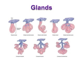
  • 31. Exocrine glands Based on number of ducts • Simple(single duct) • Compound(minor & major ducts) Based on shape of secretory piece: • Simple tubular/ alveolar • Simple branched tubular/ alveolar • Simple coiled tubular • Compound tubular/ acinar • Compound tubulo-alveolar / acinar
  • 32. • Simple: – Secretion poured to surface by un-branched duct • Compound: – Duct divides in to branches to form elaborated / complex duct system; – Each smaller terminal duct receive secretion from it’s own secretory end piece – These ducts unite to form larger ducts which finally drain on to surface Based on Number of Ducts:
  • 33. • Tubular: (Simple / Compound) – Secretory end piece is like tubule – Straight, branched or coiled Based on Shape of Secretory End Piece: Simple Straight Intestinal Crypts Simple Coiled Sweat Gland Simple Branched Uterine gland Fundic & pyloric glands of stomach Compound Cardiac glands of stomach & Brunner’s gland of duodenum
  • 34. Simple Straight Tubular Gland Intestinal Crypts
  • 35. • Alveolar: (Simple / Compound) – Secretory end piece Flask shaped with large lumen – Un-branched or branched Simple Un-branched Urethral Glands Simple Branched Sebaceous & Tarsal Glands Compound Mammary gland betn puberty & 1st Pregnancy Based on Shape of Secretory End Piece:
  • 36. • Acinar: (Mostly Compound) – Secretory end piece Round shaped with small lumen – Mostly branched Compound Acinar Pancreas Parotid Based on Shape of Secretory End Piece:
  • 37. • Tubulo – Alveolar / Acinar: – Combination of both Tubular & alveolar / Acinar – Mostly branched Compound Tubulo – Alveolar / Acinar Sublingual gland Submandibular gland Lactating mammary gland Based on Shape of Secretory End Piece:
  • 40. According to Mode of secretion: • Merocrine • Apocrine • Holocrine • Cytocrine Exocrine glands
  • 41. • Merocrine: – A.K.A.: Eccrine / Epicrine – Secretion discharged through intact cell membrane – By Exocytosis – No loss of cytoplasm Based on Mode of Secretion: E.G. Protein content of mammary gland Parotid Pancreas Typical sweat gland
  • 43. • Apocrine: – Apical portion (luminal) of cell disintegrate to discharge its secretion – Nucleus & basal portion remain intact from which cell can regenerate – Partial loss of cytoplasm Based on Mode of Secretion: E.G. Lipid content of mammary gland Atypical sweat gland (Axilla & Groin) Ceruminous gland of Ear Moll’s Gland in Eyelid Modified Sweat gland
  • 45. • Holocrine: – Entire cell disintegrate to discharge its secretion – Result in death of cell – Complete loss of cytoplasm Based on Mode of Secretion: E.G. Sebaceous gland Tarsal Glands in Eyelid
  • 47. • Cytocrine: – Cell are released as secretion Based on Mode of Secretion: E.G. Spermatozoa from Testis Ovum from Ovary Bone Marrow
  • 50. According to Nature of secretion: • Serous • Mucous • Mixed Exocrine glands Mucous Serous Mixed
  • 51. Serous Acinus / Gland • Secretion: Thin, watery, – Rich in enzymes, protein in nature • Cells: Pyramidal & Small with Indistinct boundaries • Cytoplasm: Granular (Zymogen) – Rich in rER – Stain darkly with H/E stain • Nuclei: rounded & basal / central • Size of lumen: Small • Function: Enzymatic • E.G.: Parotid & Lacrimal glands
  • 52. • Secretion: Thick Viscous – Which collects in the apical part • Cells: Low Columnar & Large with distinct boundaries • Cytoplasm: Mucinogen droplets – Stain very lightly with H/E stain & – Looks empty • Nuclei: Flattened & basal (mucoid pushes nuclei towards base) • Size of lumen: Large • Function: Protection & Lubrication • E.G.: Sublingual glands Mucous Acinus / Gland
  • 53. • Contain both serous & mucous secretory units • Sometimes serous cells form crescentic caps on mucous acini called as Serous Demilunes Mixed Acinus / Gland
  • 54. Serous Thin, watery Proteinaceous Zymogen granules Central rounded Small Lumen Indistinct Darkly stained Enzymatic action Parotid Gland Mucous Thick, viscous Mucopolysaccharides Mucinogen droplets Flat & peripheral Large Lumen Distinct Lighly stained Protection & lubrication Sublingual gland Consistency Content Cytoplasm Nucleus Lumen Cell boundaries H&E Staining Function Examples
  • 57. According to Development: • Ectodermal – Glands of skin, Mammary gland, Lacrimal gland, – Salivary glands, Pituitary gland & Chromaffin organs • Mesodermal – Suprarenal cortex, Gonads, Kidney & Spleen • Endodermal – Thyroid, Parathyroid, Thymus – Liver, Pancreas, Prostate – Urethra, Bulbo-urethral gland, Greater vestibular gland – Glands in lining epithelium of respiratory tract Exocrine glands
  • 58. Clinical • ADENOMA: Benign tumors arising in the gland • ADENOCARCINOMA: Malignant growth in the gland