Parotid gland & Facial nerve
•
279 likes•33,295 views

Detailed anatomy of parotid gland and facial nerve.

Recommended
More Related Content
What's hot
What's hot (20)
Similar to Parotid gland & Facial nerve
Similar to Parotid gland & Facial nerve (20)
More from Mehul Tandel
Recently uploaded
Recently uploaded (20)
Parotid gland & Facial nerve
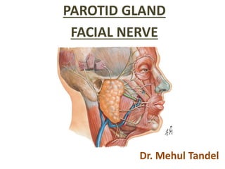
- 1. PAROTID GLAND FACIAL NERVE Dr. Mehul Tandel
- 3. • Exocrine glands • Glands with ducts • Produce saliva and pour their secretion in the oral cavity • Major (paired) • Parotid • Submandibular • Sublingual • Minor • In the tongue, palatine tonsil, palate, lips and cheeks
- 6. Objectives • INTRODUCTION • PAROTID CAPSULE • EXTERNAL FEATURES • DEVELOPMENT • RELATIONS • STRUCTURES WITHIN THE PAROTID GLAND • PAROTID DUCT • NERVE SUPPLY • LYMPHATIC DRAINAGE AND LYMPH NODES • FUNCTIONS OF PAROTID GLAND • CLINICAL ANATOMY
- 7. • Parotid region: – The largest serous salivary gland = Parotid Gland – The “queen of the face”, the facial nerve • Parotid gland: Para= Around; Otic = Ear • Parotid gland: – Average Wt - 15gm / 25gm – Irregular lobulated mass Introduction
- 8. Also projects forwards on the surface of masseter. Sternomastoid Below External acoustic meatusRamus of mandible Situation Between
- 9. • Small detached part lies on the surface of the masseter, between zygomatic arch and parotid duct :– • Accessory parotid gland or ‘Socia Parotidis’
- 10. • Occupies the deep hollow behind the ramus of the mandible. • Wedge-shaped, the base above & the apex below – behind the angle of the mandible.
- 11. Parotid Capsule 1.False Capsule 2.True Capsule
- 12. Parotid Capsule • False Capsule: Formed by investing layer of the deep cervical fascia
- 13. True Capsule: Condensation of fibrous stroma Parotid Capsule
- 14. External Features • Resembles a three sided pyramid. The apex directed downwards. • The gland has four surfaces:- 1) Superior (base of the pyramid) 2) Superficial 3) Anteromedial and 4) Posteromedial The surfaces are separted by 3 borders: a) Anterior b) Posterior and 3) Medial
- 15. Apex Resembles a three sided pyramid The apex directed downwards Transverse section Lateral View – Left Parotid
- 16. Superficial surface Posterior border Medial border Anterior border 4 Surfaces Superior Surface (Base) 3 Borders Lateral View – Left Parotid
- 17. 4 Surfaces 3 Borders Transverse section Left Parotid – Sup View
- 18. Relations - Apex • Overlaps the posterior belly of the diagastric The cervical branch of facial nerve Two divisions of the Retromandibular vein
- 19. Relations – Superior Surface / Base External acoustic meatusTemporo mandibular joint
- 20. Relations – Superficial Surface Parotid fascia Posterior fibers of the platysma and risorius Superficial Parotid Lymph Node
- 21. Grooved by posterior border of the ramus of the mandible Relations – Antero-medial Surface Styloid
- 22. Grooved by posterior border of the ramus of the mandible Relations – Postero-medial Surface Styloid
- 23. Separates Superficial surface from Antero-medial surface Relations – Anterior Border
- 24. Relations – Posterior Border Separates Superficial surface from Postero-medial surface
- 25. Relations – Medial Border Separates Antero-medial surface from Postero-medial surface Of Pharynx
- 26. Structures within the Parotid Gland Superficial temporal Artery Maxillary Artery Posterior auricular artery External carotid Arteries Deepest
- 28. Facial nerve Facial Nerve Superficial
- 30. Radiate like Goose’s Foot through Anterior Border Pes Anserinus
- 31. Parotid Duct • Ductus parotideus; Stensen’s duct • Thick walled • 5cm long and 5 mm diameter • Carries saliva to the oral cavity. • Course :
- 33. • At the anterior border of the masseter, it turns medially and pierces: (a) Buccal pad of fat. (b) Buccalpharyngeal fascia (c) Buccinator “Oblique course of the duct through the buccinator prevent infaltion during blowing.”
- 34. Opens into the vestibule of the mouth Opposite the crown of the upper 2nd molar tooth.
- 35. Nerve Supply • Parasympathetic (Secretomotor): – Auriculo temporal nerve – Produces watery secretion • Symapathetic: – Vasomotor, Mucous rich sticky secretion – Derived from plexus around the external carotid artery • Sensory: – Great auricular and auriculotemporal Nerve
- 36. Inferior Salivatory Nucleus Glossophargeal nerve (9th cranial nerve) Tympanic Branch Tympanic Plexus Pre-ganglionic fibers arise From Lesser Petrosal Nerve Otic Ganglion Post-ganglionic Fibres Auriculoemp- oral Nerve The Parotid Gland To supply Parasympathetic (Secretomotor) Pathway Relay
- 37. Glossophargeal nerve Inferior Salivatory Nucleus Tympanic Branch Tympanic Plexus Lesser Petrosal Nerve Otic Ganglion Auriculoemporal Nerve The Parotid Gland ParaSympathetic Medulla Oblangata Pons
- 38. Superior Cervical Ganglion T1 & T2 Plexus around External Carotid Artery The Parotid Gland Sympathetic
- 39. Lympatic Drainage Parotid nodes —> Upper deep cervical nodes.
- 40. Clinical Anatomy • Diseases of parotid gland A) Congenital: Aplasia or atresia:- – Any one or group of salivary glands may be absent, unilaterally or bilaterally. – Salivary loss leads to increased dental caries, burning sensation, oral infections, taste aberrations
- 41. B) Acquired: – Infective Mumps Bacterial Parotitis – Parotid Abcess – Mixed Parotid Tumor – Neurological Frey's syndrome – Sialolithiasis
- 42. • Mumps: • Viral disease • Caused by the mumps virus Paramyxovirus. • Painful swelling of the parotid gland. • Do not suppurate • Orchitis – Inflammation of testis • Pancreatitis
- 43. • Bacterial Parotitis: • It can be acute, chronic and recurrent. • Infecfion spread from mouth – Poor oral hygiene, – Particularly among elderly postoperative patient • Suppurate = Form Abcess
- 44. • Parotid Abcess: • Infection spread from mouth • Suppuration of parotid lymph node • Drain By Hilton’s Method: • Small Horizontal incision • To avoid injury of facial nerve branches
- 45. • Frey’s Syndrome: Also known as gustatory sweating or Auriculo-temporal nerve syndrome. Commonly occurs after parotid surgery or trauma. Injury of Auriculo-temporal & Great auricular nerev During healing: Secreto-motor fibers of auriculo- temporal nerve join with great auricular nerve & reach sweat glands of facial skin.
- 47. It is characterized by o Sweating o Warmth o Redness of the face As a result of salivary stimulation by the smell or taste of food
- 48. • Mixed Parotid Tumor: • Slow growing • Benign • Painless • Does not involve facial nerve
- 49. • Sialolithiasis: • Salivary calculi, or salivary stones), • Calcified mass forms within a salivary gland, • Usually in the duct of the submandibular gland • Less commonly the parotid gland.
- 50. The Cranial Cavity Base Anterior Cranial Fossa Middle Cranial Fossa Posterior Cranial Fossa
- 51. The middle cranial fossa Internal Acoustic Meatus Groove for Greater petrosal nerve Groove for Lesser petrosal nerve Petrous Part Of Temporal Bone Foramen Lacerum
- 52. Internal acoustic Meatus Greater petrosal nerve Lesser petrosal nerve
- 53. Facial Nerve
- 54. Objectives • Introduction • Functional Components • Nuclei • Course & Relations • Branches & Distributions • Ganglia Associated With Facial Nerve • Clinical Anatomy
- 55. Introduction • Seventh cranial nerve • 2nd “busiest” cranial nerve of the human body • Nerve of the second branchial arch • Mixed
- 56. Functional Components of Cranial Nerves • Total – 7, 4 – General Sensations & 3 – Special sensations • General Components: GSA – General Somatic Afferent • Touch, temperature, and pain from non-visceral structures GSE – General Somatic Efferent • Motor to skeletal muscle GVA – General Visceral Afferent • Touch (distention), temperature, and pain from the viscera GVE – General Visceral Efferent • Motor to viscera, smooth muscle, and glands
- 57. Functional Components of Cranial Nerves • Special Components: SSA – Special Somatic Afferent • Vision, hearing, and balance SSE – Doesn’t exist SVA – Special Visceral Afferent – Taste and olfaction SVE – Special Visceral Efferent / Branchial Efferent • Motor to muscles derived from the branchial arches
- 58. Functional components of Facial Nerve Special visceral Efferent / Branchial Efferent Facial Muscles, Posterior Belly Of Digastric, Stylohyoid & Stapedius
- 59. General Visceral Efferent / Parasympathetic / Secretomotor Glands of the Nose, Palate and Pharynx General Visceral Affetent
- 60. Special Visceral Afferent Taste Sensations from Anterior 2/3rd of Tongue, Palate
- 61. General Somatic Afferent Through communication with Vagus Nerve
- 63. Nuclei • 4 nuclei situated in the lower pons. 1. Motor nucleus or brachiomotor : lies deep in lower pons. 2. Superior salivatory nucleus or parasympathetic. 3. Lacrimatory nucleus – parasympathetic. 4. Nucleus of tractus solitarius – Gustatory and also receives afferent fibres from the glands.
- 64. Nuclei Motor Nucleus / Brachiomotor Superior Salivatory Nucleus Lacrimatory Nucleus Nucleus Of Tractus Solitarius Facial Nerve Pons MO MB
- 66. • The part of motor nucleus supplying upper part of face---controlled by corticonuclear fibres of both sides • The part of motor nucleus supplying lower part of face---controlled by corticonuclear fibres of opposite side (only) CENTRAL CONNECTIONS
- 68. Course and Relations Motor root Small Sensory Root Nervous Intermedius Pons MO MB
- 69. Vestibulo-cochlear Nerve Internal Acoustic Meatus Nervous Intermedius Pons Course and Relations - Intracranial
- 70. In the Internal Acoustic Meatus Divided In 3 Parts within the canal Course and Relations - Intracranial
- 71. Course and Relations - Intracranial 1st Part Genu 2nd Part 3rd Part Stylo-Mastoid Foramen
- 72. Course and Relations - Extracranial
- 73. A. Within the facial canal: 1. Greater petrosal nerve 2. The nerve to the stapedius 3. Chorda tympani B. At its exit from the stylomastoid foramen: 1. The posterior auricular 2. Digastric 3. Stylohyoid Branches and Distribution
- 74. C. Terminal branches within the parotid gland: 1. Temporal 2. Zygomatic 3. Buccal 4. Marginal mandibular 5. Cervical D. Communicating branches: With adjacent cranial and spinal nerves.
- 75. Within the facial canal: Greater Superficial Petrosal Nerve Nerve to Stapedius Chorda Tympani
- 76. Greater Superficial Petrosal Nerve Deep petrosal Nerve Nerve to Pterygoid Canal Pterygo-palatine Ganglion
- 77. Pterygo-palatine Ganglion Lacrimal Gland Nose, Palate & Pharynx
- 79. Damps down excessive vibration of Stapes Stapedius Muscle Hyperacusis
- 81. Chorda Tympani Nerve Lingual Nerve Submandibular Ganglion Submandibular & Sublingual Gland Taste Sensation from Anterior 2/3rd of Tongue
- 86. 1. Loss of Lacrimation 2. Loss of Stapedial Reflex 3. Loss of taste from Ant 2/3rd of tongue 4. Lack of Salivation 5. Facial Palsy Internal Acoustic Meatus At Genu All except loss of Lacrimation Below Genu All except loss of Lacrimation & Loss of Stapedial Reflex Below Stylomastoid Foramen Only facial Palsy
- 87. Applied • In infranuclear lesions of facial nerve (Bell’s palsy)- whole Ipsilateral face is paralyzed • Affected side is motionless • Loss of wrinkles on forehead • Eye cannot be closed • In smiling the mouth is drawn to normal side • During mastication food accumulates in vestibule of mouth • In Supranuclear lesions of facial nerve • Lower part of opposite face is paralyzed • Upper part is escaped due to • Bilateral representation of upper part in cerebral cortex
- 88. Bell’s Palsy
- 90. Communicating Branches • Facial nerve branches exchange fibers with sensory cutaneous branches of the trigeminal nerve. • For effective co-ordination
- 91. Ganglia 1. Geniculate ganglion 1. Sensory ganglion. 2. Taste fibres 2. Submandibular ganglion: 1. Parasympathetic ganglion 2. Secretomotor fibre to the submandibular and sublingual glands. 3. Pterygopalatine ganglion: 1. Parasympathetic ganglion. 2. Secretomotor fibres for the lacrimal gland