

Recommended
More Related Content
What's hot
What's hot (20)
Similar to Peritoneum Dr. Mehul Tandel
Similar to Peritoneum Dr. Mehul Tandel (20)
More from Mehul Tandel
Recently uploaded
Recently uploaded (20)
Peritoneum Dr. Mehul Tandel
- 2. • Introduction • Parts • Parietal peritoneum • Visceral peritoneum • Folds of peritoneum • Peritoneal cavity • Functions • Vertical disposition • Falciform ligament • Lesser omentum Objectives
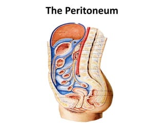
- 3. Peritoneum - Introduction Definition: • Large, thin serous membrane that lines the walls of the abdominal and pelvic cavities and cover the organs within these cavities. • Also called Serosa • Consists of 2 layers – Outer fibrous - Strength – Inner flattened mesothelial cells - lubrication
- 4. Formation Intra-parietal Closed Sac Invagination Outer Parietal Inner Visceral Folfd Of Peritoneum Folds Of Peritoneum
- 5. LAYERS: • Parietal layer: • lines the abdominal wall & pelvic cavities & under surface of diaphragm • Loosely attached – easily stripped • Somato-pleural layer of lateral plate mesoderm • Blood supply & nerve supply – same as overlyng wall • Sensitive to pain & temperature
- 6. LAYERS: • Visceral layer: • Lines outer surface of organs • Firmly adhere to it – can not be stripped • Splanchno-pleural layer of lateral plate mesoderm • Blood supply & nerve supply – same as underlying viscera = Autonomic • Sensitive to pain due to stretching, distension or ischemia of viscera
- 9. Peritoneal cavity: • Potential space between the two layers of peritoneum. • Filled with very thin film of serous fluid secreted by? • In the male – closed sac, but • In the female – open due to uterine tubes, the uterus, and the vagina
- 10. Peritoneal cavity: Divided in to: • Larger part – Greater Sac • Smaller part – Lesser Sac Communication • Epiploic Foramen / Foramen of Winslow • Small pockets or recesses – site for internal hernia
- 11. Intraperitoneal viscera: Eg: Stomach, superior part of duodenum, jejunum, ileum, cecum, vermiform appendix, transverse and sigmoid colons, spleen , ovary and uterine tube The relationship between viscera and peritoneum Intraperitoneal viscera completely surrounded by peritoneum
- 12. • Retroperitoneal viscera - Covered by peritoneum on their anterior surfaces only Eg: Kidney, suprarenal gland, pancreas, 2nd & 3rd parts of duodenum, ascending & descending colon middle and lower parts of rectum and ureter Retroperitoneal viscera
- 14. Folds of Peritoneum • Mobile organs: • Suspended by folds • Fixed: • Directly rest on Post wall • Retroperitoneal Provide passage for Vessels, Nerves & Lymphatics
- 16. Names of Folds Mes / Meso Name of organ Small Intestine / Enteron: Mesentery Large Intestine / Colon: Mesocolon Stomach Omentum / Omenta Organs – organ / Abdominal wall Ligaments
- 17. Mesentery Small Intestine / Enteron:
- 18. Mesocolon Large Intestine / Colon:
- 20. Mesoappendix
- 21. Omenta Fold of peritoneum that connect the stomach with another viscera Lessor omentum Greater omentum
- 23. Functions of Peritoneum • Movement of Viscera: – Provide slippery surface – permit free movements like? – Peristalsis, movements during respiration & Filling & evacuation of hollow viscera • Protection of Viscera: – Phagocytic cells & lymphocytes – Greater omentum – move towards infection site & seal it – Policeman of Abdomen
- 24. Functions of Peritoneum • Absorption and Dialysis: – Mesothelium – semipermeable membrane – Both secretive & absorptive – Fluid injection – Peritoneal dialysis • Healing power and Adhesions: – Mesothelial cells – Fibroblast – Abnormal adhesion - obstruction • Storage of Fat:
- 26. Development of peritoneum Dorsal Mesogastrium Ventral Mesogastrium
- 27. Development of the peritoneum
- 30. stomach Lienorenal Ligament R Gastrosplenic Ligament R R L lesser sac Lesser OmentumFalciform ligament Spleen
- 37. • Sickle shape • Extends from antero- superior surface of liver to anterior abdominal wall & undersurface of diaphragm • Free border of the ligament contains Ligamentum teres hepetis (obliterated umbilical vein) Falciform ligament of liver
- 39. • From under surface of diaphragm is reflected on to superior surface of right lobe of liver forming the upper layer of coronary ligament
- 40. • Then, it descends from sup surface of liver to ant surface then inferior surface of liver. • From post part of inferior surface peritoneum reflected on to front of right kidney & rt suprarenal gland forming the lower layer of coronary ligament.
- 41. Right triangular ligament. • Short v shaped fold. • Formed by approximation of two layers of coronary ligament on right lateral end.
- 42. Left triangular ligament Double layer. On upper border of left lobe. Anterior layer extends from left layer of falciform ligament. Posterior layer continuous with left layer of lesser omentum.
- 43. • The upper & lower layers of coronary ligament & right triangular ligament bound a large area on the post surface of the liver which has no peritoneal covering bare area
- 44. Hepato-renal pouch of Morison Right para-colic gutter Pelvic cavity
- 45. Hepato-renal pouch of Morison
- 46. Hepato-renal pouch of Morison Right Para-colic gutter Left Para-colic gutter
- 47. Lesser Omentum
- 48. Inferior surface Ant layer of Lesser Omentum
- 49. Right free margin Epiploic Foramen Porta Hepatis
- 50. Lesser omentum • Double layered fold of peritoneum which extends from lesser curvature of stomach &1st 2cm of duodenum to liver
- 51. Lessor omentum – Extends from porta hepatis to 1st part of duodenum, – It contains • Common bile duct, • Proper hepatic a. • Hepatic portal v. Hepato-gastric ligament – From porta hepatis to lesser curvature of stomach Hepato-duodenal ligament Two Parts
- 52. Lessor omentum Right free margin Epiploic Foramen
- 53. Lesser omentum - Contents Bile Duct Proper Hepatic a. Portal Vein Lymph Nodes & Lymphatics & Nerves & Plexus Left & Right Gastric V.
- 54. Ant layer of Lessor omentum Antero-superior surface Greater Omentum 4 layers & 3 margins
- 55. 1st Layer 4th Layer 2nd Layer 3rd Layer
- 56. Greater omentum • A four-layered fold of peritoneum connecting the greater curvature of stomach & 1st part of duodenum to transverse colon • Hangs down like an apron in front of coils of small intestine
- 57. Greater omentum - Contents Left Gastroepiploic vessels RightGastroepiploic vessels
- 58. Greater omentum - Functions • Storage of fat • Protect against infections – Macrophage – small dens white patches – Milky spot • Limits spread of infection – Sealing –Policeman of Abdomen
- 61. Body of Pancreas Post Abdominal wall Small Intestine Mesentery
- 62. Mesentery Broad fan shaped fold that suspends coils of Jejunum & Ilium from the posterior abdominal wall Breadth: Maximum – 20cm in center Decrease towards ends
- 63. Mesentery - Borders Attached border = Root of Mesentery Free border = Intestinal Border 15 cm 6 meters Visceral Peritoneum
- 64. Root of Mesentery Attached border – downward & right From Duodeno-jejunal flexure(Left side of L2) - Ilio-cecal junction (Upper end of Right Sacro-iliac joint)
- 65. Root of Mesentery 3rd part of Duodenum Abdominal Aorta Right Ureter Right Psoas Major Right Sacro-iliac joint Duodeno-jejunal Flexure Inferior Vena Cava
- 66. Most in lower part From root to intestinal border Near Intestinal border – oval/circular fat free translucent area Distribution of fat in Mesentery Windows Jejunum Ileum
- 67. Mesentery - Contents Jejunal and Ileal a. & v. Superior Mesenteric v. Superior mesenteric a.
- 68. Mesentery - Contents Autonomic Nerve plexus Lymphatics & Lymph nodes Fat
- 69. Mesentery – Left layer Left Para-colic gutter
- 72. Hilum Of Spleen Post Layer Lieno-renal Lig Left Kidney Ant Layer Gastro-splenic Lig Post Layer Gastro-splenic Lig Ant Layer Lieno-renal Lig
- 73. Anterior Posterior Ant Layer Gastro-splenic Lig Post Layer Lieno-renal Lig Post Layer Gastro-splenic Lig Ant Layer Lieno-renal Lig
- 74. Post Abdominal wall Sigmoid Colon Sigmoid Meso-colon
- 75. Sigmoid Meso-colon Root – Inverted V
- 76. Rectum Differ in Male & Female
- 77. Recto-vesical pouch In Males 7.5 cm above anus
- 79. In Females Recto-uterine pouch of Douglas Utero-vesical pouch 5.5 cm above anus
- 82. Apex of Urinary bladder Posterior surface of Anterior abdominal wall
- 83. Median Umbilical fold Medial Umbilical fold Lateral Umbilical fold Median Umbilical Lig Obliterated Umbilical a. Inferior Epigastric Vessels
- 84. Supra-vesical fossa Medial Inguinal fossa Lateral Inguinal fossa
- 85. Greater sac Horizontal Tracing
- 90. Lesser sac-Omental Bursa • Diverticulum of Greater sac • Behind & beyond stomach, lesser omentum & caudate lobe of liver • Act as bursa – allow expansion of stomach = Omental Bursa • Closed on all side except – epiploic foramen • Shape: – Hot water bag
- 92. Caudate lobe of liver Posterior layer of Lesser Omentum Lesser sac - Boundaries Postero-inferior surface of stomach 2nd layer of Greater Omentum Anterior Wall
- 93. Ant surface of body of pancreas Upper layer of transverse meso-colon Lesser sac - Boundaries Antero-superior surface of transverse colon 3rd layer of Greater Omentum Posterior Wall Left sura-renal gland, kidney Diaphragm
- 94. Lesser sac - Boundaries Upper Margin Diaphragm Caudate lobe of liver
- 96. Right Margin Below Transverse Colon Right free margin of Greater omentum
- 97. Epiploic Foramen Right Margin Above Transverse Colon Deficient
- 98. Epiploic Foramen Foramen of Winslow Aditus or Opening of Lesser Sac • Vertical slit – 3cm long • Communicate? • Situation: –B/H Rt free margin of lesser omentum –At T12
- 99. Epiploic Foramen Boundaries Right free margin of lesser omentum • Common bile duct • Proper hepatic a. • Hepatic portal v. • Hepatic plexus of n. • Lymphatics Anteriorly:
- 100. Epiploic Foramen Boundaries Inferior Vena Cava Posteriorly: Right kidney T12
- 101. Epiploic Foramen Boundaries Caudate Process of Liver Superiorly:
- 102. Epiploic Foramen Inferiorly: Boundaries 1st part of duodenum Hepatic artery
- 103. Divided by 2 peritoneal folds Left Gastric artery Common Hepatic artery Subdivision of Lesser Sac Right Gastro- pancreatic fold Left Gastro- pancreatic fold
- 104. Subdivision of Lesser Sac Right Gastro- pancreatic fold Left Gastro- pancreatic fold Superior Recess Inferior Recess Vestiblue Splenic Recess Omental Bursa Proper
- 105. Peritoneal subdivisions • Greater sac divided by transverse colon and transverse mesocolon into – Supracolic and – Infracolic compartments. Supra-colic compartments (subphrenic space)-lies between diaphragm and transverse colon and transverse mesocolon Supra-hepatic recess between the diaphragm & liver:- the falciform ligament divides it into right and left suprahepatic recesses 107
- 108. • Left supra-hepatic recesses – left anterior suprahepatic spaces – left posterior suprahepatic spaces • Right supra-hepatic recesses – right anterior suprahepatic spaces – right posterior suprahepatic spaces – bare area of live (extraperitoneal space) 110 Supra-hepatic recess
- 109. Rt. anterior subphrenic space Lt. anterior subphrenic space Rt. posterior subphrenic (Rt. Subhepatic)
- 110. Infrahepatic recess lies between the liver and transverse colon and transverse mesocolon-the ligamentum teres hepatic divides it into right and left infrahepatic recesses • Right infrahepatic recesses (hepatorenal pouch of morrison) • Left infrahepatic recesses – left anterior infrahepatic space – left posterior infrahepatic space = Lesser Sac 112
- 111. Morison’s pouch left subhepatic Space (Lt. posterior subphrenic)
- 112. Infracolic compartments: lies below the transverse colon and transverse mesocolon • Right paracolic gutter : – Lateral to the ascending colon – Communicates with the hepatorenal recess and the pelvic cavity – Provides a route for the spread of infection • Left paracolic gutter: – Lateral to the descending colon – Separated from spleen by the phrenicocolic ligament 114
- 113. • Right mesenteric sinus - triangular space, lies between root of mesentery, ascending colon, right 2/3 of transverse colon and transverse mesocolon • Left mesenteric sinus -lies between root of mesentery, descending colon, right 1/3 of transverse colon and transverse mesocolon, its widens below where it is continuous with the cavity of the pelvis 115
- 114. Inferior mesenteric vein in the peritoneal fold, forming the paraduodenal recess. Duodenal Recesses Close to the duodeno-jejunal junction, four small pocket like pouches of peritoneum
- 115. Cecal Recesses Folds of peritoneum close to the cecum produce three peritoneal recesses called the superior ileocecal, the inferior ileocecal, and the retrocecal recesses
- 116. Nerve supply to the peritoneum The parietal peritoneum Phrenic nerve Intercostal First lumbar nerves Obturator nerve – Pelvic Cavity The visceral peritoneum Autonomic
- 117. Applied Anatomy • Ascites: • Paracentesis • Internal abdominal hernia • Perforation of gastric ulcer • Peritoneal abscess –Hepato-renal space –Recto-uterine pouch or recto-vesical pouch • Peritonitis • Peritoneal Pain
- 118. Applied Anatomy • Pneumoperitoneum • Laparoscopy & Laparotomy • Greater omentum – policeman of abdomen • Referred pain from gut • Peritoneal Dialysis • Sub-phrenic abscess
- 119. Ascites Excessive accumulation of the peritoneal fluid within the peritoneal cavity • Peritoneal fluid excess, hydroperitoneum • Most commonly due to cirrhosis • TB • Malignancy
- 122. Internal abdominal hernia Occasionally a loop of intestine may enter into the peritoneal pouch or recesses and gets strangulated
- 123. Perforation of gastric ulcer • Posterior wall perforation • Lesser sac • If closed by adhesions at epiploic foramen – Drained by tube passed through lesser omentum
- 124. Peritoneal abscess • Hepato-renal space – m.c. site for subphrenic abscess • In supine position • Recto-uterine pouch or recto-vesical pouch • In Fowler’s position (45° angle)
- 125. Peritoneal abscess - drainage
- 126. Peritonitis • Inflammation • Localized – Pain around site of infection – Tenderness – Rigidity • Generalized – Card-board rigidity – Absent intestinal sounds
- 127. Rebound Tenderness Pressure is applied to the abdominal wall with a single finger over the site of the inflammation. The pressure is then removed by suddenly withdrawing the finger. The abdominal wall rebounds, resulting in extreme local pain, which is known as rebound tenderness
- 128. Pneumoperitoneum • Air • Perforations of stomach or intestine • Air under right dome of diaphragm
- 129. Laparoscopy
- 130. Laparotomy
- 131. Greater omentum – policeman of abdomen • Limits spread of infection – Rupture appendix – Ruptured gastric ulcer
- 132. Referred pain from gut
- 133. Peritoneal Dialysis
- 134. Sub-phrenic abscess