

Recommended
More Related Content
What's hot
What's hot (20)
Similar to Kingdom Animalia
Similar to Kingdom Animalia (20)
More from Merlyn Denesia
More from Merlyn Denesia (20)
Recently uploaded
Recently uploaded (20)
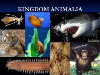
Kingdom Animalia
- 1. © 2010 Pearson Education, Inc.
- 2. © 2010 Pearson Education, Inc. What Is an Animal? • Animals are: – Eukaryotic – Multicellular – Heterotrophic organisms that obtain nutrients by ingestion – Able to digest their food within their bodies • Animal cells lack the cell walls that provide strong support in the bodies of plants and fungi.
- 3. © 2010 Pearson Education, Inc. • Most animals have: – Muscle cells – Nerve cells that control the muscles
- 4. © 2010 Pearson Education, Inc. • Most animals: – Are diploid – Reproduce sexually – Proceed through a series of typically similar developmental stages
- 5. MEIOSIS Sperm Egg Adult Key FERTILIZATION MITOSIS Eight-cell stage Internal sac Digestive tract Larva METAMORPHOSIS Haploid (n) Diploid (2n) Outer cell layer (ectoderm) Later gastrula (cross section) Future middle layer of cells (mesoderm) Inner cell layer (endoderm) Early gastrula (cross section) Blastula (cross section) Zygote (fertilized egg) Figure 17.2-8
- 6. © 2010 Pearson Education, Inc. Animal Phylogeny • Biologists categorize animals by: – General features of body structure - anatomical – More recently, using genetic data
- 7. Ancestral protist No true tissues Radial symmetry Tissues Bilateral symmetry Sponges Cnidarians Molluscs Flatworms Annelids Roundworms Arthropods Echinoderms Chordates Figure 17.5
- 8. © 2010 Pearson Education, Inc. • A second major evolutionary split is based on body symmetry. – Radial symmetry refers to animals that are identical all around a central axis. – Bilateral symmetry exists where there is only one way to split the animal into equal halves.
- 9. Radial symmetry. Parts radiate from the center, so any slice through the central axis divides into mirror images. Bilateral symmetry. Only one slice can divide left and right sides into mirror-image halves. Figure 17.6
- 10. © 2010 Pearson Education, Inc. • Animals also vary according to the presence and type of body cavity, a fluid-filled space separating the digestive tract from the outer body wall.
- 11. © 2010 Pearson Education, Inc. • There are differences in how the body cavity forms. – If the body cavity is not completely lined by tissue derived from mesoderm, it is a pseudocoelom. – A true coelom is completely lined by tissue derived from mesoderm.
- 12. (a) No body cavity (b) Pseudocoelom (c) True coelom Body covering (from ectoderm) Tissue-filled region (from mesoderm) Body covering (from ectoderm) Body covering (from ectoderm) Muscle layer (from mesoderm) Tissue layer lining coelom and suspending internal organs (from mesoderm) Digestive tract (from endoderm) Digestive tract (from endoderm) Digestive tract (from endoderm) Pseudocoelom Coelom Figure 17.7
- 13. (a) No body cavity: for example, flatworm Body covering (from ectoderm) Tissue-filled region (from mesoderm) Digestive tract (from endoderm) Figure 17.7a
- 14. (b) Pseudocoelom: a body cavity only partially lined by the mesoderm, the middle tissue layer; for example, roundworm Body covering (from ectoderm) Muscle layer (from mesoderm) Digestive tract (from endoderm) Pseudocoelom Figure 17.7b
- 15. (c) True coelom: a fluid-filled body cavity completely lined by mesoderm, for example, annelid Body covering (from ectoderm) Tissue layer lining coelom and suspending internal organs (from mesoderm)Digestive tract (from endoderm) Coelom Figure 17.7c
- 16. © 2010 Pearson Education, Inc. MAJOR INVERTEBRATE PHYLA • Invertebrates: – Are animals without backbones – Represent 95% of the animal kingdom
- 17. © 2010 Pearson Education, Inc. 17 Phylum Porifera Multicellular Body with pores (ostia) No organs or true tissues. No nervous system Adults sessile & attached to substratum. Skeleton of calcareous spicules, siliceous spicules, spongin or a combination. All aquatic, mostly marine.
- 18. © 2010 Pearson Education, Inc. Sponges • Sponges • Sponges include sessile animals that lack true tissues and that were once believed to be plants.
- 19. © 2010 Pearson Education, Inc. • The body of a sponge resembles a sac perforated with holes. • Choanocyte cells draw water through the walls of the sponge where food is collected.
- 20. Choanocyte (feeding cell) Water flow Skeletal fiber Central cavity Choanocyte in contact with an amoebocyte Amoebocyte Flagella Pores Figure 17.8
- 21. Water flow Skeletal fiber Central cavity Choanocyte in contact with an amoebocyte Amoebocyte Flagella Pores Choanocyte (feeding cell) Figure 17.8a
- 22. © 2010 Pearson Education, Inc. Cnidarians • Cnidarians (phylum Cnidaria) are characterized by: – The presence of body tissues – Radial symmetry – Tentacles with stinging cells – Has secretory organelle called “cnida” contained in a cnidocyte
- 23. © 2010 Pearson Education, Inc. • The basic body plan of a cnidarian is a sac with a gastrovascular cavity, a central digestive compartment with only one opening. • The body plan has two variations: – The sessile polyp – The floating medusa
- 24. Mouth/anus Tentacle Polyp form Gastrovascular cavity Coral Hydra Sea anemone Gastrovascular cavity Mouth/anus Tentacle Medusa form Jelly Figure 17.9
- 25. Mouth/anus Tentacle Polyp form Gastrovascular cavity Coral Hydra Sea anemone Figure 17.9a
- 28. Sea anemone Figure 17.9ac
- 31. © 2010 Pearson Education, Inc. • Cnidarians are carnivores that use tentacles, armed with cnidocytes (“stinging cells”), to capture prey.
- 33. © 2010 Pearson Education, Inc. Molluscs • Molluscs (phylum Mollusca) are represented by soft- bodied animals, usually protected by a hard shell. • Many molluscs feed by using a file-like organ called a radula to scrape up food.
- 34. © 2010 Pearson Education, Inc. • The body of a mollusc has three main parts: – A muscular foot used for movement – A visceral mass housing most of the internal organs – A mantle, which secretes the shell if present
- 36. © 2010 Pearson Education, Inc. • The three major groups of molluscs are: – Gastropods, protected by a single, spiraled shell – Most gastropods have separate sexes but some groups (mainly the Heterobranchia) are hermaphroditic
- 37. Snail (spiraled shell) Figure 17.12aa
- 38. Sea slug (no shell) Figure 17.12ab
- 39. © 2010 Pearson Education, Inc. – Bivalves, with a shell divided into two halves hinged together – Bivalves are hermaphroditic or have separate sexes
- 43. Gastropods Bivalves (hinged shell) Cephalopods (large brain and tentacles) Snail (spiraled shell) Sea slug (no shell) Scallop Octopus Squid MAJOR GROUPS OF MOLLUSCS Figure 17.12
- 44. © 2010 Pearson Education, Inc. Turritella
- 45. © 2010 Pearson Education, Inc.
- 46. © 2010 Pearson Education, Inc. Cone Shells
- 47. © 2010 Pearson Education, Inc.
- 48. © 2010 Pearson Education, Inc. Cowries (Genus: Cypraea)
- 49. © 2010 Pearson Education, Inc. Pond snail (Genus: Lymnaea)
- 50. © 2010 Pearson Education, Inc.
- 51. © 2010 Pearson Education, Inc. Tridacna
- 52. © 2010 Pearson Education, Inc. Scallop
- 53. © 2010 Pearson Education, Inc. Nautilus
- 54. © 2010 Pearson Education, Inc. Cradle Shell (Chiton)
- 55. © 2010 Pearson Education, Inc. Common Name Genus Class Turritella shell Turritella Gastropoda Cone shell Conus Gastropoda Cowries Cypraea Gastropoda Pond snail Lymnaea Gastropoda Spider shell Lambis Gastropoda Edible oyster Ostrea Bivalvia Giant clam Tridacna Bivalvia Mud clam Mya Bivalvia Scallop Pecten Bivalvia Sea mussel Mytilus Bivalvia Cuttle fish Sepia Cephalopoda Nautilus Nautilus Cephalopoda Octopus Octopus Cephalopoda Squid Loligo Loligo Cradle shell Chiton Polyplacophora
- 56. © 2010 Pearson Education, Inc. Flatworms • Flatworms (phylum Platyhelminthes) are the simplest bilateral animals. • Flatworms include forms that are: – Parasites or – Free-living in marine, freshwater, or damp habitats
- 57. Head Sucker Reproductive unit with skin removed Tapeworm Figure 17.13ba
- 59. © 2010 Pearson Education, Inc. • The gastrovascular cavity of flatworms – Is highly branched but incomplete – Provides an extensive surface area for absorption of nutrients • Nervous system -consist of pair of anterior ganglia with longitudinal nerve cords connected by transverse nerves
- 60. © 2010 Pearson Education, Inc. • Excretory system - consist of two lateral canals with branches bearing flame cells (protonephridia)
- 61. Digestive tract (gastrovascular cavity) Nerve cords Mouth Eyespots (detect light) Nervous tissue clusters (simple brain) Planarian Bilateral symmetry Figure 17.13a
- 62. Digestive tract (gastrovascular cavity) Nerve cords Mouth Eyespots (detect light) Nervous tissue clusters (simple brain) Planarian Bilateral symmetry Blood fluke Head Hooks Suckers Reproductive unit with skin removed Tapeworm Figure 17.13
- 63. © 2010 Pearson Education, Inc. Class Turbellaria -Members of this class are mostly free-living -They are bottom dwellers in freshwater and marine environments -They crawl on stones, sand, or vegetation -Turbellarians are named for the turbulence -The beating of cilia creates this turbulence in the water -Turbellarians are predators and scavengers
- 64. © 2010 Pearson Education, Inc. Class Trematoda -all trematodes (flukes) are parasitic -Most species are hermaphroditic -Most are flattened and leaf-like or ribbon-like -A circulatory system is absent -The nervous system consists of a pair of anterior ganglia, or nerve centers, and usually three pairs of lengthwise nerve cords
- 65. © 2010 Pearson Education, Inc. Class Monogenea -Members are mostly ectoparasite -have complicated attachment organs at the posterior or tail end of their bodies, often including a mixture of suckers, clamps, hooks and spines -Mostly hermaphroditic, found on the gills of fish
- 66. © 2010 Pearson Education, Inc. Class Cestoda Intestinal parasites of humans and other vertebrates.
- 67. © 2010 Pearson Education, Inc.
- 68. © 2010 Pearson Education, Inc.
- 69. © 2010 Pearson Education, Inc. Annelids • Annelids (phylum Annelida) have: – Body segmentation, a subdivision of the body along its length into a series of repeated parts – A coelom – A complete digestive tract with – Two openings, a mouth and anus – One-way movement of food
- 70. Mouth Brain Coelom Nerve cord Blood vessels Waste disposal organ Anus Accessory hearts Main heart Digestive tract Segment walls Figure 17.15
- 71. © 2010 Pearson Education, Inc.
- 72. © 2010 Pearson Education, Inc. • The three main groups of annelids are: – Earthworms, which eat their way through soil – Polychaetes, marine worms with segmental appendages for movement and gas exchange – Leeches, typically free-living carnivores but with some bloodsucking forms
- 73. © 2010 Pearson Education, Inc. Class Polychaetes Class Oligochaeta Class Hirudinea
- 74. Earthworms Giant Australian earthworm Figure 17.14a
- 75. Polychaetes Christmas tree worm Figure 17.14b
- 76. Leeches European freshwater leech Figure 17.14c
- 77. © 2010 Pearson Education, Inc. Roundworms • Roundworms (phylum Nematoda) are: – Cylindrical in shape, tapered at both ends – The most diverse and widespread of all animals • Roundworms (also called nematodes) are: – Important decomposers – Dangerous parasites in plants, humans, and other animals
- 78. (a) A free-living roundworm Figure 17.16a
- 79. (b) Parasitic roundworms in pork Figure 17.16b
- 80. (c) Canine heart infected with parasitic roundworms Figure 17.16c
- 81. © 2010 Pearson Education, Inc.
- 82. © 2010 Pearson Education, Inc. Arthropods • Arthropods (phylum Arthropoda) are named for their jointed appendages. • There are about one million arthropod species identified, mostly insects. • Arthropods are a very diverse and successful group, occurring in nearly all habitats in the biosphere.
- 83. © 2010 Pearson Education, Inc. Lectures by Chris C. Romero, updated by Edward J. Zalisko PowerPoint® Lectures for Campbell Essential Biology, Fourth Edition – Eric Simon, Jane Reece, and Jean Dickey Campbell Essential Biology with Physiology, Third Edition – Eric Simon, Jane Reece, and Jean Dickey Phylum Arthropoda Kingdom Animalia Phylum Arthropoda Subphylum Trilobita Subphylum Chelicerata Class Merostomata Class Pycnogonida Class Arachnida Subphylum Crustacea Branchiopoda, Maxilopoda, Malacostraca Subphylum Uniramia Diplopoda, Chilopoda, Pauropoda, Symphyla, Insekta Class P
- 84. © 2010 Pearson Education, Inc. General Characteristics of Arthropods • Arthropods are segmented animals with specialized segments and appendages for an efficient division of labor among body regions • Bilateral symmetry • Jointed appendages
- 85. © 2010 Pearson Education, Inc. • The body of arthropods is completely covered by an exoskeleton, an external skeleton that provides: – Protection – Points of attachment for the muscles that move appendages – Molting – shedding off of exoskeleton for growth
- 86. © 2010 Pearson Education, Inc. Compound Eyes •Bundles of up to 14,000 light sensitive units grouped together •Some on stalks that can be used like periscopes
- 87. © 2010 Pearson Education, Inc. Taxonomic Classification: Subphylum Trilobita - All extinct forms from Cambrian to Carboniferous - Body is divided into two longitudinal furrows into three lobes - Distinct head, thorax, and abdomen - Biramous appendages – limb that branches into two, and each branch consists of a series of segments attached end to end
- 88. © 2010 Pearson Education, Inc. Subphylum Chelicerata - First pair of appendages modified to form chelicerae - Mandibles - Cephalothorax and abdomen often with segments fused
- 89. © 2010 Pearson Education, Inc. Sub Phylum Chelicerata Includes Horseshoe crabs, spiders, mites Chelicerae are the only small appendages before the mouth Range from small pinchers to venomous fangs
- 90. © 2010 Pearson Education, Inc.
- 91. © 2010 Pearson Education, Inc. Class Merostomata Horseshoe Crabs Not true crabs and have not evolved for millions of years Six pairs of appendages No mandible jaw Chelicerae, Walking Legs, four pairs of Pushing Legs Telson or tail used for steering and flipping itself right side up.
- 92. © 2010 Pearson Education, Inc. Class Pycnogonida Sea Spiders Have four or more pairs of legs A large proboscis with a moth on the tip allows it to feed on soft invertebrates No respiratory system as gases diffuse through body Tiny muscles consist of one cell covered with connective tissue
- 93. © 2010 Pearson Education, Inc. • Arachnids: – Live on land – Usually have four pairs of walking legs and a specialized pair of feeding appendages – Include spiders, scorpions, ticks, and mites Class Arachnida
- 94. Two feeding appendages Leg (four pairs) Black widow spider Wood tick Dust miteScorpion Figure 17.19
- 95. Two feeding appendages Leg (four pairs) Tarantula spider (an arachnid) Figure 17.19a
- 97. © 2010 Pearson Education, Inc.
- 98. Black widow spider Figure 17.19c
- 101. © 2010 Pearson Education, Inc. • Crustaceans: – Are nearly all aquatic – Have multiple pairs of specialized appendages – Include crabs, lobsters, crayfish, shrimps, and barnacles Subphylum Crustacea
- 102. Abdomen Cephalothorax (head and thorax) Swimming appendage Antenna (sensory reception) Eyes on movable stalks Mouthparts (feeding) Walking leg Walking legs Figure 17.18
- 103. Two feeding appendages Leg (three or more pairs) Antennae BarnaclesCrayfish Pill bug Shrimp Crab Figure 17.20
- 104. Two feeding appendages Leg (three or more pairs) Antennae Crab (a crustacean) Figure 17.20a
- 105. Crab Figure 17.20b
- 106. Shrimp Figure 17.20c
- 110. © 2010 Pearson Education, Inc. • Class Diplopoda Millipedes: – Eat decaying plant matter – Have two pairs of short legs per body segment • Class Chilopoda • Centipedes: – Are terrestrial carnivores with poison claws – Have one pair of short legs per body segment Subphylum Uniramia
- 111. One pair of legs per segment Two pairs of legs per segment Millipede Centipede Figure 17.21
- 112. © 2010 Pearson Education, Inc.
- 113. © 2010 Pearson Education, Inc.
- 114. © 2010 Pearson Education, Inc.
- 115. © 2010 Pearson Education, Inc. Class Pauropoda
- 116. © 2010 Pearson Education, Inc. Class Symphyla
- 117. © 2010 Pearson Education, Inc. • Insects typically have a three-part body: – Head – Thorax – Abdomen Class Insecta
- 118. © 2010 Pearson Education, Inc. • The insect head usually bears: – A pair of sensory antennae – A pair of eyes • The mouthparts are adapted for particular kinds of eating. • Flight is one key to the great success of insects.
- 120. © 2010 Pearson Education, Inc. • Insects outnumber all other forms of life combined. • Insects live in: – Almost every terrestrial habitat – Freshwater – The air Insect Diversity
- 121. Banded Orange Heliconian Praying mantisGiraffe weevil Yellow jacket wasp Leaf beetle Leaf roller Peacock katydid Longhorn beetle Figure 17.23
- 123. Banded Orange Heliconian Figure 17.23b
- 125. Yellow jacket wasp Figure 17.23d
- 130. © 2010 Pearson Education, Inc. • Many insects undergo metamorphosis in their development. • Young insects may: – Appear to be smaller forms of the adult or – Change from a larval form to something much different as an adult Video: Butterfly Emerging
- 131. The larva (caterpillar) spends its time eating and growing, molting as it grows. After several molts, the larva becomes a pupa encased in a cocoon. Within the pupa, the larval organs break down and adult organs develop from cells that were dormant in the larva. Finally, the adult emerges from the cocoon. The butterfly flies off and reproduces, nourished mainly by calories stored when it was a caterpillar. Figure 17.24-5
- 133. © 2010 Pearson Education, Inc. Body louse
- 134. © 2010 Pearson Education, Inc.
- 135. © 2010 Pearson Education, Inc. Bed Bug
- 136. © 2010 Pearson Education, Inc.
- 137. © 2010 Pearson Education, Inc.
- 138. © 2010 Pearson Education, Inc.
- 139. © 2010 Pearson Education, Inc. Crayfish
- 140. © 2010 Pearson Education, Inc.
- 141. © 2010 Pearson Education, Inc. Lobster
- 142. © 2010 Pearson Education, Inc. Tick
- 143. © 2010 Pearson Education, Inc.
- 144. © 2010 Pearson Education, Inc. Echinoderms • Echinoderms (phylum Echinodermata): – Lack body segments – Typically show radial symmetry as adults but bilateral symmetry as larvae – Have an endoskeleton – Have a water vascular system that facilitates movement and gas exchange • Echinoderms are a very diverse group.
- 145. © 2010 Pearson Education, Inc. Pentamerous symmetry Locomotion – tube feet Digestive system – usually complete; anus is absent Respiration – By dermal branchiae, tube feet, respiratory tree and bursae Excretory organs – absent Sexes – Separate; fertilization is usually external
- 146. Sea star Sea urchin Sea cucumber Sand dollar Tube feet Figure 17.25
- 148. Sea star Figure 17.25aa
- 149. Sea star Figure 17.25ab
- 151. © 2010 Pearson Education, Inc.
- 152. © 2010 Pearson Education, Inc. Asterias rubens
- 153. © 2010 Pearson Education, Inc. Culcita
- 154. © 2010 Pearson Education, Inc. Linckia
- 155. © 2010 Pearson Education, Inc. Protoreaster
- 156. © 2010 Pearson Education, Inc. Arbacia
- 157. © 2010 Pearson Education, Inc. Diadema
- 158. © 2010 Pearson Education, Inc. Tripneustes
- 159. © 2010 Pearson Education, Inc. Holothurea
- 160. © 2010 Pearson Education, Inc.
- 161. © 2010 Pearson Education, Inc. Ophiotrix
- 162. © 2010 Pearson Education, Inc. Sand Dollar
- 163. Sea urchin Tube feet Figure 17.25b
Editor's Notes
- Figure 17.2 Life cycle of a sea star as an example of animal development (Step 8)
- Figure 17.5 An overview of animal phylogeny based on body features and genetic data
- Figure 17.6 Body symmetry
- Figure 17.7 Body plans of bilateral animals
- Figure 17.7a Body plans of bilateral animals: no body cavity
- Figure 17.7b Body plans of bilateral animals: pseudocoelom
- Figure 17.7c Body plans of bilateral animals: true coelom
- Figure 17.8 Anatomy of a sponge
- Figure 17.8a Anatomy of a sponge: art
- Figure 17.9 Polyp and medusa forms of cnidarians
- Figure 17.9a Polyp form of cnidarians
- Figure 17.9aa Polyp form of cnidarians: art
- Figure 17.9ab Polyp form of cnidarians: coral
- Figure 17.9ac Polyp form of cnidarians: sea anemone
- Figure 17.9ad Polyp form of cnidarians: hydra
- Figure 17.9b Medusa form of cnidarians
- Cnidocytes or nematocytes – used to capture prey and defense from predators
- Figure 17.10 Cnidocyte action
- Figure 17.11 The general body plan of a mollusc
- Figure 17.12aa Mollusc diversity: snail
- Figure 17.12ab Mollusc diversity: sea slug
- Figure 17.12b Mollusc diversity: bivalves
- Figure 17.12ca Mollusc diversity: octopus
- Figure 17.12cb Mollusc diversity: squid
- Figure 17.12 Mollusc diversity
- Genus: Conus
- Spider shell (Genus: Lambis)
- Genus: Mytilus
- Figure 17.13ba Flatworm diversity: tapeworm
- Figure 17.13c Flatworm diversity: blood fluke
- is a network of dead-end tubules lacking internal openings found in the phyla Platyhelminthes, Nemertea and Rotifera. The ends are called flame cells (if ciliated) or solenocytes (if flagellated); they function in osmoregulation and ionoregulation, respectively.
- Figure 17.13a Flatworm diversity: planarian
- Figure 17.13 Flatworm diversity
- flatworms
- Fasciola hepatica
- Figure 17.15 Segmented anatomy of an earthworm
- Parapodia – lateral appendages
- Figure 17.14a Annelid diversity: earthworm
- Figure 17.14b Annelid diversity: polychaete
- Figure 17.14c Annelid diversity: leech
- Figure 17.16a Roundworm diversity: free-living roundworm
- Figure 17.16b Roundworm diversity: parasitic roundworms in pork
- Figure 17.16c Roundworm diversity: parasitic roundworms in canine heart
- Amphids – chemosensory organs
- No respiratory system
- Figure 17.19 Arachnid characteristics and diversity
- Figure 17.19a Tarantula spider (an arachnid)
- Figure 17.19b Arachnid diversity: scorpion
- Figure 17.19c Arachnid diversity: black widow spider
- Figure 17.19d Arachnid diversity: dust mite
- Figure 17.19e Arachnid diversity: wood tick
- Figure 17.18 Anatomy of a lobster, a crustacean
- Figure 17.20 Crustacean characteristics and diversity
- Figure 17.20a Crab (a crustacean)
- Figure 17.20b Crustacean diversity: ghost crab
- Figure 17.20c Crustacean diversity: shrimp
- Figure 17.20d Crustacean diversity: pill bug
- Figure 17.20e Crustacean diversity: crayfish
- Figure 17.20f Crustacean diversity: barnacles
- Figure 17.21 Millipede and centipede
- Figure 17.22 Anatomy of a grasshopper, an insect
- Figure 17.23 Insect diversity
- Figure 17.23a Insect diversity: peacock katydid
- Figure 17.23b Insect diversity: Banded Orange Heliconian butterfly
- Figure 17.23c Insect diversity: giraffe weevil
- Figure 17.23d Insect diversity: yellow jacket wasp
- Figure 17.23e Insect diversity: leaf beetle
- Figure 17.23f Insect diversity: longhorn beetle
- Figure 17.23g Insect diversity: praying mantis
- Figure 17.23h Insect diversity: leaf roller moth
- Figure 17.24 Metamorphosis of a monarch butterfly (Step 5)
- Figure 17.24 Monarch butterflies
- tick
- Figure 17.25 Echinoderm diversity
- Figure 17.25a Echinoderm diversity: sea star and close up of feeding
- Figure 17.25aa Echinoderm diversity: sea star
- Figure 17.25ab Echinoderm diversity: sea star feeding
- Figure 17.25ac Echinoderm diversity: cluster of sea stars
- Sea cucumber
- Figure 17.25b Echinoderm diversity: sea urchin
- Figure 17.25c Echinoderm diversity: sea cucumber
- Figure 17.25d Echinoderm diversity: sand dollar