panorama x ray
•Download as PPT, PDF•
35 likes•16,585 views

Panoramic radiography, also called a panoramic x-ray, captures a single image of the entire mouth, including teeth, jaws, and surrounding structures. It is commonly used by dentists and oral surgeons to evaluate bones, teeth, and check for issues like tumors, cysts, or impacted teeth. The procedure involves a rotating x-ray tube that projects a beam through the patient's head and onto a rotating film or detector. It is painless, fast, and provides a wider view than intraoral x-rays. While it does not show the same detail as other imaging tests, panoramic x-rays are useful for initial evaluation of dental problems.

Recommended
More Related Content
What's hot
What's hot (20)
Viewers also liked
Viewers also liked (15)
Similar to panorama x ray
Similar to panorama x ray (20)
More from Mo7ammed Nabil Al Ali
Recently uploaded
Recently uploaded (20)
panorama x ray
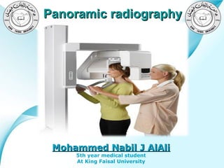
- 1. Panoramic radiography Mohammed Nabil J AlAli 5th year medical student Powerpoint Templates At King Faisal University Page 1
- 2. Outlines - What is Panoramic X-ray? - What are some common uses of the procedure? - How should I prepare? - What are the components ? - How does the procedure work? - What will I experience during and after the procedure? - What are the benefits vs. risks? - What are the limitations of Panoramic X-ray? - Images . Powerpoint Templates Page 2
- 3. Panoramic radiography Panoramic means “Wide View” Powerpoint Templates Page 3
- 4. What is Panoramic X-ray? • Panoramic radiography, also called panoramic x-ray, is a two-dimensional (2-D) dental x-ray examination that captures the entire mouth in a single image, including the teeth, upper and lower jaws, surrounding structures and tissues. • The jaw is a curved structure similar to that of a horse shoe. However, the panoramic x-ray produces a flat image of the curved structure. It is typically set to provide details of the bones and teeth. • A non-invasive medical test . • The film for a panoramic x-ray is contained inside of the machine. Powerpoint Templates Page 4
- 6. What are some common uses of the procedure? • Commonly performed examination by dentists and oral surgeons in everyday practice and is an important diagnostic tool. • It covers a wider area than a conventional intraoral x-ray and, as a result, provides valuable information about the nasal area, maxillary sinuses, tooth positioning and gum and bone irregularities. A panoramic x-ray can also reveal the presence of an existing issue or potential problem such as: • • • • • • Advanced periodontal disease Oral cysts Tumors and oral cancer Impacted teeth Temporo-mandibular joint disorder (TMJ) Powerpoint Templates Sinusitis Page 6
- 7. How should I prepare? • A panoramic x-ray examination requires no • You will be asked to - wear a lead apron. - Remove your jewelry from the region being imaged, eye glasses and any metal objects that might interfere with the x-ray images. • Women should always inform their dentist or oral surgeon if there is any possibility that they are pregnant. If an x-ray is necessary, minimize radiation exposure to the baby. Powerpoint Templates special preparation. Page 7
- 8. What are the components? 1. X-ray unit. 2. Intensifying screens 3. Screen film 4. Cassette Powerpoint Templates Page 8
- 9. How does the procedure work? • an x-ray machine produces a small burst of radiation that passes through the body, recording an image on photographic film or a special detector. • the x-ray tube rotates in a semicircle around the patient's head, starting at one side of the jaw and ending at the other side. • Rather than relying on film placed inside the mouth, a panoramic x-ray machine projects a beam through the patient onto film or a detector rotating oppositePowerpoint Templates the x-ray tube. Page 9
- 10. What will I experience during and after the procedure? • A panoramic x-ray exam is painless, fast and easy and may be recommended over intraoral x-rays for patients that have a sensitive gag reflex. Powerpoint Templates Page 10
- 11. What are the benefits vs. risks? Benefits • • No radiation remains in a patient's body after an x-ray examination. X-rays usually have no side effects in the typical diagnostic range for this exam. • Panoramic x-rays can be used for very young children since the film does not have to be placed inside the mouth. Risks • Women should always inform their dentist or oral surgeon if there is any possibility that they are pregnant. Powerpoint Templates Page 11
- 12. What are the limitations of Panoramic X-ray? • A panoramic x-ray does not provide precise and detailed information about each individual tooth or soft tissues, such as the muscles. • It is generally used as an initial evaluation of the bones and teeth. If further evaluation is needed, a computed tomography (CT) scan or magnetic resonance imaging (MRI) may be ordered. Powerpoint Templates Page 12
- 15. Projection of OP Powerpoint Templates Page 15
- 17. Hard Tissue Inf. Orbital canal and foramen Ant. wall of Maxillary sinus Articular eminence Ptregomaxillary Fissure Hard palate Floor of Maxillary sinus Nasal fossa Inf. orbital rim Panoramic Innominate line (Infra temporal surface of Zyg. bone Man. fossa Ext. Auditory meatus Mandibular condyle Lat. ptreg. plate Zygomatic bone Zyg. process of Maxilla Coronoid process C- Spine Ext. oblique ridge Mental foramen Inf. border of Mandible Powerpoint Templates Hyoid bone Inf. Alveolar canal Page 17
- 18. Soft tissues on OP Powerpoint Templates Page 18
- 19. Soft tissue (edentulous) Middle meatus Inf. nasal meatus Soft palate Inf. nasal concha (turbinate) Post. Wall of the pharynx Dorsal surface of the tongue Upper lip Lower lip Ghost image of opposite Man. Powerpoint Templates Page 19
- 30. Positioning errors ( lips and tongue ) Powerpoint Templates Page 30
Editor's Notes
- What is Panoramic X-ray? Panoramic radiography, also called panoramic x-ray, is a two-dimensional (2-D) dental x-ray examination that captures the entire mouth in a single image, including the teeth, upper and lower jaws, surrounding structures and tissues. The jaw is a curved structure similar to that of a horse shoe. However, the panoramic x-ray produces a flat image of the curved structure. It is typically set to provide details of the bones and teeth. An x-ray (radiograph) is a noninvasive medical test that helps physicians diagnose and treat medical conditions. Imaging with x-rays involves exposing a part of the body to a small dose ofionizing radiation to produce pictures of the inside of the body. X-rays are the oldest and most frequently used form of medical imaging. Unlike a traditional intraoral x-ray where the film is placed inside of the mouth, the film for a panoramic x-ray is contained inside of the machine.
- What are some common uses of the procedure? A panoramic x-ray is a commonly performed examination by dentists and oral surgeons in everyday practice and is an important diagnostic tool. It covers a wider area than a conventional intraoral x-ray and, as a result, provides valuable information about the nasal area, maxillary sinuses, tooth positioning and gum and bone irregularities. This examination is also used to plan treatment for full and partial dentures, braces, extractions and implants. A panoramic x-ray can also reveal the presence of an existing issue or potential problem such as: advanced periodontal disease oral cysts tumors and oral cancer impacted teeth temporomandibular joint disorder (TMJ) sinusitis
- How should I prepare? A panoramic x-ray examination requires no special preparation. You will be asked to wear a lead apron as a safety precaution to protect the rest of your body from any radiation exposure that may scatter from the panoramic x-ray beam. You may also be asked to remove your jewelry from the region being imaged, eye glasses and any metal objects that might interfere with the x-ray images. Women should always inform their dentist or oral surgeon if there is any possibility that they are pregnant. Many imaging tests are not performed during pregnancy so as not to expose the fetus to radiation. If an x-ray is necessary, precautions will be taken to minimize radiation exposure to the baby. See the Safety page for more information about pregnancy and x-rays.
- What does the equipment look like? A panoramic x-ray machine consists of an x-ray tube mounted on a horizontal arm. X-ray film or a detector are mounted on another horizontal arm on the opposite side of the patient. For a sharper, clearer image, the patient's head is positioned with chin, forehead and side rests. The patient is also provided with a bite blocker to prop open the oral cavity.
- How does the procedure work? X-rays are a form of radiation like light or radio waves. X-rays pass through most objects, including the body. Once it is carefully aimed at the part of the body being examined, an x-ray machine produces a small burst of radiation that passes through the body, recording an image on photographic film or a special detector. During a panoramic x-ray examination, the x-ray tube rotates in a semicircle around the patient's head, starting at one side of the jaw and ending at the other side. Rather than relying on film placed inside the mouth, a panoramic x-ray machine projects a beam through the patient onto film or a detector rotating opposite the x-ray tube. Until recently, x-ray images were maintained as hard film copy (much like a photographic negative). Today, most images are digital files that are stored electronically. These stored images are easily accessible and are frequently compared to current x-ray images for diagnosis and disease management. The digital format also allows the dentist to adjust and change the contrast, brightness and darkness of the image for better visualization of certain structures and tissues. Images on film cannot be adjusted or changed.
- What will I experience during and after the procedure? A panoramic x-ray exam is painless, fast and easy and may be recommended over intraoral x-rays for patients that have a sensitive gag reflex.
- What are the benefits vs. risks? Benefits No radiation remains in a patient's body after an x-ray examination. X-rays usually have no side effects in the typical diagnostic range for this exam. Panoramic x-rays can be used for very young children since the film does not have to be placed inside the mouth. Risks Women should always inform their dentist or oral surgeon if there is any possibility that they are pregnant. See the Safety page for more information about pregnancy and x-rays.
- What are the limitations of Panoramic X-ray? A panoramic x-ray does not provide precise and detailed information about each individual tooth or soft tissues, such as the muscles. It is generally used as an initial evaluation of the bones and teeth. If further evaluation is needed, a computed tomography (CT) scan or magnetic resonance imaging (MRI) may be ordered. Frequently, there is also a degree of distortion on the panoramic x-ray, therefore it may be difficult to obtain accurate measurements.