SlideShare a Scribd company logo
1 of 80
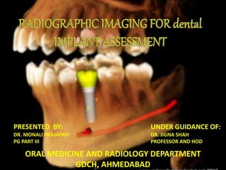
PRESENTED BY:
DR. MONALI PRAJAPATI
PG PART III
UNDER GUIDANCE OF:
DR. JIGNA SHAH
PROFESSOR AND HOD
ORAL MEDICINE AND RADIOLOGY DEPARTMENT
GDCH, AHMEDABAD
In the present era when it comes to oral rehabilitation, a wide array of options are available
to restore the missing teeth using the fixed or removable prosthesis. The advent of implants
in the field of dentistry has given the dental professionals a viable option to provide the
patients with nearly a third set of dentition.
Figure -2Figure -1
Figure -3 Figure -4
1.5mm
3mm
Bone Density Description Tactile Analog Typical Anatomical
Location
D1 Dense Cortical Oak or maple wood Anterior mandible
D2 Porous cortical and
coarse trabecular
White pine or spruce wood Anterior mandible
Posterior mandible
Anterior maxilla
D3 Porous cortical (thin) and
fine trabecular
Balsa wood Anterior maxilla
Posterior maxilla
Posterior mandible
D4 Fine trabecular Styrofoam Posterior maxilla
ORAL AND
MAXILLOFACIAL
RADIOLOGISTS
IMPLANTOLOGIST
S
MAXIMISE DIAGNOSTIC EFFICIENCY
MINIMISE RADIATION RISK
 To decide if implant treatment is appropriate for the patient,
 To detect any possible pathological conditions,
 To ascertain height, buccolingual width, and angulation of
alveolar process,
 To identify the location of vital anatomical structures such as
the inferior alveolar nerve and maxillary sinus,
 To ascertain bone quantity,
 To decide the length and width of the implant to be placed
 PHASE 1: PRE-PROSTHETIC IMPLANT IMAGING
 PHASE 2: SURGICAL AND INTERVENTIONAL
IMPLANT IMAGING
 PHASE 3: POST-PROSTHETIC IMPLANT IMAGING
 PLANAR TWO DIMENSIONAL IMAGING
 QUASI THREE DIMENSIONAL IMAGING
 THREE DIMENSIONAL IMAGING
DETERMINE
IMPLANT
ORIENTATION
DETERMINE
IMPLANT POSITION
IDENTIFY
DISEASE
DETERMINE BONE
QUANTITY
DETERMINE
BONE
QUALITY
MAXILLARY SINUS
LINING
VERTICAL HEIGHT
ORIENTATION
MESIODISTAL WIDTH
FOR SINGLE IMPLANT SITE
Residual bone cyst Dense bone islands
Sites with acute infection
including exudate or pus flow are
considered high-risk, and can
cause post-surgical
complications if implants are
inserted in these infected sites.
FLOOR OF NASAL CAVITY NASOPALATINE CANAL INCISIVE CANAL
MAXILLARY SINUS
INCICIVE FORAMEN
MENTAL FORAMEN
MANDIBULAR CANAL
A reduced vertical bone
height at an adjacent
root surface (>6mm)
affect the height of the
Interdental papilla
following implant
therapy.
COMPUTER-ASSISTED MEASUREMENTS, RULERS, CALIPERS, AND SUPRABONY
THREAD EVALUATION
DISTORTION
LACKS THIRD
DIMENSION
FI NE TRABECULAR
PLATES AND MULTIPLE
SMALL TRABECULAR
SPACES
GENERALLY SHOWING LARGE
MARROW SPACES AND SPARSE
TRABECULATION
COARSER TRABECULAR
PLATES AND LARGER
MARROW SPACES
CRESTAL BONE LOSS EVALUATION CAN BE BENEFITTED USING
DIGITAL PERIAPICAL RADIOGRAPHY
ASSESS ALVEOLAR CREST
The mandibular occlusal radiograph shows the
widest width of bone (i.e., the symphysis) versus the
width at the crest, which is where diagnostic
information is needed most .
SPATIAL RELATION OF OCCLUSION
AND ESTHETICS
CROSS-SECTION OF CANINE AND
LATERAL INCISOR REGION
VERTICAL DIMENSION
RELATION OF LINGUAL PLATE WITH PATIENTS SKELETAL ANATOMY CAN BE
DETERMINED
(IMPLANTS ARE USUALLY PLACE ADJACENT TO LINGUAL PLATE IN ANTERIOR REGION)
THESE TECHNIQUE PRODUCES A NUMBER OF CLOSELY SPACED
TOMOGRAPHIC IMAGES
VERTICAL HEIGHT MESIODISTAL WIDTH
ORIENTATION PERIODONTAL CONDITION
PATHOLOGY VITAL ANATOMIC
STRUCTURES
WHEN PANORAMIC AND THE PERIAPICAL IMAGES
ARE THE ONLY DIAGNOSTIC TOOL TO ASSESS
AVAILABLE BONE HEIGHT
ZONE OF SAFETY
DEMERITS
 Distortions inherent in the panoramic systems
 Fixed vertical magnification of upto 10%
 Uncertain horizontal magnification upto 20%
RADIOGRAPHIC MARKER
(a)Cropped panoramic radiograph
demonstrating excellent bone height in
the lower right molar region. (b)
Reformatted cross-sectional CT images
showing reasonable bone height but the
ridge is narrow bucco-lingually.
CROSS SECTIONAL DIAGRAM
OF THE MANDIBLE
SHOWING THAT
STRUCTURES THAT ARE
MORE LINGUAL ARE
PROJECTED HIGHER ON THE
FILM THAN STRUCTURES
THAT ARE MORE BUCCAL
DOES NOT DEMONSTRATE BONE
QUALITY
NO SPATIAL RELATIONSHIP BETWEEN
STRUCTURES CAN BE ESTABLISHED
Dentascan imaging provides programmed
reformation, organization and display of the imaging
study.
Cancellous bone density Hounsfield unit
D1 > 1250HU
D2 850-1250HU
D3 350-850HU
D4 150-350HU
D5 <150HU
DEMERITS
HIGH RADIATION DOSE
TECHNIQUE SENSITIVE
EXPENSIVE
EXPOSURE TIME – 36seconds
SINGLE EXPOSURE REQUIRED
LESS RADIATION EXPOSURE
EXPOSES BOTH ARCHES SIMULTANEOUSLY
LESS SCATTER
If the facial wall is thin (≤ 1 mm), this bone will resorb
within 4 to 8 weeks leading to a horizontal, crater-
shaped bone defect and a loss of bone height on the
facial aspect.
EVALUATES THE
SURGICAL SITES
DURING AND
IMMEDIATELY
AFTER SURGERY
OPTIMAL
POSITIONIN
G AND
ORIENTATIO
N OF
DENTAL
IMPLANTS
TO
ASCERTAIN
THE HEALING
TO ENSURE
APPROPRIATE
ABUTMENT
POSITIONING
AND
PROSTHESIS
FABRICATION
ORIENTATION OF
IMPLANT
SEATING OF PROSTHESIS
INVERSION OF GRAY
SCALE TO EVALUATE
OSSEOUS QUALITY AND
LOCATION OF VITAL
STRUTURES
EDGE ENHANCEMENT,” WHICH IS THE ABILITY TO DETECT SPACE
BETWEEN THE IMPLANT AND THE SURROUNDING BONE
ALLOW VIEWING OF ANY SUBTLE CHANGES IN BONE DENSITY
AROUND THE IMPLANT INTERFACE.
BONE LOSS AROUND A ROOT-
FORM DENTAL IMPLANT (THIN
RADIOLUCENT BAND
SURROUNDING THE IMPLANT),
INDICATING FAILURE OF
OSSEOUS INTEGRATION.
PERIAPICAL VIEW OF A
FRACTURED
ENDOSSEOUS IMPLANT.
A panoramic radiograph used for
postoperative assessment of multiple
successfully restored rootform implants.
The cross-sectional reformatted CBCT images reveal nonrestorable ectopic
placement of the existing implants with lingual cortical perforation and extension
into the lingual tissues.
DETERMINE
CRESTAL BONE
LEVELS
ASSESS THE BONE
ADJACENT TO THE
DENTAL IMPLANT
CLOSE
APPOSITION OF
THE BONE TO THE
SURFACE OF
EACH IMPLANT.
MINOR AMOUNT OF SAUCERIZATION IS PRESENT AT THE
ALVEOLAR CREST ADJACENT TO THE DISTAL FIXTURE
BITEWING RADIOGRAPH
FACTORS TWO DIMENSIONAL
IMAGING
THREE DIMENSIONAL
IMAGING
MESIODISTAL WIDTH ASSESSED ASSESSED
BUCCOLINGUAL WIDTH NOT ASSESSED ASSESSED
VERTICAL HEIGHT ASSESSED ASSESSED
SPATIAL RELATION WITH
ANATOMIC STRUCTURE
NOT ASSESED ASSESSED
RADIATION LESS MORE
MAGNIFICATION MORE LESS
AVAILABITY AND
CONVENIENCE
EASY, CONVENIENT DIFFICULT
BONE DENSITY CAN NOT BE EVALUATED CAN BE EVALUATED
PERIAPICAL RADIOGRAPHY
ADVANTAGES DISADVANTAGES INDICATIONS
• Low radiation dose
• Minimal magnification
with proper technique
• High resolution
• Inexpensive
• Distortion and magnification
• Minimal site evaluation
• Difficulty in film placement
• Lack of cross-sectional imaging
• Bucco-lingual width can not be
measured
• Spatial relation can not be
established
• Bone density can not be
evaluated
• Single implant site
(anterior, middle,
posterior
maxilla/mandible)
• Alignment and
orientation during
surgery
(interventional
phase)
• Post- prosthetic stage
evaluation
OCCLUSAL RADIOGRAPHY
ADVANTAGES DISADVANTAGES INDICATIONS
• Low radiation dose
• High resolution
• Inexpensive
• spatial relation can not be
established
• Bone density can not be
evaluated
Of little value
CEPHALOMETERIC IMAGING
• Height / width in
anterior region
• Low magnification
• Skeletal relationship
• Crown/ implant ratio
in anterior region
• Relation of lingual
cortical plate to
skeletal structure can
be established
• Availability
• Image information limited to
midline
• Reduced resolution
• Single implant site
evaluation
• Anterior
maxilla/mandible
region
• Symphysis bone graft
evaluation
PANORAMIC RADIOGRAPHY
ADVANTAGES DISADVANTAGES INDICATIONS
• Single image of maxilla
and mandible
obtained
• Convenience, ease,
and speed in
performance
• Distortion and magnification
• Lack of cross-sectional imaging
• Bucco-lingual width can not be
measured
• Spatial relation can not be
established
• Bone density can not be
evaluated
• Single implant site
(middle, posterior
maxilla/mandible)
• Multiple implant site
• Implant overdenture
site
• Alignment and
orientation during
surgery
(interventional
phase)
• Post- prosthetic
stage evaluation
DENTASCAN/ CBCT
ADVANTAGES DISADVANTAGES INDICATIONS
• Negligible
magnification
• High contrast
• Axial, coronal sagittal
views
• Buccolingial width
determined
• Spatial relation can be
established
• Interactive treatment
planning
• High radiation exposure
• Cost
• Technique sensitive
• Single implant site (anterior,
middle, posterior
maxilla/mandible)
• Multiple implant site
• Implant overdenture site
• Unless any complication, not
advisable for interventional
and post prosthetic
evaluation
• Bone density
 Carl E. Misch, Conmtemporary implant dentistry, 3rd edition
 Textbook Of Dental And Maxillofacial Radiology By Freny Karjodkar
 White & Pharoah, 6th Edition
 Lingeshwar D, Dhanasekar B, Aparna -Diagnostic Imaging In Implant Dentistry In International Journal Of Oral
Implantology And Clinical Research, September-December 2010;1(3):147-153
 Aishwarya Nagarajan, Rajapriya Perumalsamy, Ramakrishnan Thyagarajan, Ambalavanan Namasivayam- Diagnostic
Imaging For Dental Implant Therapy In Journal Of Clinical Imaging Science | Vol. 4 | Dental Suppl 2 | Oct-Dec 2014
 Dale A. Miles* And Ronald K. Shelle- Pre-Surgicalimplant Site Assessment ,Part I - Precise And Practical
Radiographic Stent Construction For Cone Beam Ct Imaging
 Martin J. Bourgeois Dds, M.Ed., Dip. Oral Rad. 20000701, Radiology: PreSurgical Radiographic Imaging For
The Placement Of Dental Implants
 S. J. J. Mccrea- Pre-Operative Radiographs For Dental Implants – Are Selection Criteria Being Followed British Dental
Journal Volume 204 No. 12 Jun 28 2008
 Maxillofacial Radiology On Selection Criteria For The Use Of Radiology In Dental Implantology With Emphasis On
Cone Beam Computed Tomography By Donald A. Tyndall, Dds, Msph, Phd,Scott D. Ganz, Dmd,Jeffery B. Price, Dds,
Ms,Charles Hildebolt, Dds, Phd,Sotirios Tetradis, Dds, Phd,And William C. Scarfe, Bds, Ms Position Statement Of The
American Academy Of Oral .OOO, Vol. 113 No. 6 June 2012
RADIOGRAPHIC IMAGING FOR DENTAL IMPLANT ASSESSMENT

More Related Content

What's hot

Surgical implant placement guides/ dentistry dental implants
Surgical implant placement guides/ dentistry dental implantsSurgical implant placement guides/ dentistry dental implants
Surgical implant placement guides/ dentistry dental implantsIndian dental academy
 
Basic implant surgery
Basic implant surgeryBasic implant surgery
Basic implant surgeryNitika Jain
 
Radiographic considerations in dental implants
Radiographic considerations in dental implantsRadiographic considerations in dental implants
Radiographic considerations in dental implantsDr. Ishaan Adhaulia
 
recent advances in implant dentistry
recent advances in implant dentistryrecent advances in implant dentistry
recent advances in implant dentistryDr.Pallavi Chavan
 
Twin occlusion prosthesis in a class 3
Twin occlusion prosthesis in a class 3Twin occlusion prosthesis in a class 3
Twin occlusion prosthesis in a class 3Nishu Priya
 
Recent advances in prosthdontics
Recent advances in prosthdonticsRecent advances in prosthdontics
Recent advances in prosthdonticsJoel Koshy
 
Computer guided implant surgery
Computer guided implant surgeryComputer guided implant surgery
Computer guided implant surgeryUS Dental
 
Provisional restoration in fixed partial denture
Provisional restoration in fixed partial dentureProvisional restoration in fixed partial denture
Provisional restoration in fixed partial denturebhuvanesh4668
 
Paralleling and bisecting radiographic techniques
Paralleling and bisecting  radiographic techniquesParalleling and bisecting  radiographic techniques
Paralleling and bisecting radiographic techniquesDr. Ritu Gupta
 
Prosthetic options in implant dentistry
Prosthetic options in implant dentistryProsthetic options in implant dentistry
Prosthetic options in implant dentistryNAMITHA ANAND
 
Gingival Retraction
Gingival Retraction Gingival Retraction
Gingival Retraction Harshil Modi
 
Esthetic considerations in implant placement
Esthetic considerations in implant placementEsthetic considerations in implant placement
Esthetic considerations in implant placementRAVINDER NARWAL
 
PLATFORM SWITCHING
PLATFORM SWITCHINGPLATFORM SWITCHING
PLATFORM SWITCHINGshari kurup
 
dental implant impression technique
dental implant impression technique dental implant impression technique
dental implant impression technique Sara Zaky
 

What's hot (20)

Surgical implant placement guides/ dentistry dental implants
Surgical implant placement guides/ dentistry dental implantsSurgical implant placement guides/ dentistry dental implants
Surgical implant placement guides/ dentistry dental implants
 
Basic implant surgery
Basic implant surgeryBasic implant surgery
Basic implant surgery
 
Radiographic considerations in dental implants
Radiographic considerations in dental implantsRadiographic considerations in dental implants
Radiographic considerations in dental implants
 
recent advances in implant dentistry
recent advances in implant dentistryrecent advances in implant dentistry
recent advances in implant dentistry
 
Twin occlusion prosthesis in a class 3
Twin occlusion prosthesis in a class 3Twin occlusion prosthesis in a class 3
Twin occlusion prosthesis in a class 3
 
Pontics
PonticsPontics
Pontics
 
Loading protocols in implant
Loading protocols in implantLoading protocols in implant
Loading protocols in implant
 
Recent advances in prosthdontics
Recent advances in prosthdonticsRecent advances in prosthdontics
Recent advances in prosthdontics
 
Computer guided implant surgery
Computer guided implant surgeryComputer guided implant surgery
Computer guided implant surgery
 
Temporization in fixed prosthodontics
Temporization in fixed prosthodonticsTemporization in fixed prosthodontics
Temporization in fixed prosthodontics
 
Provisional restoration in fixed partial denture
Provisional restoration in fixed partial dentureProvisional restoration in fixed partial denture
Provisional restoration in fixed partial denture
 
Osseointegration
OsseointegrationOsseointegration
Osseointegration
 
Apexification
ApexificationApexification
Apexification
 
Paralleling and bisecting radiographic techniques
Paralleling and bisecting  radiographic techniquesParalleling and bisecting  radiographic techniques
Paralleling and bisecting radiographic techniques
 
Computer guided
Computer guidedComputer guided
Computer guided
 
Prosthetic options in implant dentistry
Prosthetic options in implant dentistryProsthetic options in implant dentistry
Prosthetic options in implant dentistry
 
Gingival Retraction
Gingival Retraction Gingival Retraction
Gingival Retraction
 
Esthetic considerations in implant placement
Esthetic considerations in implant placementEsthetic considerations in implant placement
Esthetic considerations in implant placement
 
PLATFORM SWITCHING
PLATFORM SWITCHINGPLATFORM SWITCHING
PLATFORM SWITCHING
 
dental implant impression technique
dental implant impression technique dental implant impression technique
dental implant impression technique
 

Similar to RADIOGRAPHIC IMAGING FOR DENTAL IMPLANT ASSESSMENT

summaryofcbct-161216070820.pptx
summaryofcbct-161216070820.pptxsummaryofcbct-161216070820.pptx
summaryofcbct-161216070820.pptxpoojamuley7
 
Radiographs in prosthodontics/dental crown &bridge course by Indian dental ac...
Radiographs in prosthodontics/dental crown &bridge course by Indian dental ac...Radiographs in prosthodontics/dental crown &bridge course by Indian dental ac...
Radiographs in prosthodontics/dental crown &bridge course by Indian dental ac...Indian dental academy
 
per flap with vest deep.pptx
per flap with vest deep.pptxper flap with vest deep.pptx
per flap with vest deep.pptxmayankgupta672202
 
Diagnostic aids in implant seminar in detail
Diagnostic aids in implant seminar in detailDiagnostic aids in implant seminar in detail
Diagnostic aids in implant seminar in detailNikitaChhabariya
 
Radiography in conservative dentistry
Radiography in conservative dentistryRadiography in conservative dentistry
Radiography in conservative dentistryEdward Kaliisa
 
Maxillary and jaw lesions
Maxillary and jaw lesionsMaxillary and jaw lesions
Maxillary and jaw lesionsSangeeta Jha
 
Maxillofacial imaging in implant treatment planning.pptx
Maxillofacial imaging in implant treatment planning.pptxMaxillofacial imaging in implant treatment planning.pptx
Maxillofacial imaging in implant treatment planning.pptxKaushikDutta233455
 
Selectionofpatientfordentalimplant 190308171548
Selectionofpatientfordentalimplant 190308171548Selectionofpatientfordentalimplant 190308171548
Selectionofpatientfordentalimplant 190308171548Dr. Shailee Swarup
 
Radiographs in endodontics
Radiographs in endodonticsRadiographs in endodontics
Radiographs in endodonticsIAU Dent
 
Traumatic injuries of teeth
Traumatic injuries of teethTraumatic injuries of teeth
Traumatic injuries of teethKUMARAVEL SM
 
Radiology in dentistry
Radiology in dentistry Radiology in dentistry
Radiology in dentistry Masuma Ryzvee
 
Fenestration and dehiscence
Fenestration and dehiscenceFenestration and dehiscence
Fenestration and dehiscenceAhmed Baattiah
 
Decision making in implants.ppt
Decision making in implants.pptDecision making in implants.ppt
Decision making in implants.pptmalti19
 

Similar to RADIOGRAPHIC IMAGING FOR DENTAL IMPLANT ASSESSMENT (20)

summaryofcbct-161216070820.pptx
summaryofcbct-161216070820.pptxsummaryofcbct-161216070820.pptx
summaryofcbct-161216070820.pptx
 
Radiographs in prosthodontics/dental crown &bridge course by Indian dental ac...
Radiographs in prosthodontics/dental crown &bridge course by Indian dental ac...Radiographs in prosthodontics/dental crown &bridge course by Indian dental ac...
Radiographs in prosthodontics/dental crown &bridge course by Indian dental ac...
 
Radiographs in prosthodontics
Radiographs in prosthodonticsRadiographs in prosthodontics
Radiographs in prosthodontics
 
diagnostic aids in implant.pptx
diagnostic aids in implant.pptxdiagnostic aids in implant.pptx
diagnostic aids in implant.pptx
 
per flap with vest deep.pptx
per flap with vest deep.pptxper flap with vest deep.pptx
per flap with vest deep.pptx
 
Diagnostic aids in implant seminar in detail
Diagnostic aids in implant seminar in detailDiagnostic aids in implant seminar in detail
Diagnostic aids in implant seminar in detail
 
Radiography in conservative dentistry
Radiography in conservative dentistryRadiography in conservative dentistry
Radiography in conservative dentistry
 
Maxillary and jaw lesions
Maxillary and jaw lesionsMaxillary and jaw lesions
Maxillary and jaw lesions
 
Maxillofacial imaging in implant treatment planning.pptx
Maxillofacial imaging in implant treatment planning.pptxMaxillofacial imaging in implant treatment planning.pptx
Maxillofacial imaging in implant treatment planning.pptx
 
Single tooth implants
Single tooth implantsSingle tooth implants
Single tooth implants
 
Selectionofpatientfordentalimplant 190308171548
Selectionofpatientfordentalimplant 190308171548Selectionofpatientfordentalimplant 190308171548
Selectionofpatientfordentalimplant 190308171548
 
Selection of patient for dental implant
Selection of patient for dental implantSelection of patient for dental implant
Selection of patient for dental implant
 
Radiographs in endodontics
Radiographs in endodonticsRadiographs in endodontics
Radiographs in endodontics
 
Traumatic injuries of teeth
Traumatic injuries of teethTraumatic injuries of teeth
Traumatic injuries of teeth
 
RESECTIVE OSSEOUS SURGERY.ppt
RESECTIVE OSSEOUS SURGERY.pptRESECTIVE OSSEOUS SURGERY.ppt
RESECTIVE OSSEOUS SURGERY.ppt
 
Radiology in dentistry
Radiology in dentistry Radiology in dentistry
Radiology in dentistry
 
Diagnosis and treatment planning
Diagnosis and treatment planningDiagnosis and treatment planning
Diagnosis and treatment planning
 
Diagnosis and treatment planning part 1
Diagnosis and treatment planning part 1Diagnosis and treatment planning part 1
Diagnosis and treatment planning part 1
 
Fenestration and dehiscence
Fenestration and dehiscenceFenestration and dehiscence
Fenestration and dehiscence
 
Decision making in implants.ppt
Decision making in implants.pptDecision making in implants.ppt
Decision making in implants.ppt
 

More from Dr. Monali Prajapati

More from Dr. Monali Prajapati (7)

Tongue disorders
Tongue disordersTongue disorders
Tongue disorders
 
Pigmented lesions of oral cavity
Pigmented lesions of oral cavityPigmented lesions of oral cavity
Pigmented lesions of oral cavity
 
Tumor markers
Tumor markersTumor markers
Tumor markers
 
Diabetes and its oral complication
Diabetes and its oral complicationDiabetes and its oral complication
Diabetes and its oral complication
 
LYMPH NODES OF HEAD AND NECK AND DIFFERENTIAL DIAGNOSIS OF CERVICAL LYMPHA...
LYMPH NODES OF HEAD AND NECK  AND  DIFFERENTIAL DIAGNOSIS OF  CERVICAL LYMPHA...LYMPH NODES OF HEAD AND NECK  AND  DIFFERENTIAL DIAGNOSIS OF  CERVICAL LYMPHA...
LYMPH NODES OF HEAD AND NECK AND DIFFERENTIAL DIAGNOSIS OF CERVICAL LYMPHA...
 
Soft tissue calcification orofacial region
Soft tissue calcification orofacial regionSoft tissue calcification orofacial region
Soft tissue calcification orofacial region
 
Vitamin D
Vitamin DVitamin D
Vitamin D
 

Recently uploaded

Incoming and Outgoing Shipments in 3 STEPS Using Odoo 17
Incoming and Outgoing Shipments in 3 STEPS Using Odoo 17Incoming and Outgoing Shipments in 3 STEPS Using Odoo 17
Incoming and Outgoing Shipments in 3 STEPS Using Odoo 17Celine George
 
Roles & Responsibilities in Pharmacovigilance
Roles & Responsibilities in PharmacovigilanceRoles & Responsibilities in Pharmacovigilance
Roles & Responsibilities in PharmacovigilanceSamikshaHamane
 
GRADE 4 - SUMMATIVE TEST QUARTER 4 ALL SUBJECTS
GRADE 4 - SUMMATIVE TEST QUARTER 4 ALL SUBJECTSGRADE 4 - SUMMATIVE TEST QUARTER 4 ALL SUBJECTS
GRADE 4 - SUMMATIVE TEST QUARTER 4 ALL SUBJECTSJoshuaGantuangco2
 
AMERICAN LANGUAGE HUB_Level2_Student'sBook_Answerkey.pdf
AMERICAN LANGUAGE HUB_Level2_Student'sBook_Answerkey.pdfAMERICAN LANGUAGE HUB_Level2_Student'sBook_Answerkey.pdf
AMERICAN LANGUAGE HUB_Level2_Student'sBook_Answerkey.pdfphamnguyenenglishnb
 
Full Stack Web Development Course for Beginners
Full Stack Web Development Course  for BeginnersFull Stack Web Development Course  for Beginners
Full Stack Web Development Course for BeginnersSabitha Banu
 
ECONOMIC CONTEXT - LONG FORM TV DRAMA - PPT
ECONOMIC CONTEXT - LONG FORM TV DRAMA - PPTECONOMIC CONTEXT - LONG FORM TV DRAMA - PPT
ECONOMIC CONTEXT - LONG FORM TV DRAMA - PPTiammrhaywood
 
Field Attribute Index Feature in Odoo 17
Field Attribute Index Feature in Odoo 17Field Attribute Index Feature in Odoo 17
Field Attribute Index Feature in Odoo 17Celine George
 
HỌC TỐT TIẾNG ANH 11 THEO CHƯƠNG TRÌNH GLOBAL SUCCESS ĐÁP ÁN CHI TIẾT - CẢ NĂ...
HỌC TỐT TIẾNG ANH 11 THEO CHƯƠNG TRÌNH GLOBAL SUCCESS ĐÁP ÁN CHI TIẾT - CẢ NĂ...HỌC TỐT TIẾNG ANH 11 THEO CHƯƠNG TRÌNH GLOBAL SUCCESS ĐÁP ÁN CHI TIẾT - CẢ NĂ...
HỌC TỐT TIẾNG ANH 11 THEO CHƯƠNG TRÌNH GLOBAL SUCCESS ĐÁP ÁN CHI TIẾT - CẢ NĂ...Nguyen Thanh Tu Collection
 
4.18.24 Movement Legacies, Reflection, and Review.pptx
4.18.24 Movement Legacies, Reflection, and Review.pptx4.18.24 Movement Legacies, Reflection, and Review.pptx
4.18.24 Movement Legacies, Reflection, and Review.pptxmary850239
 
What is Model Inheritance in Odoo 17 ERP
What is Model Inheritance in Odoo 17 ERPWhat is Model Inheritance in Odoo 17 ERP
What is Model Inheritance in Odoo 17 ERPCeline George
 
ENGLISH6-Q4-W3.pptxqurter our high choom
ENGLISH6-Q4-W3.pptxqurter our high choomENGLISH6-Q4-W3.pptxqurter our high choom
ENGLISH6-Q4-W3.pptxqurter our high choomnelietumpap1
 
Proudly South Africa powerpoint Thorisha.pptx
Proudly South Africa powerpoint Thorisha.pptxProudly South Africa powerpoint Thorisha.pptx
Proudly South Africa powerpoint Thorisha.pptxthorishapillay1
 
Gas measurement O2,Co2,& ph) 04/2024.pptx
Gas measurement O2,Co2,& ph) 04/2024.pptxGas measurement O2,Co2,& ph) 04/2024.pptx
Gas measurement O2,Co2,& ph) 04/2024.pptxDr.Ibrahim Hassaan
 
Difference Between Search & Browse Methods in Odoo 17
Difference Between Search & Browse Methods in Odoo 17Difference Between Search & Browse Methods in Odoo 17
Difference Between Search & Browse Methods in Odoo 17Celine George
 
INTRODUCTION TO CATHOLIC CHRISTOLOGY.pptx
INTRODUCTION TO CATHOLIC CHRISTOLOGY.pptxINTRODUCTION TO CATHOLIC CHRISTOLOGY.pptx
INTRODUCTION TO CATHOLIC CHRISTOLOGY.pptxHumphrey A Beña
 
Keynote by Prof. Wurzer at Nordex about IP-design
Keynote by Prof. Wurzer at Nordex about IP-designKeynote by Prof. Wurzer at Nordex about IP-design
Keynote by Prof. Wurzer at Nordex about IP-designMIPLM
 

Recently uploaded (20)

OS-operating systems- ch04 (Threads) ...
OS-operating systems- ch04 (Threads) ...OS-operating systems- ch04 (Threads) ...
OS-operating systems- ch04 (Threads) ...
 
Incoming and Outgoing Shipments in 3 STEPS Using Odoo 17
Incoming and Outgoing Shipments in 3 STEPS Using Odoo 17Incoming and Outgoing Shipments in 3 STEPS Using Odoo 17
Incoming and Outgoing Shipments in 3 STEPS Using Odoo 17
 
Roles & Responsibilities in Pharmacovigilance
Roles & Responsibilities in PharmacovigilanceRoles & Responsibilities in Pharmacovigilance
Roles & Responsibilities in Pharmacovigilance
 
GRADE 4 - SUMMATIVE TEST QUARTER 4 ALL SUBJECTS
GRADE 4 - SUMMATIVE TEST QUARTER 4 ALL SUBJECTSGRADE 4 - SUMMATIVE TEST QUARTER 4 ALL SUBJECTS
GRADE 4 - SUMMATIVE TEST QUARTER 4 ALL SUBJECTS
 
Model Call Girl in Tilak Nagar Delhi reach out to us at 🔝9953056974🔝
Model Call Girl in Tilak Nagar Delhi reach out to us at 🔝9953056974🔝Model Call Girl in Tilak Nagar Delhi reach out to us at 🔝9953056974🔝
Model Call Girl in Tilak Nagar Delhi reach out to us at 🔝9953056974🔝
 
YOUVE GOT EMAIL_FINALS_EL_DORADO_2024.pptx
YOUVE GOT EMAIL_FINALS_EL_DORADO_2024.pptxYOUVE GOT EMAIL_FINALS_EL_DORADO_2024.pptx
YOUVE GOT EMAIL_FINALS_EL_DORADO_2024.pptx
 
AMERICAN LANGUAGE HUB_Level2_Student'sBook_Answerkey.pdf
AMERICAN LANGUAGE HUB_Level2_Student'sBook_Answerkey.pdfAMERICAN LANGUAGE HUB_Level2_Student'sBook_Answerkey.pdf
AMERICAN LANGUAGE HUB_Level2_Student'sBook_Answerkey.pdf
 
Full Stack Web Development Course for Beginners
Full Stack Web Development Course  for BeginnersFull Stack Web Development Course  for Beginners
Full Stack Web Development Course for Beginners
 
ECONOMIC CONTEXT - LONG FORM TV DRAMA - PPT
ECONOMIC CONTEXT - LONG FORM TV DRAMA - PPTECONOMIC CONTEXT - LONG FORM TV DRAMA - PPT
ECONOMIC CONTEXT - LONG FORM TV DRAMA - PPT
 
Field Attribute Index Feature in Odoo 17
Field Attribute Index Feature in Odoo 17Field Attribute Index Feature in Odoo 17
Field Attribute Index Feature in Odoo 17
 
HỌC TỐT TIẾNG ANH 11 THEO CHƯƠNG TRÌNH GLOBAL SUCCESS ĐÁP ÁN CHI TIẾT - CẢ NĂ...
HỌC TỐT TIẾNG ANH 11 THEO CHƯƠNG TRÌNH GLOBAL SUCCESS ĐÁP ÁN CHI TIẾT - CẢ NĂ...HỌC TỐT TIẾNG ANH 11 THEO CHƯƠNG TRÌNH GLOBAL SUCCESS ĐÁP ÁN CHI TIẾT - CẢ NĂ...
HỌC TỐT TIẾNG ANH 11 THEO CHƯƠNG TRÌNH GLOBAL SUCCESS ĐÁP ÁN CHI TIẾT - CẢ NĂ...
 
4.18.24 Movement Legacies, Reflection, and Review.pptx
4.18.24 Movement Legacies, Reflection, and Review.pptx4.18.24 Movement Legacies, Reflection, and Review.pptx
4.18.24 Movement Legacies, Reflection, and Review.pptx
 
What is Model Inheritance in Odoo 17 ERP
What is Model Inheritance in Odoo 17 ERPWhat is Model Inheritance in Odoo 17 ERP
What is Model Inheritance in Odoo 17 ERP
 
ENGLISH6-Q4-W3.pptxqurter our high choom
ENGLISH6-Q4-W3.pptxqurter our high choomENGLISH6-Q4-W3.pptxqurter our high choom
ENGLISH6-Q4-W3.pptxqurter our high choom
 
Proudly South Africa powerpoint Thorisha.pptx
Proudly South Africa powerpoint Thorisha.pptxProudly South Africa powerpoint Thorisha.pptx
Proudly South Africa powerpoint Thorisha.pptx
 
Gas measurement O2,Co2,& ph) 04/2024.pptx
Gas measurement O2,Co2,& ph) 04/2024.pptxGas measurement O2,Co2,& ph) 04/2024.pptx
Gas measurement O2,Co2,& ph) 04/2024.pptx
 
Difference Between Search & Browse Methods in Odoo 17
Difference Between Search & Browse Methods in Odoo 17Difference Between Search & Browse Methods in Odoo 17
Difference Between Search & Browse Methods in Odoo 17
 
INTRODUCTION TO CATHOLIC CHRISTOLOGY.pptx
INTRODUCTION TO CATHOLIC CHRISTOLOGY.pptxINTRODUCTION TO CATHOLIC CHRISTOLOGY.pptx
INTRODUCTION TO CATHOLIC CHRISTOLOGY.pptx
 
TataKelola dan KamSiber Kecerdasan Buatan v022.pdf
TataKelola dan KamSiber Kecerdasan Buatan v022.pdfTataKelola dan KamSiber Kecerdasan Buatan v022.pdf
TataKelola dan KamSiber Kecerdasan Buatan v022.pdf
 
Keynote by Prof. Wurzer at Nordex about IP-design
Keynote by Prof. Wurzer at Nordex about IP-designKeynote by Prof. Wurzer at Nordex about IP-design
Keynote by Prof. Wurzer at Nordex about IP-design
 

RADIOGRAPHIC IMAGING FOR DENTAL IMPLANT ASSESSMENT

  • 1. PRESENTED BY: DR. MONALI PRAJAPATI PG PART III UNDER GUIDANCE OF: DR. JIGNA SHAH PROFESSOR AND HOD ORAL MEDICINE AND RADIOLOGY DEPARTMENT GDCH, AHMEDABAD
  • 2. In the present era when it comes to oral rehabilitation, a wide array of options are available to restore the missing teeth using the fixed or removable prosthesis. The advent of implants in the field of dentistry has given the dental professionals a viable option to provide the patients with nearly a third set of dentition.
  • 3.
  • 4.
  • 6.
  • 8.
  • 9. Bone Density Description Tactile Analog Typical Anatomical Location D1 Dense Cortical Oak or maple wood Anterior mandible D2 Porous cortical and coarse trabecular White pine or spruce wood Anterior mandible Posterior mandible Anterior maxilla D3 Porous cortical (thin) and fine trabecular Balsa wood Anterior maxilla Posterior maxilla Posterior mandible D4 Fine trabecular Styrofoam Posterior maxilla
  • 12.  To decide if implant treatment is appropriate for the patient,  To detect any possible pathological conditions,  To ascertain height, buccolingual width, and angulation of alveolar process,  To identify the location of vital anatomical structures such as the inferior alveolar nerve and maxillary sinus,  To ascertain bone quantity,  To decide the length and width of the implant to be placed
  • 13.  PHASE 1: PRE-PROSTHETIC IMPLANT IMAGING  PHASE 2: SURGICAL AND INTERVENTIONAL IMPLANT IMAGING  PHASE 3: POST-PROSTHETIC IMPLANT IMAGING
  • 14.  PLANAR TWO DIMENSIONAL IMAGING  QUASI THREE DIMENSIONAL IMAGING  THREE DIMENSIONAL IMAGING
  • 16.
  • 18. Residual bone cyst Dense bone islands
  • 19. Sites with acute infection including exudate or pus flow are considered high-risk, and can cause post-surgical complications if implants are inserted in these infected sites.
  • 20. FLOOR OF NASAL CAVITY NASOPALATINE CANAL INCISIVE CANAL MAXILLARY SINUS
  • 22. A reduced vertical bone height at an adjacent root surface (>6mm) affect the height of the Interdental papilla following implant therapy.
  • 23. COMPUTER-ASSISTED MEASUREMENTS, RULERS, CALIPERS, AND SUPRABONY THREAD EVALUATION
  • 24.
  • 26.
  • 28. FI NE TRABECULAR PLATES AND MULTIPLE SMALL TRABECULAR SPACES GENERALLY SHOWING LARGE MARROW SPACES AND SPARSE TRABECULATION COARSER TRABECULAR PLATES AND LARGER MARROW SPACES
  • 29. CRESTAL BONE LOSS EVALUATION CAN BE BENEFITTED USING DIGITAL PERIAPICAL RADIOGRAPHY
  • 31.
  • 32. The mandibular occlusal radiograph shows the widest width of bone (i.e., the symphysis) versus the width at the crest, which is where diagnostic information is needed most .
  • 33.
  • 34. SPATIAL RELATION OF OCCLUSION AND ESTHETICS CROSS-SECTION OF CANINE AND LATERAL INCISOR REGION VERTICAL DIMENSION RELATION OF LINGUAL PLATE WITH PATIENTS SKELETAL ANATOMY CAN BE DETERMINED (IMPLANTS ARE USUALLY PLACE ADJACENT TO LINGUAL PLATE IN ANTERIOR REGION)
  • 35.
  • 36. THESE TECHNIQUE PRODUCES A NUMBER OF CLOSELY SPACED TOMOGRAPHIC IMAGES
  • 37.
  • 38. VERTICAL HEIGHT MESIODISTAL WIDTH ORIENTATION PERIODONTAL CONDITION PATHOLOGY VITAL ANATOMIC STRUCTURES
  • 39.
  • 40. WHEN PANORAMIC AND THE PERIAPICAL IMAGES ARE THE ONLY DIAGNOSTIC TOOL TO ASSESS AVAILABLE BONE HEIGHT ZONE OF SAFETY
  • 41. DEMERITS  Distortions inherent in the panoramic systems  Fixed vertical magnification of upto 10%  Uncertain horizontal magnification upto 20%
  • 43. (a)Cropped panoramic radiograph demonstrating excellent bone height in the lower right molar region. (b) Reformatted cross-sectional CT images showing reasonable bone height but the ridge is narrow bucco-lingually.
  • 44. CROSS SECTIONAL DIAGRAM OF THE MANDIBLE SHOWING THAT STRUCTURES THAT ARE MORE LINGUAL ARE PROJECTED HIGHER ON THE FILM THAN STRUCTURES THAT ARE MORE BUCCAL
  • 45.
  • 46. DOES NOT DEMONSTRATE BONE QUALITY NO SPATIAL RELATIONSHIP BETWEEN STRUCTURES CAN BE ESTABLISHED
  • 47.
  • 48.
  • 49.
  • 50. Dentascan imaging provides programmed reformation, organization and display of the imaging study.
  • 51.
  • 52. Cancellous bone density Hounsfield unit D1 > 1250HU D2 850-1250HU D3 350-850HU D4 150-350HU D5 <150HU
  • 54.
  • 55. EXPOSURE TIME – 36seconds SINGLE EXPOSURE REQUIRED LESS RADIATION EXPOSURE EXPOSES BOTH ARCHES SIMULTANEOUSLY LESS SCATTER
  • 56.
  • 57.
  • 58.
  • 59. If the facial wall is thin (≤ 1 mm), this bone will resorb within 4 to 8 weeks leading to a horizontal, crater- shaped bone defect and a loss of bone height on the facial aspect.
  • 60. EVALUATES THE SURGICAL SITES DURING AND IMMEDIATELY AFTER SURGERY OPTIMAL POSITIONIN G AND ORIENTATIO N OF DENTAL IMPLANTS TO ASCERTAIN THE HEALING TO ENSURE APPROPRIATE ABUTMENT POSITIONING AND PROSTHESIS FABRICATION
  • 62. INVERSION OF GRAY SCALE TO EVALUATE OSSEOUS QUALITY AND LOCATION OF VITAL STRUTURES
  • 63. EDGE ENHANCEMENT,” WHICH IS THE ABILITY TO DETECT SPACE BETWEEN THE IMPLANT AND THE SURROUNDING BONE ALLOW VIEWING OF ANY SUBTLE CHANGES IN BONE DENSITY AROUND THE IMPLANT INTERFACE.
  • 64. BONE LOSS AROUND A ROOT- FORM DENTAL IMPLANT (THIN RADIOLUCENT BAND SURROUNDING THE IMPLANT), INDICATING FAILURE OF OSSEOUS INTEGRATION. PERIAPICAL VIEW OF A FRACTURED ENDOSSEOUS IMPLANT.
  • 65. A panoramic radiograph used for postoperative assessment of multiple successfully restored rootform implants.
  • 66.
  • 67. The cross-sectional reformatted CBCT images reveal nonrestorable ectopic placement of the existing implants with lingual cortical perforation and extension into the lingual tissues.
  • 68. DETERMINE CRESTAL BONE LEVELS ASSESS THE BONE ADJACENT TO THE DENTAL IMPLANT
  • 69. CLOSE APPOSITION OF THE BONE TO THE SURFACE OF EACH IMPLANT. MINOR AMOUNT OF SAUCERIZATION IS PRESENT AT THE ALVEOLAR CREST ADJACENT TO THE DISTAL FIXTURE
  • 71.
  • 72.
  • 73. FACTORS TWO DIMENSIONAL IMAGING THREE DIMENSIONAL IMAGING MESIODISTAL WIDTH ASSESSED ASSESSED BUCCOLINGUAL WIDTH NOT ASSESSED ASSESSED VERTICAL HEIGHT ASSESSED ASSESSED SPATIAL RELATION WITH ANATOMIC STRUCTURE NOT ASSESED ASSESSED RADIATION LESS MORE MAGNIFICATION MORE LESS AVAILABITY AND CONVENIENCE EASY, CONVENIENT DIFFICULT BONE DENSITY CAN NOT BE EVALUATED CAN BE EVALUATED
  • 74. PERIAPICAL RADIOGRAPHY ADVANTAGES DISADVANTAGES INDICATIONS • Low radiation dose • Minimal magnification with proper technique • High resolution • Inexpensive • Distortion and magnification • Minimal site evaluation • Difficulty in film placement • Lack of cross-sectional imaging • Bucco-lingual width can not be measured • Spatial relation can not be established • Bone density can not be evaluated • Single implant site (anterior, middle, posterior maxilla/mandible) • Alignment and orientation during surgery (interventional phase) • Post- prosthetic stage evaluation
  • 75. OCCLUSAL RADIOGRAPHY ADVANTAGES DISADVANTAGES INDICATIONS • Low radiation dose • High resolution • Inexpensive • spatial relation can not be established • Bone density can not be evaluated Of little value CEPHALOMETERIC IMAGING • Height / width in anterior region • Low magnification • Skeletal relationship • Crown/ implant ratio in anterior region • Relation of lingual cortical plate to skeletal structure can be established • Availability • Image information limited to midline • Reduced resolution • Single implant site evaluation • Anterior maxilla/mandible region • Symphysis bone graft evaluation
  • 76. PANORAMIC RADIOGRAPHY ADVANTAGES DISADVANTAGES INDICATIONS • Single image of maxilla and mandible obtained • Convenience, ease, and speed in performance • Distortion and magnification • Lack of cross-sectional imaging • Bucco-lingual width can not be measured • Spatial relation can not be established • Bone density can not be evaluated • Single implant site (middle, posterior maxilla/mandible) • Multiple implant site • Implant overdenture site • Alignment and orientation during surgery (interventional phase) • Post- prosthetic stage evaluation
  • 77. DENTASCAN/ CBCT ADVANTAGES DISADVANTAGES INDICATIONS • Negligible magnification • High contrast • Axial, coronal sagittal views • Buccolingial width determined • Spatial relation can be established • Interactive treatment planning • High radiation exposure • Cost • Technique sensitive • Single implant site (anterior, middle, posterior maxilla/mandible) • Multiple implant site • Implant overdenture site • Unless any complication, not advisable for interventional and post prosthetic evaluation • Bone density
  • 78.
  • 79.  Carl E. Misch, Conmtemporary implant dentistry, 3rd edition  Textbook Of Dental And Maxillofacial Radiology By Freny Karjodkar  White & Pharoah, 6th Edition  Lingeshwar D, Dhanasekar B, Aparna -Diagnostic Imaging In Implant Dentistry In International Journal Of Oral Implantology And Clinical Research, September-December 2010;1(3):147-153  Aishwarya Nagarajan, Rajapriya Perumalsamy, Ramakrishnan Thyagarajan, Ambalavanan Namasivayam- Diagnostic Imaging For Dental Implant Therapy In Journal Of Clinical Imaging Science | Vol. 4 | Dental Suppl 2 | Oct-Dec 2014  Dale A. Miles* And Ronald K. Shelle- Pre-Surgicalimplant Site Assessment ,Part I - Precise And Practical Radiographic Stent Construction For Cone Beam Ct Imaging  Martin J. Bourgeois Dds, M.Ed., Dip. Oral Rad. 20000701, Radiology: PreSurgical Radiographic Imaging For The Placement Of Dental Implants  S. J. J. Mccrea- Pre-Operative Radiographs For Dental Implants – Are Selection Criteria Being Followed British Dental Journal Volume 204 No. 12 Jun 28 2008  Maxillofacial Radiology On Selection Criteria For The Use Of Radiology In Dental Implantology With Emphasis On Cone Beam Computed Tomography By Donald A. Tyndall, Dds, Msph, Phd,Scott D. Ganz, Dmd,Jeffery B. Price, Dds, Ms,Charles Hildebolt, Dds, Phd,Sotirios Tetradis, Dds, Phd,And William C. Scarfe, Bds, Ms Position Statement Of The American Academy Of Oral .OOO, Vol. 113 No. 6 June 2012