SlideShare a Scribd company logo
1 of 70
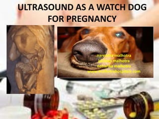
ULTRASOUND AS A WATCH DOG
      FOR PREGNANCY


                narendra malhotra
                 jaideep malhotra
                neharika malhotra
             www.malhotrahospitals.com
http://en.wikipedia.org/wiki/watch dog
 • Watchdog may refer to:
 Dog
 Guard dog,
   a dog that barks to alert its owners of
   an intruder's presence

 WIKTIONARY
 Noun
 watchdog (plural watchdogs)
 a guard dog
 a person or organization that monitors and publicizes the behavior of
 others (individuals, corporations, governments) to discover undesirable
 activity.
PREGNANCY IS THE OWNER

  AND THE “GREAT OBSTETRICAL SYNDROME” IS THE
                   INTRUDER

       AND ULTRASOUND THE WATCH DOG

AND WE OBSTETRICIANS ARE THE DOG TRAINERS

so it becomes a very doggy-bitchy story and lecture
The Challenge of
   Obstetrics
Obstetrical Disease

          •      Preterm labor

          •      Preterm Rupture of membranes

          •      Pre-eclampsia

          •      SGA/IUGR

          •      Fetal Death

In addition to the above ;first trimester preg failure,early anomalies ,late anomalies
The History of Obstetrics

• A search for a single test to predict each
  obstetrical disorder has failed.

• The discovery of an effective treatment
  and preventive strategy has not been
  successful.
Diagnostic tools
Only one single
diagnostic tool has
proven to be the only
tool which can predict
problems and watch the
pregnancy like a watch
dog and indicate the
intruders of the “great
obstetrical syndrome”
Treatments available today

   Disease                          Treatment

Preterm labor                       Tocolysis

                                  Expectant
Preterm PROM
                                 management
                              Antihypertensive
Pre-eclampsia
                                   agents

     IUGR                            Delivery
FAILED PREGNANCY AND LETHAL ANOMALIES……TERMINATION
“Great Obstetrical Syndromes”

•   Multiple etiologies
•   Long pre-clinical phase
•   Fetal diseases
•   Clinical manifestations are adaptive
•   Symptomatic treatment is ineffective
•   Genetic/environmental factors
Small for Gestational Age
         Environmental     Infection/
                         Inflammation


  Genetic                          Endocrine


                                         Maternal
Nutritional


Placental                               Unknown
Umbilical
vessels      Chorionic     Chorionic
              vessels        plate                Amnion


                                           Placental
 Uteroplacental    Basal   Spiral           septum
     veins         plate   artery


                             Sadler TW Lagman’s Medical Embryology 1990
OBSTETRICAL ULTRASOUND HELPS
PICK UP ALL THESE PROBLEMS EARLY
• EARLY SCAN TO DETECT PREGNANCY AND
  RULE OUT ECTOPIC
• FETAL CARDIAC ACTIVITY/VIABILITY SCAN
• CHORIONICITY IN MULTIPLE GESTATION
• 11-14 WEEKS GENETIC SCAN
• 20 WEEKS ANOMALY SCAN
• 24 WEEKS DOPPLER
• THIRD TRIMESTER GROWTH AND LIQUOR
Definite signs of Early Pregnancy
                     Failure
• Absence of cardiac activity in an embryo
               -Embryonic demise
• Absence of yolk sac/embryo in a large GS
               -Blighted ovum


            FAILED PREGNANCY
Definite signs of Early Pregnancy
                  Failure
What is the descriminatory size for safe diagnosis?

      Mean Sac diameter
      CRL
GUIDELINES FOR DIAGNOSIS
           OF EARLY PREGNANCY FAILURE
                           Society of                      American College
Royal College of           Obstetricians and               of Radiologists
Obstetricians              Gynaecologists of               (ACR) 2000
and                        Canada (SOGC) 2005
Gynaecologists                                             • CRL > 5mm with no
                           • CRL > 5mm with no visible     visible cardiac activity
(RCOG) 2006                cardiac activity, >9mm(TAS)
                                                           • MSD > 16mm without a
• CRL ≥ 6mm with no        • MSD > 8mm without a visible   visible embryo or yolk sac
visible cardiac activity   yolk sac, 20mm (TAS)

• MSD ≥ 20mm without
                                                           AIUM, 2007
                           • MSD > 16mm without a          • CRL > 5mm (TVS) with no
a visible embryo or        visible embryo, (25mm (TAS)
yolk sac                                                   visible cardiac activity

                              LEVEL 11-2 a
GUIDELINES FOR DIAGNOSIS
          OF EARLY PREGNANCY FAILURE
                             Australian Society            Practice in the
Hongkong College                                           Philippines
of Obstetricians             for Ulltrasound in
and Gynaecologists           Medicine (ASUM)
(HKCOG) 2004
                                                           • CRL > 5mm with no
• CRL > 5mm (TVS), >9mm      • CRL > 6mm with no
                                                           visible cardiac activity
(TAS) with no visible        visible cardiac activity
cardiac activity                                           • MSD > 18mm without a
                             • MSD > 20mm without a
                                                           visible embryo or yolk sac
• MSD ≥ 20mm without a       visible embryo or yolk sac
                                                            OB-GYN USG for practicing
visible embryo or yolk sac                                  Clinician 2nd Ed


         FOGSI GUIDELINES A FEW YEARS BACK
         MSD >20without YS/E :CRL >6mm without cardiac activity
         IFUMB/ICMU and ICOG
RECOMMENDATIONS


Empty GS = an MSD of 25 mm with out yolk sac
                                 or embryo

Embryonic demise= A CRL of 7mm with
                                 no cardiac activity

Wait for 7-10 days before a repeat scan if results are
            below the descriminatory level.
Down syndrome screening
•   NT (11-13+6wk), PAPP-A, beta-hCG
•   Best at 12 wk for anomaly as well
•   One-stop
•   90% sensitivity at 5% FP rate
•   Addition of Doppler assessment of blood
    flow in the ductus venosus and across the
    tricuspid valve together with above can
    identify more than 95% of all major
    aneuploidies for a FP rate of less than 3%.
Why 13+6 wk?
• To provide women with affected
  fetuses the option of first- rather
  than second-trimester
  termination,
• The incidence of abnormal
  accumulation of nuchal fluid in
  chromosomally abnormal
  fetuses decreases after 13 weeks
• The success rate for taking a
  measurement decreases after 13
  weeks because the fetus
  becomes vertical, making it
  more difficult to obtain the
  appropriate image.
Other aneuploidies
                        NT          Beta-hCG    PAPP-A

Trisomy 21              increased   increased   decreased

Trisomy 18              increased   decreased   decreased

Trisomy 13              increased   decreased   decreased

Turner syndrome         increased   normal      decreased

Tripoloidy (paternal)               increased   decreased mildy
Cardiac defect
• Major cardiac
  defect in 7.6% of
  chromosomally
  normal fetuses
  with NT>=3.5mm
• Indication for fetal
  echocardiography
• Detailed cardiac
  scan at 14 wk
OTHER MARKERS BY USG

 ICT


                        DV

                 NB
CORD DIAMETER
                             WIDE ILIAC BONES


                          FACIAL ANGLE
STRUCTURAL ANOMALIES
ULTRASOUND IS A GOOD WATCH DOG
 FOR FIRST TRIMESTER PREGNANCY
       PROBLEM PREDICTION
The mid-trimester fetal ultrasound scan

   Who should have one: everyone

              ……all pregnant women should
              be offered an ultrasound scan
              for the detection of fetal
              anomalies and pregnancy
              complications…….if problems
              in unselected low risk have
              also to be picked up….(no
              clear evidence on usefulness)
The mid-trimester fetal ultrasound scan

  When should the scan be performed?
• “18-22 weeks”
• Earlier scans date better
• Earlier scans require equipment, expertise and
  time
• Later scans see better
• Later scans see more
• Local legislation
The mid-trimester fetal ultrasound scan

  And now coming to the core stuff!

           •Fetal biometry
            and well being
           •Anatomical
            survey
The mid-trimester fetal ultrasound scan

    Fetal biometry and well being
   • Fetal biometry
   • Amniotic fluid assessment
   • Fetal movement
   • Doppler ultrasonography
   • Multiple gestation
Fetal biometry:parameters


• Biparietal diameter (BPD)
• Head circumference (HC)
• Abdominal circumference (AC)
• Femur (diaphysis) length (FL)
• Cerebellar transverse diameter
Fetal biometry

                  Parameters
• Standardised manner and strict quality criteria
• Audit of results
• Still images to document the measurements
Fetal well being

             Estimated fetal weight
• The     degree of deviation from normal at this
    early stage of pregnancy that would justify
    action (e.g. follow-up scan to assess fetal growth
    or fetal chromosomal analysis) has not been
    firmly established
•   if gestational age is determined at an earlier
    scan, EFW can be compared to dedicated
    normal, preferably local, reference ranges for
    this parameter
Fetal well being

                                                            Amniotic fluid assessment
• Amniotic                               fluid volume can be estimated
                             subjectively     or  by    using   sonographic
                             measurements
•                            Subjective estimation is not inferior to the
                             quantitative measurement techniques (e.g.
                             deepest pocket, amniotic fluid index) when
                       270
                       250
                             performed by experienced examiners
Amniotic Fluid Index


                       230
                       210
                       190
                       170
                       150
                                                                                                •   Patients with deviations from normal
                       130
                       110                                                                          should have more detailed anatomical
                        90
                        70                                                                          evaluation and clinical follow-up
                             16   18   20   22   24   26    28    30   32   34   36   38   40
                                                           Week
Fetal well being

                    Fetal movement
•   There are no specific movement
    patterns at this stage of pregnancy
•   Temporary absence or reduction of
    fetal movements during the scan
    should not be considered as a risk
•   Abnormal positioning or unusually
    restricted or persistently absent
    fetal movements may suggest
    abnormal fetal conditions such as
    arthrogryposis
Fetal well being

                Fetal movement
• The biophysical profile is not considered part of
    a routine mid-trimester scan!
•   Fetal brain is not yet mature enough to control
    sympathetic and parasympathetic of fetal heart!
Fetal well being
            Doppler ultrasonography
• The      application   of     Doppler
    techniques     is  not     currently
    recommended as part of the routine
    second-trimester         ultrasound
    examination
•   There is insufficient evidence to
    support universal use of uterine or
    umbilical artery Doppler evaluation
    for the screening of low-risk
    pregnancies
Fetal well being

                   Multiple gestations
•   visualization of the placental cord
    insertion
•   distinguishing features (gender,
    unique markers, position in
    uterus)
•   determination of chorionicity is
    sometimes feasible in the second
    trimester if there are clearly two
    separate placental masses and
    discordant genders. Chorionicity
    is much better evaluated before
    14–15 weeks (lambda sign or T-
    sign).
The anatomical survey in second trimester

                                      At a glance
Head Intact cranium                             Abdomen
           Cavum septi pellucidi                     Stomach in normal position
           Midline falx                                    Bowel not dilated
           Thalami                                         Both kidneys present
           Cerebral ventricles                             Cord insertion site
           Cerebellum                           Skeletal
           Cisterna magna                            No spinal defects or masses (transverse and
Face Both orbits present                        sagittal)
           Median facial profile                           Arms and hands present, normal
           Mouth present                        relationships
           Upper lip intact                                Legs and feet present, normal relationships
Neck Absence of masses (e.g. cystic hygroma)    Placenta
                                                           Position
Chest/Heart                                          No masses present
    Normal shape/size of chest and lungs                   Accessory lobe
         Heart activity present                 Umbilical cord
         Four-chamber view of heart in normal        Three-vessel cord
position                                        Genitalia
         Aortic and pulmonary outflow tracts         Male or female
         No evidence of diaphragmatic hernia
Placenta
      Guidelines for maturity and position
                                                                                    +
                                                                           +
                                         +
          +                                  +
           +


•   Women with a history of uterine surgery and low anterior placenta or placenta previa are
    at risk for placental attachment disorders. In these cases, the placenta should be examined
    for findings of accreta, the most sensitive of which are the presence of multiple irregular
    placental lacunae that show arterial or mixed flow
•    Abnormal appearance of the uterine wall–bladder wall interface is quite specific for
    accreta, but is seen in few cases. Loss of the echolucent space between an anterior
    placenta and the uterine wall is neither a sensitive nor a specific marker for placenta
    accreta
Maternal anatomy

                         Guidelines
•   Currently, there is sufficient
    evidence to recommend
    routine cervical length
    measurements        with     a
    transvaginal scan at the mid
    trimester even in an
    unselected population
•   Uterine      fibroids     and
    adnexal masses should be
    documented
THIRD TRIMESTER SCAN
    Great obstetrical syndrome

•      Preterm labor

•      Preterm
       Rupture of
       membranes

•      Pre-eclampsia

•      SGA/IUGR

•      Fetal Death


              Fetal growth restriction
FGR may be
     Symmetrical                      Asymmetrical
                 and body
Fetal brain (BPD & HC)         Fetal brain (BPD & HC) and
(AC) and long bones are        long bones are large when
proportionately small.         compared to the AC (liver).

may occur when the fetus       may occur when the fetus
experiences a problem during   experiences a problem during
early development.             later development

Chromosomal malformation
Constituently Small (Small     Hypoxemic hypoxia
Mother Small Baby )            utero placental insufficiency
Accurate fetal biometry
     to measure the size of
     fetus – AC & EFW


Predictive tools rule
        of 2
Accurate fetal biometry
  to measure the size of
  fetus – AC & EFW

                            Accurate measurement
                            of fetal growth - Indian
Predictive tools            &      customized
   rule of 2                fetal growth charts
Accurate fetal biometry
     to measure the size of
     fetus – AC & EFW

                               Accurate measurement
                               of fetal growth - Indian
Predictive tools rule          & customized         fetal
        of 2                   growth charts


          Accurate knowledge of fetal
          physiology and intrauterine
          environment -               fetal
          Doppler study & AFI ACHARYA
                         DR.PRASHANT
• Biometric tests (tests to measure size)

     • Biometric tests are designed to
         predict size and growth
                AC, EFW
USG TOOLS – HOW EFFECTIVE ?

• Ratio measures, such as head to
  abdominal circumference (HC/AC)
  and femoral length to abdominal
  circumference (FL/AC) ratios are
  poorer than AC or EFW alone in
  predicting IUGR
Ask for serial measurements
           and plot the findings in
           growth chart –       not
           single USG reading


2/3/2012              DR.PRASHANT ACHARYA   47
2/3/2012   DR.PRASHANT ACHARYA   48
PROPOSED INDIAN CUSTOMISED GROWTH CHART


• The term 40 weeks birth       • The term 40 weeks birth
  weight of fetus is 3051 for     weight of fetus is 3455 for
  normal primi patient            normal primi patient
  having average weight 52        having average weight 64
  Kgs & average height of         Kgs & average height of
  152 cms                         163 cms
Role of Doppler study in FGR
• In diagnosing fetal
  hypoxia by detecting
  redistribution of
  fetal blood flow
• Helps in deciding
  the Timing of        Doppler detects flow of RBCs in any
  delivery             vessel - Quantity and speed


• Measuring AFI
Uterine artery Doppler waveform


• Impaired placentation


• P I P I P (OBS SYNDROME)

50-67% Positive predictive value

>95 % negative predictive value
Impaired placentation
Uterine artery
   Doppler
                                                             Good
                                                         diastolic flow
                      ď‚žHigh resistance ,
                      persistence of diastolic
                      notching and even absent end
                      systolic forward flow can
                      persist through out pregnancy
                      if PLACENTATION IS
                      INADEQUATE
                      ď‚žIn 50-67 % is associated       Early
                                                      diastolic
                                                      notch               Poor diastolic
                      with complication of                                    flow

                      P I P I P &PPIH


DR.PRASHANT ACHARYA                    2/3/2012                               52
Prediction & Prevention of
FGR by

   UTERINE ARTERY DOPPLER
 P I P I P GREAT OBSTETRICAL SYNDROME

    Preeclampsia
    IUD
    Prematurity
    IUGR
    Placental abruption
UMBILICAL    ARTERY     FLOW
characteristic saw-tooth appearance of arterial flow in
one direction and continuous umbilical venous blood flow
in the other.


                                                      54
Absent / Reversed end diastolic volume
                        (AEDV/REDV)
• AEDV/REDV + PREMATURITY = high chances of HMD
• neonatal complications are Asphyxia ,ICH are increased


 2/3/2012                 DR.PRASHANT ACHARYA
When Umbilical artery Doppler
     parameters are altered


Multi vessel Doppler
                             Hypoxia and
examination (MCA and         re
DV)                          distributation


Bio Physical Profile Score   CNS
Umbilical artery Doppler
• When end diastolic
  flow is present ,
  delay delivery until
  at least 35 weeks,
  provided other
  surveillance
  findings are
  normal.
MCA abnormality expressed by the
              compromised fetus
Brain sparing effect –                                  Fetal anemia
Cerebral perfusion
increased ( RI & PI
decreased)


     MCA supplies blood to Brain - the most important
     organ of body-

     MCA PSV evaluates speed of fetal RBCs flow which
     has  DIRECT application in FETAL ANEMIA
Middle cerebral artery
   An early stage in fetal
  adaptation to hypoxemia - central
  redistribution of blood flow
 ( brain-sparing reflex)

increased blood flow to protect the
  brain, heart, and adrenals

reduced flow to the peripheral and
  placental circulations
FETAL AORTA


 Aortic Isthmus

Descending aorta
• The aortic isthmus PI is increased and
     absolute velocities (especially the TAMXV)
     are reduced in intrauterine growth-restricted
     fetuses


Aortic isthmus blood flow velocimetry provides important information on fetal cardiovascular
function, i.e. individual performance of ventricles, relative changes in upper (including brain)
and lower (including placenta) body resistances and fetal oxygenation, and has the potential
to become a valuable clinical tool for fetal evaluation
FETAL DECENDING AORTA
• it is important to be aware of the fact that
  the brief reversal of flow during end-systole,
  which is a normal finding in the third
  trimester, can give falsely high PI values
DUCTUS VENOSUS (DV)
    INFERIOR VENA CAVA       FORAMEN OVALE

RIGHT HEPATIC VEIN
  MIDDLE HEPATIC VEIN        LEFT HEPATIC VEIN
                         DUCTUS VENOSUS
PORTAL VEIN


                                   UMBILICAL VEIN
RA

                  RV


                   HV
             DV


        RA

                  RV


                   HV
             DV


Growth Retardation
Ductus venosus (DV)
                      Final
                      verdict


•   Sensitive to fetal oxygenation status
•   Dilates as fetal hypoxia worsens
•   In severe hypoxia –reversal of (a) wave
•   Immediate delivery of Fetus- in abnormal
    DV flow
• Reflects VOLUME
                    MANAGEMENT by RIGHT
                    atrium and is responsive to
                    fetal oxygenation
                  • Dilates as fetal hypoxia
                    worsens
                  • In severe hypoxia –reversal
                    of (a) wave ,due to atrial
                    contraction s/o cardiac
                    failure and decompansation
                    due to increase in severe
                    after load
                  • Needs immediate delivery of
                    Fetus


2/3/2012   DR.PRASHANT ACHARYA                66
Amniotic Fluid Index
• Reduction in AFI is Supporting evidence of a
  hostile intrauterine environment

• Amniotic fluid volume monitoring is very
  helpful in monitoring the physiological status
  of the fetus rather than the anatomical
  growth.
BPPS or Modified BPPS ??

• VAST
• AFI
• Instant, Easy and cost
  effective
• Helps in delivering the fetus at
  optimal gestational age
3D 4 D AS WATCH DOGS
Take Home Message
• Worldwide,   it is likely that much of the
  ultrasonography currently performed is carried
  out by individuals with little or no formal
  training(hence misinterpretations)
• Performed       with      proper    guidelines
  ROUTINE USG IN PREGNANCY                   can
  predict many problems and be a good watch dog
  for fetal and maternal wellbeing

More Related Content

What's hot

Methods of pregnancy diagnosis in sheep and goat
Methods of pregnancy diagnosis in sheep and goatMethods of pregnancy diagnosis in sheep and goat
Methods of pregnancy diagnosis in sheep and goatSulake Fadhil
 
First trimester ultrasound Dr. Muhammad Bin Zulfiqar
First trimester ultrasound Dr. Muhammad Bin ZulfiqarFirst trimester ultrasound Dr. Muhammad Bin Zulfiqar
First trimester ultrasound Dr. Muhammad Bin ZulfiqarDr. Muhammad Bin Zulfiqar
 
Umblical & uterine artery Doppler
Umblical & uterine artery DopplerUmblical & uterine artery Doppler
Umblical & uterine artery DopplerAboubakr Elnashar
 
Pregnancy markers for early pregnancy diagnosis
Pregnancy  markers for early pregnancy diagnosisPregnancy  markers for early pregnancy diagnosis
Pregnancy markers for early pregnancy diagnosisVarij Nayan
 
Intrauterine growth restriction
Intrauterine growth restrictionIntrauterine growth restriction
Intrauterine growth restrictionBharti Gahtori
 
Current approach for pregnancy diagnosis in animals
Current approach for pregnancy diagnosis in animalsCurrent approach for pregnancy diagnosis in animals
Current approach for pregnancy diagnosis in animalsGangaram Chaudhary
 
Multifetal pregnancy fetal reduction
Multifetal  pregnancy fetal reductionMultifetal  pregnancy fetal reduction
Multifetal pregnancy fetal reductionSantosh Gupta
 
Obstetrical Ultrasound
Obstetrical UltrasoundObstetrical Ultrasound
Obstetrical UltrasoundLa Lura White
 
Basics of early pregnancy scan.pptx
Basics of early pregnancy scan.pptxBasics of early pregnancy scan.pptx
Basics of early pregnancy scan.pptxPoonam Loomba
 
Ultrasonography of the ovary
Ultrasonography of the ovaryUltrasonography of the ovary
Ultrasonography of the ovaryAboubakr Elnashar
 
Aneuploidy screening Aneuploidy screening
Aneuploidy screening  Aneuploidy screening Aneuploidy screening  Aneuploidy screening
Aneuploidy screening Aneuploidy screening Dr Praman Kushwah
 
BASIC OBSTETRIC ULTRASOUND TRAINING
BASIC OBSTETRIC ULTRASOUND TRAININGBASIC OBSTETRIC ULTRASOUND TRAINING
BASIC OBSTETRIC ULTRASOUND TRAININGStephenVictorCobbina1
 
Practical approaches for treatment of Repeat Breeding Syndrome in Dairy cattle
Practical approaches for treatment of Repeat Breeding Syndrome in Dairy cattlePractical approaches for treatment of Repeat Breeding Syndrome in Dairy cattle
Practical approaches for treatment of Repeat Breeding Syndrome in Dairy cattleAMOLYEDE
 
Imaging in obstetrics & gynaecology (part 1- Gynaecological scans in benign c...
Imaging in obstetrics & gynaecology (part 1- Gynaecological scans in benign c...Imaging in obstetrics & gynaecology (part 1- Gynaecological scans in benign c...
Imaging in obstetrics & gynaecology (part 1- Gynaecological scans in benign c...drmcbansal
 
First trimester scan
First trimester scanFirst trimester scan
First trimester scanArchana Tandon
 
Usg in third trimester, placenta and amniotic fluid
Usg in third trimester, placenta and amniotic fluidUsg in third trimester, placenta and amniotic fluid
Usg in third trimester, placenta and amniotic fluidDouble M
 
NT SCAN (11-13.6 weeks) & Serum BIOPROFILE
NT SCAN (11-13.6 weeks) & Serum BIOPROFILENT SCAN (11-13.6 weeks) & Serum BIOPROFILE
NT SCAN (11-13.6 weeks) & Serum BIOPROFILEDrNisheethOza
 

What's hot (20)

Methods of pregnancy diagnosis in sheep and goat
Methods of pregnancy diagnosis in sheep and goatMethods of pregnancy diagnosis in sheep and goat
Methods of pregnancy diagnosis in sheep and goat
 
First trimester ultrasound Dr. Muhammad Bin Zulfiqar
First trimester ultrasound Dr. Muhammad Bin ZulfiqarFirst trimester ultrasound Dr. Muhammad Bin Zulfiqar
First trimester ultrasound Dr. Muhammad Bin Zulfiqar
 
Genetic sonogram
Genetic sonogramGenetic sonogram
Genetic sonogram
 
Umblical & uterine artery Doppler
Umblical & uterine artery DopplerUmblical & uterine artery Doppler
Umblical & uterine artery Doppler
 
Pregnancy test in cow
Pregnancy test in cowPregnancy test in cow
Pregnancy test in cow
 
Pregnancy markers for early pregnancy diagnosis
Pregnancy  markers for early pregnancy diagnosisPregnancy  markers for early pregnancy diagnosis
Pregnancy markers for early pregnancy diagnosis
 
Intrauterine growth restriction
Intrauterine growth restrictionIntrauterine growth restriction
Intrauterine growth restriction
 
Current approach for pregnancy diagnosis in animals
Current approach for pregnancy diagnosis in animalsCurrent approach for pregnancy diagnosis in animals
Current approach for pregnancy diagnosis in animals
 
Nuchal translucency
Nuchal translucencyNuchal translucency
Nuchal translucency
 
Multifetal pregnancy fetal reduction
Multifetal  pregnancy fetal reductionMultifetal  pregnancy fetal reduction
Multifetal pregnancy fetal reduction
 
Obstetrical Ultrasound
Obstetrical UltrasoundObstetrical Ultrasound
Obstetrical Ultrasound
 
Basics of early pregnancy scan.pptx
Basics of early pregnancy scan.pptxBasics of early pregnancy scan.pptx
Basics of early pregnancy scan.pptx
 
Ultrasonography of the ovary
Ultrasonography of the ovaryUltrasonography of the ovary
Ultrasonography of the ovary
 
Aneuploidy screening Aneuploidy screening
Aneuploidy screening  Aneuploidy screening Aneuploidy screening  Aneuploidy screening
Aneuploidy screening Aneuploidy screening
 
BASIC OBSTETRIC ULTRASOUND TRAINING
BASIC OBSTETRIC ULTRASOUND TRAININGBASIC OBSTETRIC ULTRASOUND TRAINING
BASIC OBSTETRIC ULTRASOUND TRAINING
 
Practical approaches for treatment of Repeat Breeding Syndrome in Dairy cattle
Practical approaches for treatment of Repeat Breeding Syndrome in Dairy cattlePractical approaches for treatment of Repeat Breeding Syndrome in Dairy cattle
Practical approaches for treatment of Repeat Breeding Syndrome in Dairy cattle
 
Imaging in obstetrics & gynaecology (part 1- Gynaecological scans in benign c...
Imaging in obstetrics & gynaecology (part 1- Gynaecological scans in benign c...Imaging in obstetrics & gynaecology (part 1- Gynaecological scans in benign c...
Imaging in obstetrics & gynaecology (part 1- Gynaecological scans in benign c...
 
First trimester scan
First trimester scanFirst trimester scan
First trimester scan
 
Usg in third trimester, placenta and amniotic fluid
Usg in third trimester, placenta and amniotic fluidUsg in third trimester, placenta and amniotic fluid
Usg in third trimester, placenta and amniotic fluid
 
NT SCAN (11-13.6 weeks) & Serum BIOPROFILE
NT SCAN (11-13.6 weeks) & Serum BIOPROFILENT SCAN (11-13.6 weeks) & Serum BIOPROFILE
NT SCAN (11-13.6 weeks) & Serum BIOPROFILE
 

Similar to USG WATCH DOG IN PREGNANCY

Prediction of early pregnancy outcomes.
Prediction of early pregnancy outcomes.Prediction of early pregnancy outcomes.
Prediction of early pregnancy outcomes.NARENDRA MALHOTRA
 
Abnormal first trimester scan
Abnormal first trimester scanAbnormal first trimester scan
Abnormal first trimester scanMahmoud Abdel-Aleem
 
18 - 22 Weeks Anomaly Scan
18 - 22 Weeks Anomaly Scan18 - 22 Weeks Anomaly Scan
18 - 22 Weeks Anomaly ScanDrNisheethOza
 
Ultrasonography Scans in Pregnancy
Ultrasonography Scans in PregnancyUltrasonography Scans in Pregnancy
Ultrasonography Scans in PregnancyDrNisheethOza
 
Abnormal first trimester scan
Abnormal first trimester scanAbnormal first trimester scan
Abnormal first trimester scanMahmoud Abdel-Aleem
 
Recurrent Pregnancy Loss Sharing Personal Experience (10 years)
Recurrent Pregnancy Loss Sharing Personal Experience (10 years)    Recurrent Pregnancy Loss Sharing Personal Experience (10 years)
Recurrent Pregnancy Loss Sharing Personal Experience (10 years) Lifecare Centre
 
First trimester ultrasound
First trimester ultrasoundFirst trimester ultrasound
First trimester ultrasoundRoshan Valentine
 
First trimester ultrasound
First trimester ultrasoundFirst trimester ultrasound
First trimester ultrasoundobsgynhsnz
 
Common Diseases In Well Baby Clinic .pptx
Common Diseases In Well Baby Clinic .pptxCommon Diseases In Well Baby Clinic .pptx
Common Diseases In Well Baby Clinic .pptxAbdulaziz Bagasi
 
assement of fetal well being
assement of fetal well beingassement of fetal well being
assement of fetal well beingpangebaaz
 
Sridhar prenatal diagnosis
Sridhar prenatal diagnosisSridhar prenatal diagnosis
Sridhar prenatal diagnosisSridhar Mulaka
 
Screening of high risk pregnancy
Screening of high risk pregnancyScreening of high risk pregnancy
Screening of high risk pregnancySanthosh Antony
 
Cervical Screening and Colposcopy Update April 2019
Cervical Screening and Colposcopy Update April 2019Cervical Screening and Colposcopy Update April 2019
Cervical Screening and Colposcopy Update April 2019Kervindran Mohanasundaram
 
Common algorithm of the management of Infertility
Common algorithm of the management of InfertilityCommon algorithm of the management of Infertility
Common algorithm of the management of InfertilityRupal Shah
 
ultrasound in 1st TM.pdf
ultrasound in 1st TM.pdfultrasound in 1st TM.pdf
ultrasound in 1st TM.pdfRabbirraaJiisaa
 
antepartum fetal surveillance - raw.pptx
antepartum fetal surveillance -  raw.pptxantepartum fetal surveillance -  raw.pptx
antepartum fetal surveillance - raw.pptxKLVK19
 
Adnexal mass in pregnancy
Adnexal mass in pregnancyAdnexal mass in pregnancy
Adnexal mass in pregnancyNiranjan Chavan
 
e1bf47c4-0d1b-4286-ba6e3d3c098ace6d.pdf
e1bf47c4-0d1b-4286-ba6e3d3c098ace6d.pdfe1bf47c4-0d1b-4286-ba6e3d3c098ace6d.pdf
e1bf47c4-0d1b-4286-ba6e3d3c098ace6d.pdfDany443291
 
Intra uterine growth restriction
Intra uterine growth restrictionIntra uterine growth restriction
Intra uterine growth restrictionDr. Shantala Vadeyar
 

Similar to USG WATCH DOG IN PREGNANCY (20)

Prediction of early pregnancy outcomes.
Prediction of early pregnancy outcomes.Prediction of early pregnancy outcomes.
Prediction of early pregnancy outcomes.
 
Abnormal first trimester scan
Abnormal first trimester scanAbnormal first trimester scan
Abnormal first trimester scan
 
18 - 22 Weeks Anomaly Scan
18 - 22 Weeks Anomaly Scan18 - 22 Weeks Anomaly Scan
18 - 22 Weeks Anomaly Scan
 
Ultrasonography Scans in Pregnancy
Ultrasonography Scans in PregnancyUltrasonography Scans in Pregnancy
Ultrasonography Scans in Pregnancy
 
Abnormal first trimester scan
Abnormal first trimester scanAbnormal first trimester scan
Abnormal first trimester scan
 
Recurrent Pregnancy Loss Sharing Personal Experience (10 years)
Recurrent Pregnancy Loss Sharing Personal Experience (10 years)    Recurrent Pregnancy Loss Sharing Personal Experience (10 years)
Recurrent Pregnancy Loss Sharing Personal Experience (10 years)
 
First trimester ultrasound
First trimester ultrasoundFirst trimester ultrasound
First trimester ultrasound
 
First trimester ultrasound
First trimester ultrasoundFirst trimester ultrasound
First trimester ultrasound
 
Common Diseases In Well Baby Clinic .pptx
Common Diseases In Well Baby Clinic .pptxCommon Diseases In Well Baby Clinic .pptx
Common Diseases In Well Baby Clinic .pptx
 
assement of fetal well being
assement of fetal well beingassement of fetal well being
assement of fetal well being
 
Sridhar prenatal diagnosis
Sridhar prenatal diagnosisSridhar prenatal diagnosis
Sridhar prenatal diagnosis
 
Screening of high risk pregnancy
Screening of high risk pregnancyScreening of high risk pregnancy
Screening of high risk pregnancy
 
Cervical Screening and Colposcopy Update April 2019
Cervical Screening and Colposcopy Update April 2019Cervical Screening and Colposcopy Update April 2019
Cervical Screening and Colposcopy Update April 2019
 
Common algorithm of the management of Infertility
Common algorithm of the management of InfertilityCommon algorithm of the management of Infertility
Common algorithm of the management of Infertility
 
ultrasound in 1st TM.pdf
ultrasound in 1st TM.pdfultrasound in 1st TM.pdf
ultrasound in 1st TM.pdf
 
antepartum fetal surveillance - raw.pptx
antepartum fetal surveillance -  raw.pptxantepartum fetal surveillance -  raw.pptx
antepartum fetal surveillance - raw.pptx
 
Every fetus at risk
Every fetus at riskEvery fetus at risk
Every fetus at risk
 
Adnexal mass in pregnancy
Adnexal mass in pregnancyAdnexal mass in pregnancy
Adnexal mass in pregnancy
 
e1bf47c4-0d1b-4286-ba6e3d3c098ace6d.pdf
e1bf47c4-0d1b-4286-ba6e3d3c098ace6d.pdfe1bf47c4-0d1b-4286-ba6e3d3c098ace6d.pdf
e1bf47c4-0d1b-4286-ba6e3d3c098ace6d.pdf
 
Intra uterine growth restriction
Intra uterine growth restrictionIntra uterine growth restriction
Intra uterine growth restriction
 

More from NARENDRA MALHOTRA

12_Prenatal_diagnotic_tests.pdf
12_Prenatal_diagnotic_tests.pdf12_Prenatal_diagnotic_tests.pdf
12_Prenatal_diagnotic_tests.pdfNARENDRA MALHOTRA
 
FETAL GROWTH ASSESSMENT CONCEPT OF F.G.R. & PLOTTING GROWTH CHARTS
FETAL GROWTH ASSESSMENT CONCEPT OF F.G.R. & PLOTTING GROWTH CHARTSFETAL GROWTH ASSESSMENT CONCEPT OF F.G.R. & PLOTTING GROWTH CHARTS
FETAL GROWTH ASSESSMENT CONCEPT OF F.G.R. & PLOTTING GROWTH CHARTSNARENDRA MALHOTRA
 
Adnexal Masses in Reproductive Age
Adnexal Masses in Reproductive AgeAdnexal Masses in Reproductive Age
Adnexal Masses in Reproductive AgeNARENDRA MALHOTRA
 
FOGSI'S ACHIEVER COUPLE (2) (1).pdf
FOGSI'S ACHIEVER COUPLE (2) (1).pdfFOGSI'S ACHIEVER COUPLE (2) (1).pdf
FOGSI'S ACHIEVER COUPLE (2) (1).pdfNARENDRA MALHOTRA
 
Reviewing the burden of haemorrhoids in pregnancy
Reviewing the burden of haemorrhoids in pregnancyReviewing the burden of haemorrhoids in pregnancy
Reviewing the burden of haemorrhoids in pregnancyNARENDRA MALHOTRA
 
Fogsi uniform-consents
Fogsi uniform-consentsFogsi uniform-consents
Fogsi uniform-consentsNARENDRA MALHOTRA
 
Ujala Cygnus Rainbow Hospital Magazine 2021
Ujala Cygnus Rainbow Hospital Magazine 2021Ujala Cygnus Rainbow Hospital Magazine 2021
Ujala Cygnus Rainbow Hospital Magazine 2021NARENDRA MALHOTRA
 
Pocketbook do-dont-1.pdf
Pocketbook do-dont-1.pdfPocketbook do-dont-1.pdf
Pocketbook do-dont-1.pdfNARENDRA MALHOTRA
 
Ferrous ascorbate current clinical place in management of ida
Ferrous ascorbate current clinical place in management of idaFerrous ascorbate current clinical place in management of ida
Ferrous ascorbate current clinical place in management of idaNARENDRA MALHOTRA
 
MINDFUL DIGITAL PROGRAM
MINDFUL DIGITAL PROGRAMMINDFUL DIGITAL PROGRAM
MINDFUL DIGITAL PROGRAMNARENDRA MALHOTRA
 
Role of prenatal probiotics in preterm birth
Role of prenatal probiotics in preterm birthRole of prenatal probiotics in preterm birth
Role of prenatal probiotics in preterm birthNARENDRA MALHOTRA
 
3 D Ultrasound in reproductive medicine
3 D Ultrasound in reproductive medicine3 D Ultrasound in reproductive medicine
3 D Ultrasound in reproductive medicineNARENDRA MALHOTRA
 
3D-4D ULTRASOUND IN UTERINE SEPTUM EVALUATION
3D-4D ULTRASOUND  IN UTERINE SEPTUM EVALUATION3D-4D ULTRASOUND  IN UTERINE SEPTUM EVALUATION
3D-4D ULTRASOUND IN UTERINE SEPTUM EVALUATIONNARENDRA MALHOTRA
 
VACCINATE PREGNANT WOMEN & SAVE TWO LIVES
VACCINATE PREGNANT WOMEN & SAVE TWO LIVESVACCINATE PREGNANT WOMEN & SAVE TWO LIVES
VACCINATE PREGNANT WOMEN & SAVE TWO LIVESNARENDRA MALHOTRA
 
FOGSI POSITION STATEMENT COVID VACCINATION FOR PREGNANT & BREASTFEEDING WOMEN
FOGSI POSITION STATEMENT COVID VACCINATION FOR PREGNANT & BREASTFEEDING WOMENFOGSI POSITION STATEMENT COVID VACCINATION FOR PREGNANT & BREASTFEEDING WOMEN
FOGSI POSITION STATEMENT COVID VACCINATION FOR PREGNANT & BREASTFEEDING WOMENNARENDRA MALHOTRA
 
Rainbow insights magazine 2020
Rainbow insights magazine 2020Rainbow insights magazine 2020
Rainbow insights magazine 2020NARENDRA MALHOTRA
 

More from NARENDRA MALHOTRA (20)

12_Prenatal_diagnotic_tests.pdf
12_Prenatal_diagnotic_tests.pdf12_Prenatal_diagnotic_tests.pdf
12_Prenatal_diagnotic_tests.pdf
 
FETAL GROWTH ASSESSMENT CONCEPT OF F.G.R. & PLOTTING GROWTH CHARTS
FETAL GROWTH ASSESSMENT CONCEPT OF F.G.R. & PLOTTING GROWTH CHARTSFETAL GROWTH ASSESSMENT CONCEPT OF F.G.R. & PLOTTING GROWTH CHARTS
FETAL GROWTH ASSESSMENT CONCEPT OF F.G.R. & PLOTTING GROWTH CHARTS
 
Adnexal Masses in Reproductive Age
Adnexal Masses in Reproductive AgeAdnexal Masses in Reproductive Age
Adnexal Masses in Reproductive Age
 
FOGSI'S ACHIEVER COUPLE (2) (1).pdf
FOGSI'S ACHIEVER COUPLE (2) (1).pdfFOGSI'S ACHIEVER COUPLE (2) (1).pdf
FOGSI'S ACHIEVER COUPLE (2) (1).pdf
 
Jeevan nidhi magazine
Jeevan nidhi magazineJeevan nidhi magazine
Jeevan nidhi magazine
 
Reviewing the burden of haemorrhoids in pregnancy
Reviewing the burden of haemorrhoids in pregnancyReviewing the burden of haemorrhoids in pregnancy
Reviewing the burden of haemorrhoids in pregnancy
 
Fogsi uniform-consents
Fogsi uniform-consentsFogsi uniform-consents
Fogsi uniform-consents
 
Ujala Cygnus Rainbow Hospital Magazine 2021
Ujala Cygnus Rainbow Hospital Magazine 2021Ujala Cygnus Rainbow Hospital Magazine 2021
Ujala Cygnus Rainbow Hospital Magazine 2021
 
Pocketbook do-dont-1.pdf
Pocketbook do-dont-1.pdfPocketbook do-dont-1.pdf
Pocketbook do-dont-1.pdf
 
Ferrous ascorbate current clinical place in management of ida
Ferrous ascorbate current clinical place in management of idaFerrous ascorbate current clinical place in management of ida
Ferrous ascorbate current clinical place in management of ida
 
MINDFUL DIGITAL PROGRAM
MINDFUL DIGITAL PROGRAMMINDFUL DIGITAL PROGRAM
MINDFUL DIGITAL PROGRAM
 
Role of prenatal probiotics in preterm birth
Role of prenatal probiotics in preterm birthRole of prenatal probiotics in preterm birth
Role of prenatal probiotics in preterm birth
 
Adnexal Masses
Adnexal MassesAdnexal Masses
Adnexal Masses
 
3 D Ultrasound in reproductive medicine
3 D Ultrasound in reproductive medicine3 D Ultrasound in reproductive medicine
3 D Ultrasound in reproductive medicine
 
ENDOMETRIOSIS
ENDOMETRIOSISENDOMETRIOSIS
ENDOMETRIOSIS
 
3D-4D ULTRASOUND IN UTERINE SEPTUM EVALUATION
3D-4D ULTRASOUND  IN UTERINE SEPTUM EVALUATION3D-4D ULTRASOUND  IN UTERINE SEPTUM EVALUATION
3D-4D ULTRASOUND IN UTERINE SEPTUM EVALUATION
 
VACCINATE PREGNANT WOMEN & SAVE TWO LIVES
VACCINATE PREGNANT WOMEN & SAVE TWO LIVESVACCINATE PREGNANT WOMEN & SAVE TWO LIVES
VACCINATE PREGNANT WOMEN & SAVE TWO LIVES
 
FOGSI POSITION STATEMENT COVID VACCINATION FOR PREGNANT & BREASTFEEDING WOMEN
FOGSI POSITION STATEMENT COVID VACCINATION FOR PREGNANT & BREASTFEEDING WOMENFOGSI POSITION STATEMENT COVID VACCINATION FOR PREGNANT & BREASTFEEDING WOMEN
FOGSI POSITION STATEMENT COVID VACCINATION FOR PREGNANT & BREASTFEEDING WOMEN
 
Aub ieta -lucknow
Aub   ieta -lucknowAub   ieta -lucknow
Aub ieta -lucknow
 
Rainbow insights magazine 2020
Rainbow insights magazine 2020Rainbow insights magazine 2020
Rainbow insights magazine 2020
 

Recently uploaded

epilepsy and status epilepticus for undergraduate.pptx
epilepsy and status epilepticus  for undergraduate.pptxepilepsy and status epilepticus  for undergraduate.pptx
epilepsy and status epilepticus for undergraduate.pptxMohamed Rizk Khodair
 
PULMONARY EMBOLISM AND ITS MANAGEMENTS.pdf
PULMONARY EMBOLISM AND ITS MANAGEMENTS.pdfPULMONARY EMBOLISM AND ITS MANAGEMENTS.pdf
PULMONARY EMBOLISM AND ITS MANAGEMENTS.pdfDolisha Warbi
 
Hematology and Immunology - Leukocytes Functions
Hematology and Immunology - Leukocytes FunctionsHematology and Immunology - Leukocytes Functions
Hematology and Immunology - Leukocytes FunctionsMedicoseAcademics
 
LUNG TUMORS AND ITS CLASSIFICATIONS.pdf
LUNG TUMORS AND ITS  CLASSIFICATIONS.pdfLUNG TUMORS AND ITS  CLASSIFICATIONS.pdf
LUNG TUMORS AND ITS CLASSIFICATIONS.pdfDolisha Warbi
 
SYNDESMOTIC INJURY- ANATOMICAL REPAIR.pptx
SYNDESMOTIC INJURY- ANATOMICAL REPAIR.pptxSYNDESMOTIC INJURY- ANATOMICAL REPAIR.pptx
SYNDESMOTIC INJURY- ANATOMICAL REPAIR.pptxdrashraf369
 
call girls in green park DELHI 🔝 >༒9540349809 🔝 genuine Escort Service 🔝✔️✔️
call girls in green park  DELHI 🔝 >༒9540349809 🔝 genuine Escort Service 🔝✔️✔️call girls in green park  DELHI 🔝 >༒9540349809 🔝 genuine Escort Service 🔝✔️✔️
call girls in green park DELHI 🔝 >༒9540349809 🔝 genuine Escort Service 🔝✔️✔️saminamagar
 
call girls in aerocity DELHI 🔝 >༒9540349809 🔝 genuine Escort Service 🔝✔️✔️
call girls in aerocity DELHI 🔝 >༒9540349809 🔝 genuine Escort Service 🔝✔️✔️call girls in aerocity DELHI 🔝 >༒9540349809 🔝 genuine Escort Service 🔝✔️✔️
call girls in aerocity DELHI 🔝 >༒9540349809 🔝 genuine Escort Service 🔝✔️✔️saminamagar
 
Biomechanics- Shoulder Joint!!!!!!!!!!!!
Biomechanics- Shoulder Joint!!!!!!!!!!!!Biomechanics- Shoulder Joint!!!!!!!!!!!!
Biomechanics- Shoulder Joint!!!!!!!!!!!!ibtesaam huma
 
Informed Consent Empowering Healthcare Decision-Making.pptx
Informed Consent Empowering Healthcare Decision-Making.pptxInformed Consent Empowering Healthcare Decision-Making.pptx
Informed Consent Empowering Healthcare Decision-Making.pptxSasikiranMarri
 
POST NATAL EXERCISES AND ITS IMPACT.pptx
POST NATAL EXERCISES AND ITS IMPACT.pptxPOST NATAL EXERCISES AND ITS IMPACT.pptx
POST NATAL EXERCISES AND ITS IMPACT.pptxvirengeeta
 
world health day presentation ppt download
world health day presentation ppt downloadworld health day presentation ppt download
world health day presentation ppt downloadAnkitKumar311566
 
History and Development of Pharmacovigilence.pdf
History and Development of Pharmacovigilence.pdfHistory and Development of Pharmacovigilence.pdf
History and Development of Pharmacovigilence.pdfSasikiranMarri
 
VarSeq 2.6.0: Advancing Pharmacogenomics and Genomic Analysis
VarSeq 2.6.0: Advancing Pharmacogenomics and Genomic AnalysisVarSeq 2.6.0: Advancing Pharmacogenomics and Genomic Analysis
VarSeq 2.6.0: Advancing Pharmacogenomics and Genomic AnalysisGolden Helix
 
call girls in paharganj DELHI 🔝 >༒9540349809 🔝 genuine Escort Service 🔝✔️✔️
call girls in paharganj DELHI 🔝 >༒9540349809 🔝 genuine Escort Service 🔝✔️✔️call girls in paharganj DELHI 🔝 >༒9540349809 🔝 genuine Escort Service 🔝✔️✔️
call girls in paharganj DELHI 🔝 >༒9540349809 🔝 genuine Escort Service 🔝✔️✔️saminamagar
 
SWD (Short wave diathermy)- Physiotherapy.ppt
SWD (Short wave diathermy)- Physiotherapy.pptSWD (Short wave diathermy)- Physiotherapy.ppt
SWD (Short wave diathermy)- Physiotherapy.pptMumux Mirani
 
Wessex Health Partners Wessex Integrated Care, Population Health, Research & ...
Wessex Health Partners Wessex Integrated Care, Population Health, Research & ...Wessex Health Partners Wessex Integrated Care, Population Health, Research & ...
Wessex Health Partners Wessex Integrated Care, Population Health, Research & ...Wessex Health Partners
 
PULMONARY EDEMA AND ITS MANAGEMENT.pdf
PULMONARY EDEMA AND  ITS  MANAGEMENT.pdfPULMONARY EDEMA AND  ITS  MANAGEMENT.pdf
PULMONARY EDEMA AND ITS MANAGEMENT.pdfDolisha Warbi
 
Radiation Dosimetry Parameters and Isodose Curves.pptx
Radiation Dosimetry Parameters and Isodose Curves.pptxRadiation Dosimetry Parameters and Isodose Curves.pptx
Radiation Dosimetry Parameters and Isodose Curves.pptxDr. Dheeraj Kumar
 
Case Report Peripartum Cardiomyopathy.pptx
Case Report Peripartum Cardiomyopathy.pptxCase Report Peripartum Cardiomyopathy.pptx
Case Report Peripartum Cardiomyopathy.pptxNiranjan Chavan
 

Recently uploaded (20)

epilepsy and status epilepticus for undergraduate.pptx
epilepsy and status epilepticus  for undergraduate.pptxepilepsy and status epilepticus  for undergraduate.pptx
epilepsy and status epilepticus for undergraduate.pptx
 
PULMONARY EMBOLISM AND ITS MANAGEMENTS.pdf
PULMONARY EMBOLISM AND ITS MANAGEMENTS.pdfPULMONARY EMBOLISM AND ITS MANAGEMENTS.pdf
PULMONARY EMBOLISM AND ITS MANAGEMENTS.pdf
 
Hematology and Immunology - Leukocytes Functions
Hematology and Immunology - Leukocytes FunctionsHematology and Immunology - Leukocytes Functions
Hematology and Immunology - Leukocytes Functions
 
LUNG TUMORS AND ITS CLASSIFICATIONS.pdf
LUNG TUMORS AND ITS  CLASSIFICATIONS.pdfLUNG TUMORS AND ITS  CLASSIFICATIONS.pdf
LUNG TUMORS AND ITS CLASSIFICATIONS.pdf
 
Epilepsy
EpilepsyEpilepsy
Epilepsy
 
SYNDESMOTIC INJURY- ANATOMICAL REPAIR.pptx
SYNDESMOTIC INJURY- ANATOMICAL REPAIR.pptxSYNDESMOTIC INJURY- ANATOMICAL REPAIR.pptx
SYNDESMOTIC INJURY- ANATOMICAL REPAIR.pptx
 
call girls in green park DELHI 🔝 >༒9540349809 🔝 genuine Escort Service 🔝✔️✔️
call girls in green park  DELHI 🔝 >༒9540349809 🔝 genuine Escort Service 🔝✔️✔️call girls in green park  DELHI 🔝 >༒9540349809 🔝 genuine Escort Service 🔝✔️✔️
call girls in green park DELHI 🔝 >༒9540349809 🔝 genuine Escort Service 🔝✔️✔️
 
call girls in aerocity DELHI 🔝 >༒9540349809 🔝 genuine Escort Service 🔝✔️✔️
call girls in aerocity DELHI 🔝 >༒9540349809 🔝 genuine Escort Service 🔝✔️✔️call girls in aerocity DELHI 🔝 >༒9540349809 🔝 genuine Escort Service 🔝✔️✔️
call girls in aerocity DELHI 🔝 >༒9540349809 🔝 genuine Escort Service 🔝✔️✔️
 
Biomechanics- Shoulder Joint!!!!!!!!!!!!
Biomechanics- Shoulder Joint!!!!!!!!!!!!Biomechanics- Shoulder Joint!!!!!!!!!!!!
Biomechanics- Shoulder Joint!!!!!!!!!!!!
 
Informed Consent Empowering Healthcare Decision-Making.pptx
Informed Consent Empowering Healthcare Decision-Making.pptxInformed Consent Empowering Healthcare Decision-Making.pptx
Informed Consent Empowering Healthcare Decision-Making.pptx
 
POST NATAL EXERCISES AND ITS IMPACT.pptx
POST NATAL EXERCISES AND ITS IMPACT.pptxPOST NATAL EXERCISES AND ITS IMPACT.pptx
POST NATAL EXERCISES AND ITS IMPACT.pptx
 
world health day presentation ppt download
world health day presentation ppt downloadworld health day presentation ppt download
world health day presentation ppt download
 
History and Development of Pharmacovigilence.pdf
History and Development of Pharmacovigilence.pdfHistory and Development of Pharmacovigilence.pdf
History and Development of Pharmacovigilence.pdf
 
VarSeq 2.6.0: Advancing Pharmacogenomics and Genomic Analysis
VarSeq 2.6.0: Advancing Pharmacogenomics and Genomic AnalysisVarSeq 2.6.0: Advancing Pharmacogenomics and Genomic Analysis
VarSeq 2.6.0: Advancing Pharmacogenomics and Genomic Analysis
 
call girls in paharganj DELHI 🔝 >༒9540349809 🔝 genuine Escort Service 🔝✔️✔️
call girls in paharganj DELHI 🔝 >༒9540349809 🔝 genuine Escort Service 🔝✔️✔️call girls in paharganj DELHI 🔝 >༒9540349809 🔝 genuine Escort Service 🔝✔️✔️
call girls in paharganj DELHI 🔝 >༒9540349809 🔝 genuine Escort Service 🔝✔️✔️
 
SWD (Short wave diathermy)- Physiotherapy.ppt
SWD (Short wave diathermy)- Physiotherapy.pptSWD (Short wave diathermy)- Physiotherapy.ppt
SWD (Short wave diathermy)- Physiotherapy.ppt
 
Wessex Health Partners Wessex Integrated Care, Population Health, Research & ...
Wessex Health Partners Wessex Integrated Care, Population Health, Research & ...Wessex Health Partners Wessex Integrated Care, Population Health, Research & ...
Wessex Health Partners Wessex Integrated Care, Population Health, Research & ...
 
PULMONARY EDEMA AND ITS MANAGEMENT.pdf
PULMONARY EDEMA AND  ITS  MANAGEMENT.pdfPULMONARY EDEMA AND  ITS  MANAGEMENT.pdf
PULMONARY EDEMA AND ITS MANAGEMENT.pdf
 
Radiation Dosimetry Parameters and Isodose Curves.pptx
Radiation Dosimetry Parameters and Isodose Curves.pptxRadiation Dosimetry Parameters and Isodose Curves.pptx
Radiation Dosimetry Parameters and Isodose Curves.pptx
 
Case Report Peripartum Cardiomyopathy.pptx
Case Report Peripartum Cardiomyopathy.pptxCase Report Peripartum Cardiomyopathy.pptx
Case Report Peripartum Cardiomyopathy.pptx
 

USG WATCH DOG IN PREGNANCY

  • 1. ULTRASOUND AS A WATCH DOG FOR PREGNANCY narendra malhotra jaideep malhotra neharika malhotra www.malhotrahospitals.com
  • 2. http://en.wikipedia.org/wiki/watch dog • Watchdog may refer to: Dog Guard dog, a dog that barks to alert its owners of an intruder's presence WIKTIONARY Noun watchdog (plural watchdogs) a guard dog a person or organization that monitors and publicizes the behavior of others (individuals, corporations, governments) to discover undesirable activity.
  • 3. PREGNANCY IS THE OWNER AND THE “GREAT OBSTETRICAL SYNDROME” IS THE INTRUDER AND ULTRASOUND THE WATCH DOG AND WE OBSTETRICIANS ARE THE DOG TRAINERS so it becomes a very doggy-bitchy story and lecture
  • 4. The Challenge of Obstetrics
  • 5. Obstetrical Disease • Preterm labor • Preterm Rupture of membranes • Pre-eclampsia • SGA/IUGR • Fetal Death In addition to the above ;first trimester preg failure,early anomalies ,late anomalies
  • 6. The History of Obstetrics • A search for a single test to predict each obstetrical disorder has failed. • The discovery of an effective treatment and preventive strategy has not been successful.
  • 7. Diagnostic tools Only one single diagnostic tool has proven to be the only tool which can predict problems and watch the pregnancy like a watch dog and indicate the intruders of the “great obstetrical syndrome”
  • 8. Treatments available today Disease Treatment Preterm labor Tocolysis Expectant Preterm PROM management Antihypertensive Pre-eclampsia agents IUGR Delivery FAILED PREGNANCY AND LETHAL ANOMALIES……TERMINATION
  • 9. “Great Obstetrical Syndromes” • Multiple etiologies • Long pre-clinical phase • Fetal diseases • Clinical manifestations are adaptive • Symptomatic treatment is ineffective • Genetic/environmental factors
  • 10. Small for Gestational Age Environmental Infection/ Inflammation Genetic Endocrine Maternal Nutritional Placental Unknown
  • 11. Umbilical vessels Chorionic Chorionic vessels plate Amnion Placental Uteroplacental Basal Spiral septum veins plate artery Sadler TW Lagman’s Medical Embryology 1990
  • 12. OBSTETRICAL ULTRASOUND HELPS PICK UP ALL THESE PROBLEMS EARLY • EARLY SCAN TO DETECT PREGNANCY AND RULE OUT ECTOPIC • FETAL CARDIAC ACTIVITY/VIABILITY SCAN • CHORIONICITY IN MULTIPLE GESTATION • 11-14 WEEKS GENETIC SCAN • 20 WEEKS ANOMALY SCAN • 24 WEEKS DOPPLER • THIRD TRIMESTER GROWTH AND LIQUOR
  • 13. Definite signs of Early Pregnancy Failure • Absence of cardiac activity in an embryo -Embryonic demise • Absence of yolk sac/embryo in a large GS -Blighted ovum FAILED PREGNANCY
  • 14. Definite signs of Early Pregnancy Failure What is the descriminatory size for safe diagnosis? Mean Sac diameter CRL
  • 15. GUIDELINES FOR DIAGNOSIS OF EARLY PREGNANCY FAILURE Society of American College Royal College of Obstetricians and of Radiologists Obstetricians Gynaecologists of (ACR) 2000 and Canada (SOGC) 2005 Gynaecologists • CRL > 5mm with no • CRL > 5mm with no visible visible cardiac activity (RCOG) 2006 cardiac activity, >9mm(TAS) • MSD > 16mm without a • CRL ≥ 6mm with no • MSD > 8mm without a visible visible embryo or yolk sac visible cardiac activity yolk sac, 20mm (TAS) • MSD ≥ 20mm without AIUM, 2007 • MSD > 16mm without a • CRL > 5mm (TVS) with no a visible embryo or visible embryo, (25mm (TAS) yolk sac visible cardiac activity LEVEL 11-2 a
  • 16. GUIDELINES FOR DIAGNOSIS OF EARLY PREGNANCY FAILURE Australian Society Practice in the Hongkong College Philippines of Obstetricians for Ulltrasound in and Gynaecologists Medicine (ASUM) (HKCOG) 2004 • CRL > 5mm with no • CRL > 5mm (TVS), >9mm • CRL > 6mm with no visible cardiac activity (TAS) with no visible visible cardiac activity cardiac activity • MSD > 18mm without a • MSD > 20mm without a visible embryo or yolk sac • MSD ≥ 20mm without a visible embryo or yolk sac OB-GYN USG for practicing visible embryo or yolk sac Clinician 2nd Ed FOGSI GUIDELINES A FEW YEARS BACK MSD >20without YS/E :CRL >6mm without cardiac activity IFUMB/ICMU and ICOG
  • 17. RECOMMENDATIONS Empty GS = an MSD of 25 mm with out yolk sac or embryo Embryonic demise= A CRL of 7mm with no cardiac activity Wait for 7-10 days before a repeat scan if results are below the descriminatory level.
  • 18. Down syndrome screening • NT (11-13+6wk), PAPP-A, beta-hCG • Best at 12 wk for anomaly as well • One-stop • 90% sensitivity at 5% FP rate • Addition of Doppler assessment of blood flow in the ductus venosus and across the tricuspid valve together with above can identify more than 95% of all major aneuploidies for a FP rate of less than 3%.
  • 19. Why 13+6 wk? • To provide women with affected fetuses the option of first- rather than second-trimester termination, • The incidence of abnormal accumulation of nuchal fluid in chromosomally abnormal fetuses decreases after 13 weeks • The success rate for taking a measurement decreases after 13 weeks because the fetus becomes vertical, making it more difficult to obtain the appropriate image.
  • 20. Other aneuploidies NT Beta-hCG PAPP-A Trisomy 21 increased increased decreased Trisomy 18 increased decreased decreased Trisomy 13 increased decreased decreased Turner syndrome increased normal decreased Tripoloidy (paternal) increased decreased mildy
  • 21. Cardiac defect • Major cardiac defect in 7.6% of chromosomally normal fetuses with NT>=3.5mm • Indication for fetal echocardiography • Detailed cardiac scan at 14 wk
  • 22. OTHER MARKERS BY USG ICT DV NB CORD DIAMETER WIDE ILIAC BONES FACIAL ANGLE
  • 24. ULTRASOUND IS A GOOD WATCH DOG FOR FIRST TRIMESTER PREGNANCY PROBLEM PREDICTION
  • 25. The mid-trimester fetal ultrasound scan Who should have one: everyone ……all pregnant women should be offered an ultrasound scan for the detection of fetal anomalies and pregnancy complications…….if problems in unselected low risk have also to be picked up….(no clear evidence on usefulness)
  • 26. The mid-trimester fetal ultrasound scan When should the scan be performed? • “18-22 weeks” • Earlier scans date better • Earlier scans require equipment, expertise and time • Later scans see better • Later scans see more • Local legislation
  • 27. The mid-trimester fetal ultrasound scan And now coming to the core stuff! •Fetal biometry and well being •Anatomical survey
  • 28. The mid-trimester fetal ultrasound scan Fetal biometry and well being • Fetal biometry • Amniotic fluid assessment • Fetal movement • Doppler ultrasonography • Multiple gestation
  • 29. Fetal biometry:parameters • Biparietal diameter (BPD) • Head circumference (HC) • Abdominal circumference (AC) • Femur (diaphysis) length (FL) • Cerebellar transverse diameter
  • 30. Fetal biometry Parameters • Standardised manner and strict quality criteria • Audit of results • Still images to document the measurements
  • 31. Fetal well being Estimated fetal weight • The degree of deviation from normal at this early stage of pregnancy that would justify action (e.g. follow-up scan to assess fetal growth or fetal chromosomal analysis) has not been firmly established • if gestational age is determined at an earlier scan, EFW can be compared to dedicated normal, preferably local, reference ranges for this parameter
  • 32. Fetal well being Amniotic fluid assessment • Amniotic fluid volume can be estimated subjectively or by using sonographic measurements • Subjective estimation is not inferior to the quantitative measurement techniques (e.g. deepest pocket, amniotic fluid index) when 270 250 performed by experienced examiners Amniotic Fluid Index 230 210 190 170 150 • Patients with deviations from normal 130 110 should have more detailed anatomical 90 70 evaluation and clinical follow-up 16 18 20 22 24 26 28 30 32 34 36 38 40 Week
  • 33. Fetal well being Fetal movement • There are no specific movement patterns at this stage of pregnancy • Temporary absence or reduction of fetal movements during the scan should not be considered as a risk • Abnormal positioning or unusually restricted or persistently absent fetal movements may suggest abnormal fetal conditions such as arthrogryposis
  • 34. Fetal well being Fetal movement • The biophysical profile is not considered part of a routine mid-trimester scan! • Fetal brain is not yet mature enough to control sympathetic and parasympathetic of fetal heart!
  • 35. Fetal well being Doppler ultrasonography • The application of Doppler techniques is not currently recommended as part of the routine second-trimester ultrasound examination • There is insufficient evidence to support universal use of uterine or umbilical artery Doppler evaluation for the screening of low-risk pregnancies
  • 36. Fetal well being Multiple gestations • visualization of the placental cord insertion • distinguishing features (gender, unique markers, position in uterus) • determination of chorionicity is sometimes feasible in the second trimester if there are clearly two separate placental masses and discordant genders. Chorionicity is much better evaluated before 14–15 weeks (lambda sign or T- sign).
  • 37. The anatomical survey in second trimester At a glance Head Intact cranium Abdomen Cavum septi pellucidi Stomach in normal position Midline falx Bowel not dilated Thalami Both kidneys present Cerebral ventricles Cord insertion site Cerebellum Skeletal Cisterna magna No spinal defects or masses (transverse and Face Both orbits present sagittal) Median facial profile Arms and hands present, normal Mouth present relationships Upper lip intact Legs and feet present, normal relationships Neck Absence of masses (e.g. cystic hygroma) Placenta Position Chest/Heart No masses present Normal shape/size of chest and lungs Accessory lobe Heart activity present Umbilical cord Four-chamber view of heart in normal Three-vessel cord position Genitalia Aortic and pulmonary outflow tracts Male or female No evidence of diaphragmatic hernia
  • 38. Placenta Guidelines for maturity and position + + + + + + • Women with a history of uterine surgery and low anterior placenta or placenta previa are at risk for placental attachment disorders. In these cases, the placenta should be examined for findings of accreta, the most sensitive of which are the presence of multiple irregular placental lacunae that show arterial or mixed flow • Abnormal appearance of the uterine wall–bladder wall interface is quite specific for accreta, but is seen in few cases. Loss of the echolucent space between an anterior placenta and the uterine wall is neither a sensitive nor a specific marker for placenta accreta
  • 39. Maternal anatomy Guidelines • Currently, there is sufficient evidence to recommend routine cervical length measurements with a transvaginal scan at the mid trimester even in an unselected population • Uterine fibroids and adnexal masses should be documented
  • 40. THIRD TRIMESTER SCAN Great obstetrical syndrome • Preterm labor • Preterm Rupture of membranes • Pre-eclampsia • SGA/IUGR • Fetal Death Fetal growth restriction
  • 41. FGR may be Symmetrical Asymmetrical and body Fetal brain (BPD & HC) Fetal brain (BPD & HC) and (AC) and long bones are long bones are large when proportionately small. compared to the AC (liver). may occur when the fetus may occur when the fetus experiences a problem during experiences a problem during early development. later development Chromosomal malformation Constituently Small (Small Hypoxemic hypoxia Mother Small Baby ) utero placental insufficiency
  • 42. Accurate fetal biometry to measure the size of fetus – AC & EFW Predictive tools rule of 2
  • 43. Accurate fetal biometry to measure the size of fetus – AC & EFW Accurate measurement of fetal growth - Indian Predictive tools & customized rule of 2 fetal growth charts
  • 44. Accurate fetal biometry to measure the size of fetus – AC & EFW Accurate measurement of fetal growth - Indian Predictive tools rule & customized fetal of 2 growth charts Accurate knowledge of fetal physiology and intrauterine environment - fetal Doppler study & AFI ACHARYA DR.PRASHANT
  • 45. • Biometric tests (tests to measure size) • Biometric tests are designed to predict size and growth AC, EFW
  • 46. USG TOOLS – HOW EFFECTIVE ? • Ratio measures, such as head to abdominal circumference (HC/AC) and femoral length to abdominal circumference (FL/AC) ratios are poorer than AC or EFW alone in predicting IUGR
  • 47. Ask for serial measurements and plot the findings in growth chart – not single USG reading 2/3/2012 DR.PRASHANT ACHARYA 47
  • 48. 2/3/2012 DR.PRASHANT ACHARYA 48
  • 49. PROPOSED INDIAN CUSTOMISED GROWTH CHART • The term 40 weeks birth • The term 40 weeks birth weight of fetus is 3051 for weight of fetus is 3455 for normal primi patient normal primi patient having average weight 52 having average weight 64 Kgs & average height of Kgs & average height of 152 cms 163 cms
  • 50. Role of Doppler study in FGR • In diagnosing fetal hypoxia by detecting redistribution of fetal blood flow • Helps in deciding the Timing of Doppler detects flow of RBCs in any delivery vessel - Quantity and speed • Measuring AFI
  • 51. Uterine artery Doppler waveform • Impaired placentation • P I P I P (OBS SYNDROME) 50-67% Positive predictive value >95 % negative predictive value
  • 52. Impaired placentation Uterine artery Doppler Good diastolic flow ď‚žHigh resistance , persistence of diastolic notching and even absent end systolic forward flow can persist through out pregnancy if PLACENTATION IS INADEQUATE ď‚žIn 50-67 % is associated Early diastolic notch Poor diastolic with complication of flow P I P I P &PPIH DR.PRASHANT ACHARYA 2/3/2012 52
  • 53. Prediction & Prevention of FGR by UTERINE ARTERY DOPPLER P I P I P GREAT OBSTETRICAL SYNDROME Preeclampsia IUD Prematurity IUGR Placental abruption
  • 54. UMBILICAL ARTERY FLOW characteristic saw-tooth appearance of arterial flow in one direction and continuous umbilical venous blood flow in the other. 54
  • 55. Absent / Reversed end diastolic volume (AEDV/REDV) • AEDV/REDV + PREMATURITY = high chances of HMD • neonatal complications are Asphyxia ,ICH are increased 2/3/2012 DR.PRASHANT ACHARYA
  • 56. When Umbilical artery Doppler parameters are altered Multi vessel Doppler Hypoxia and examination (MCA and re DV) distributation Bio Physical Profile Score CNS
  • 57. Umbilical artery Doppler • When end diastolic flow is present , delay delivery until at least 35 weeks, provided other surveillance findings are normal.
  • 58. MCA abnormality expressed by the compromised fetus Brain sparing effect – Fetal anemia Cerebral perfusion increased ( RI & PI decreased) MCA supplies blood to Brain - the most important organ of body- MCA PSV evaluates speed of fetal RBCs flow which has DIRECT application in FETAL ANEMIA
  • 59. Middle cerebral artery An early stage in fetal adaptation to hypoxemia - central redistribution of blood flow ( brain-sparing reflex) increased blood flow to protect the brain, heart, and adrenals reduced flow to the peripheral and placental circulations
  • 60. FETAL AORTA Aortic Isthmus Descending aorta
  • 61. • The aortic isthmus PI is increased and absolute velocities (especially the TAMXV) are reduced in intrauterine growth-restricted fetuses Aortic isthmus blood flow velocimetry provides important information on fetal cardiovascular function, i.e. individual performance of ventricles, relative changes in upper (including brain) and lower (including placenta) body resistances and fetal oxygenation, and has the potential to become a valuable clinical tool for fetal evaluation
  • 62. FETAL DECENDING AORTA • it is important to be aware of the fact that the brief reversal of flow during end-systole, which is a normal finding in the third trimester, can give falsely high PI values
  • 63. DUCTUS VENOSUS (DV) INFERIOR VENA CAVA FORAMEN OVALE RIGHT HEPATIC VEIN MIDDLE HEPATIC VEIN LEFT HEPATIC VEIN DUCTUS VENOSUS PORTAL VEIN UMBILICAL VEIN
  • 64. RA RV HV DV RA RV HV DV Growth Retardation
  • 65. Ductus venosus (DV) Final verdict • Sensitive to fetal oxygenation status • Dilates as fetal hypoxia worsens • In severe hypoxia –reversal of (a) wave • Immediate delivery of Fetus- in abnormal DV flow
  • 66. • Reflects VOLUME MANAGEMENT by RIGHT atrium and is responsive to fetal oxygenation • Dilates as fetal hypoxia worsens • In severe hypoxia –reversal of (a) wave ,due to atrial contraction s/o cardiac failure and decompansation due to increase in severe after load • Needs immediate delivery of Fetus 2/3/2012 DR.PRASHANT ACHARYA 66
  • 67. Amniotic Fluid Index • Reduction in AFI is Supporting evidence of a hostile intrauterine environment • Amniotic fluid volume monitoring is very helpful in monitoring the physiological status of the fetus rather than the anatomical growth.
  • 68. BPPS or Modified BPPS ?? • VAST • AFI • Instant, Easy and cost effective • Helps in delivering the fetus at optimal gestational age
  • 69. 3D 4 D AS WATCH DOGS
  • 70. Take Home Message • Worldwide, it is likely that much of the ultrasonography currently performed is carried out by individuals with little or no formal training(hence misinterpretations) • Performed with proper guidelines ROUTINE USG IN PREGNANCY can predict many problems and be a good watch dog for fetal and maternal wellbeing