Limb length discrepency
•Download as PPT, PDF•
266 likes•73,357 views

limb length discrepency its causes and management plan.

Recommended
More Related Content
What's hot
What's hot (20)
Viewers also liked
Viewers also liked (9)
Similar to Limb length discrepency
Similar to Limb length discrepency (20)
Recently uploaded
Recently uploaded (20)
Limb length discrepency
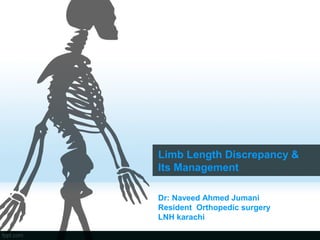
- 1. Limb Length Discrepancy & Its Management Dr: Naveed Ahmed Jumani Resident Orthopedic surgery LNH karachi
- 2. Definition • Limb length discrepancy or anisomelia, is defined as a condition in which the paired lower extremity limbs have a noticeably unequal length.
- 3. Introduction • Limb-length equality in the lower extremity is not only a cosmetic concern but also a functional concern. • limb-length inequalities of 0.5 to 2.0 cm are common in the normal, asymptomatic population. • Limb-length inequality of more than 2.5 cm has traditionally been considered significant, with an increased likelihood of knee, hip, and lumbar spine pain;
- 4. • The short leg gait is awkward • increases energy expenditure because of the excessive vertical rise and fall of the pelvis • back pain from long-standing significant discrepancies. • Compensatory scoliosis and decreased spinal mobility.
- 5. Classification • Structural (SLLD) or anatomical: – Differences in leg length resulting from inequalities in bony structure. – An acutal shortening or lengthening of the skeletal system occurs between the head of the femur and the ankle joint mortise, which may have a congenital or acquired cause. • Functional (FLLD) or apparent: – factors other than actual bone shortening or lengthening make one leg shorter or longer than the other, a functional inequality occurs – Unilateral asymmetry of the lower extremity without any concomitant shortening of the osseous components of the lower limb
- 6. Classification • McCaw and Bates (1991) report the following classification: – LLD has been classified according to the magnitude of the inequality, generally expressed in cm or mm, and described as mild, moderate, or severe. • Mild Less than 3 cm • Moderate 3-6 cm • Severe More than 6 cm
- 7. Etiology of Undergrowth • Congenital limb deficiency : – Congenital femoral deficiency, – congenital fibular deficiency, – tibial hemimelia. • Asymmetrical neurological disorders: – Hemiplegic CP, – poliomyelitis, – Hemimyelomeningocele. • Traumatic causes: – malunion, – growth plate arrest. • Hemiatrophy: Idiopathic, Russel-Silver syndrome.
- 8. • Other causes: – infection, tumor, – post-irradiation, – Blount’s disease, – LCPD, – unilateral clubfoot, – congenital pseudarthrosis of the tibia
- 9. Etiology of Overgrowth • Post-traumatic overgrowth: – femur shaft fracture, – tibia shaft fracture. • Soft tissue overgrowth syndrome: – Gigantism with neurofibromatosis, – Klippel-Treunaunay syndrome, – Beckwith-Wiedemann syndrome, – Proteus syndrome • Chronic inflammatory arthritis – Rheumatoid Arthritis • Idiopathic hemihypertrophy.
- 10. Mechanism of compensation • The child with LLD usually compensates better than the adult. • The child can compensate for minor degrees of LLD by walking on the toes of shot leg. • The adult seldom compensate that manner - tend to heel -toe gait:vaulting gait & excessive pelvic motion and tilt.
- 11. Mechanism of compensation • Check for specific compensation used by the patient to level out the difference in height. – Pronation in the ankle of the longer leg – Plantar flexion in the shorter leg – Knee and hip extension of the shorter or flexion in the longer leg – If the leg is left uncompensated, the anterior and posterior iliac spine on the side of the short leg can be lower which may result in a sacral base unleveling and/or scoliosis. – Increased muscle activity in several muscle groups
- 12. Effect in the Spine • Low back pain and late degenerative arthritis: controversial. • LLD - causes increased incidence of scoliosis. • Severity of the problem: – related to the severity of LLD – uncompensated or uncorrected – Onset of age.
- 13. Effect on Hip • Pelvic obliquity • Relatively uncovered hip of long leg and increased coverage of the hip of the short leg. • A longer leg might be a predisposing factor in Osteoarthritis (OA). • With length increasing, femoral head contact/ weight bearing area is decreasing. • Combined with an increased tone in hip abductors caused by elongated distance between origin and insertion and an increased ground reaction force puts the longer leg at risk.
- 14. Central edge angle • Decreased in CE angle on the long leg side () • decreased in the load bearing area • causes late degenerative arthritis.
- 15. Effect in Gait cycle
- 16. Effects on long bones • Greater incidence of stress fractures in the tibia, metatarsals and femur of the longer leg appears consistent with the greater forces emitted through the longer leg.
- 17. Evaluation • History • Examination • Imaging –Radiographs –Bone age –Scanogram
- 18. History • Congenital or developmental. • Determine cause • Determine deformity • Onset and mode of deformity
- 19. EXAMINATION
- 20. Physical exam • Wood block test: – with the patient standing, add blocks under the short leg until the pelvis is level, then measure the blocks to determine the discrepancy. – block testing is considered the best initial screening method. – Add blocks (known height) until the pelvis is level
- 22. Physical exam • Leg-length measurement: • Apparent length: – from the umbilicus to the medial malleolus • True length: – from the ASIS to the medial malleolus
- 24. Radiographs (Measure Length Discrepancy) • Telorengenogram • Orthoroentgenogram • Scanogram (x-ray/ CT)
- 25. teleoroentgenogram Length of x-ray shadow • An X-ray photograph taken at a distance of usually six feet with resultant practical parallelism of the rays and production of shadows of natural size.
- 27. Orthoroentgenogram Length of x-ray shadow A A′ A″ a a′ a″ b c d
- 28. Orthoroentgenogram • It is a radiographic study used to evaluate anatomic leg length and calculate leg- length discrepancies. • This study utilises a long ruler placed on the film, and three radiographs including bilateral hips, knees and ankles
- 29. SCANOGRAM Roentgen ray tube Slit diaphragm Slit-like roentgen ray beam Cassette
- 30. SCANOGRAM • low-radiation technique similar to an orthoroentgenogram utilizing three exposures
- 31. CT scan • Software measures distances – accurate to 0.2 mm – legs must be in same position – fast
- 32. Skeletal Age • 1. Greulich- Pyle Atlas – Xray Left hand (non dominant) – correlated with Green- Anderson table LL – less accurate < 6 – improved accuracy by focusing on hand bones rather than carpal bones • 2. Tanner- Whitehouse Atlas – more refined – 20 landmarks graded L Hand – more accurate – can't use as not correlated with LL
- 34. Growth • Proximal Femur – 3mm / year – 15% leg • Distal Femur – 9mm / year – 37% leg – 70% of femur • Growth Cessation – 14-15 Girls – 16-17 Boys • Proximal Tibia – 6mm / year – 28% – 60% tibia • Distal Tibia – 3mm / year – 20%
- 35. Prediction of Growth • Note that all methods have an inherent error of 12 months • gives accuracy to 1.5 cm • Need > 3 measures 4/12 apart for all methods • If inadequate data wait till older or wait till skeletally mature • If acquired event caused LLD, can plot onto graph
- 37. 2. Green & Anderson tables • Growth remaining method – uses skeletal age – requires graph – estimates growth potential in distal femur and proximal tibia at various skeletal ages – separate charts for girls and boys
- 38. 3. Moseley • Straight - Line Graph Method – uses Green & Anderson data – applied to a chart • At least 3 measurements each time – 1. Length long leg – 2. Length short leg – 3. Skeletal age • Do so 3 times separated by 3-6 months – accuracy improves with increased plotting.
- 39. Moseley chart
- 40. 4. Paley multiplier • State of the art: – take LLD for boy or girl – multiplier for chronological or skeletal age – predicts LLD at maturity
- 42. Paley multiplier • Congenital Limb Length Discrepancy ∆m = ∆ x M – ∆:Current Limb-length discrepancy – ∆m: Limb discrepancy at skeletal maturity – M:multiplier). • Example:current lld is 4cm in Congenital hemihypertrophy at 10 yrs age • Using value of 1.310 according to Multiplier chart at age of 10 in tibia • 4 x 1.310 = 5.24 cm(lld at maturity)
- 43. • Developmental LLD Leg-length discrepancy • ∆m = ∆ + (IXG) • I=1 -(S – S’)/(L – L’) • G=L(M-1) • G= amount of growth remaining • I=amount of growth inhibition • L= current length of long limb • L’=length of long limb as measured on previous radiographs • Lm length =length of femur or tibia at skeletal maturity of femur or tibia at skeletal maturity • M=multiplier • S= current length of short limb • S’ =length o f short limb as measured on previous radiographs • ∆ = current limb-length discrepancy • ∆m=limb length discrepancy at skeletal maturity
- 44. Example • Femur length(cm) right (abnormal) left (normal) • previous 24 26 at age of 8yrs • Current 26 29 at age of 10 yrs • I=1 -(S – S’)/(L – L’) I =1-(26-24/29-26) = 1-2/3=0.33(amount of growth inhibition) • G=L(M-1) G=29(1.310-1)=29 x 0.310=8.99(amount of growth remaining) • ∆m = ∆ + (IXG) ∆m = 3 +(0.33 x 8.99)=3 + 2.97 = 5.97 cm(lld at skeletal maturity)
- 45. Time of Epiphysiodesis • Lm=L x M • L ε = Lm – G ε • M ε =Lm/Lε – Lm= length of femur or tibia at skeletal maturity – L= current length of long limb – M=multiplier – Lε =desired length of bone to undergo epiphysiodesis at time of epiphysiodesis – ε=desire d correction following epiphysiodesis – Gε=amount of femoral or tibial growth remaining at age of epiphysiodesis(G ε= ε/0.71 for femur and ε/0.57 for tibia) – Mε=multiplier at age of epiphysiodesis
- 46. • Femur length(cm) right (abnormal) left (normal) • previous 24 26 at age of 8yrs • Current 26 29 at age of 10 yrs • Lm = L(29) X M(1.31) = 37.99(length at maturity) • Lε = Lm(37.99 ) – Gε(3/0.71) =37.99-4.22 = 33.77(desired length of bone to undergo epiphysiodesis at time of epiphysiodesis) • Mε=Lm(37.99)/Lε(33 .77) =1.125(multiplier at age of epiphysiodesis)
- 47. Leg-Length Discrepancy Guidelines for Management Discrepancy Management (CM) <2 No treatment or shoe lift 2-5 Growth Modulation(Shoe lift epiphyiodesis shortening) 5-12.5 Consider bone-lengthening >12.5 Combinations of above or amputation
- 48. Shoe lift • Patient who do not wish or are not appropriate for surgery. • Lift higher than 5 cm poorly tolerated.
- 49. Prosthetic fitting • Significant discrepancies, deformed functionally useless feet • Discrepancies greater than 15-20cm and femoral length less than 50% • Fibular hemimelia with unstable ankle • PFFD: A/K prosthesis or BK prosthesis with Van Nes rotationplasty • Optimal age: syme amputation- end of 1yr Rotation plasty: 3 yr
- 50. Epiphysiodesis • Very low morbidity and complication rate. • Slowing growth rate of long leg and allowing short leg to catch up. • Suitable for sufficient data to enable a confident prediction of discrepancy at maturity. • Tibial epiphysiodesis should be accompanied by arrest of proximal fibular physis if tibial shortening is greater than 2.5cm.
- 51. Epiphysiodesis • Phemister technique (JBJS 1933) • To stop the growth with open destruction of physis at correct time to achieve equal limbs.
- 53. PERCUTANEOUS EPIPHYSIODESIS PETS(Percutaneous Epiphysiodesis using Trans epiphyseal Screws ) (Metaizeau JP, et al, JPO,1998)
- 54. TENSION PLATE EPIPHYSIODESIS • this technique is largely reserved for hemiepiphysiodesis in angular corrections, • it can be used for complete epiphysiodesis if implants are used on both sides of the physis. • This technique also has the advantage of potential growth resumption with implant removal; however, restoration of normal growth often is unpredictable after implant removal, and careful timing of epiphysiodesis is still important
- 55. TENSION PLATE EPIPHYSIODESIS • Most of these plating systems are nonlocking, which allows some degree of screw divergence within the plate as the physis continues to grow. It is likely that growth arrest does not occur until maximal screw divergence is reached. Therefore, it is advisable to place the screws in a divergent fashion at the time of implantation to allow growth arrest to occur as quickly as possible
- 58. Problems of Epiphysiodesis • Undercorrection – growth or angulation • Overcorrection – growth or angulation • Rebound phenomenon (staples or screws) • Failure of growth restoration • Staple breakage or bending
- 59. Shortening operation • Mature patient • Tibia< 4cm, Femur< 5cm • Neurovascular complication is higher in tibia, fasciotomy is advisable.
- 64. Limb lengthening operation Codvilla(1905) – first described limb lengthening • Compere & Sofield (1936) • Anderson (1952) • Wagner (1978) • De Bastiani (1986) • Ilizarov (1989)
- 65. Limb lengthening operation • Limb lengthening procedures (LLP) are used to replace missing bones and/or to straighten deformed bones. • Can be performed on both children and adults with limb length discrepancies (< 6cms) and angular deformities due to birth defects, injuries or diseases.
- 66. Limb lengthening operation • Device for gradual lengthening – Unilateral fixator – Circular ring fixator (Ilizarov, Taylor spatial frame ) • Combined internal and external fixation – (Lengthening over IM Nailing) • Totally implantable lengthening device – Albizzia nail – ISKD(inter medullary skeletal kinetic device) – Fitbone
- 67. One stage lengthening • Transiliac:(MILLIS AND HALL) – Shortening < 3cm – Acetabular dysplasia
- 69. Distraction Epiphysiolysis • Chonodrodiastasis (Gelbke,1951, De Bastiani,1986) • Separation of the epiphyseal plate • Immature patient • Risk of septic arthritis • Painful stiffness of the joint • Premature closure of the physis
- 70. Tibia Lengthening • (DEBASTIANI ET AL) Orthofix lengthening devices • The reconstruction system with three screws in each segment • The telescopic device with three screws in each segment. • The Garches device with three screws in each segment.
- 72. Tibia Lengthening • Ilizarov Technique and its variations are commonly used. • Principle: – It is based on the principle of distraction osteogenesis. – A bone that has been cut during surgery are gradually pulled apart (distraction) – Leads to new bone formation (osteogenesis) at the site of lengthening
- 74. Femur lengthening COLE, PALEY, AND DAHL)
- 75. Immediate lengthening • When performing acute lengthening, the orthopaedic surgeon makes a cut in the bone, slides it and maintains the length and position with an internal device (i.e., screws or metal plates). • Or the surgeon may cut the bone, spread the two sections apart, and insert a graft and internal metal devices to maintain the length. • Surrounding muscles, nerves and blood vessels do not tolerate a lot of stretching. So acute lengthening can only achieve limited increases. For example, forearm bones (radius or ulna) and foot bones (metatarsals) are lengthened by this method when only a small gain in length is needed.
- 76. Gradual lengthening - Distraction Osteogenesis- • Ilizarov technique – 1) Corticotomy: preserve endosteal & periosteal blood supply – 2) Ilizarov Ring fixator: permit micro-axial motion – 3) Latency period: 7-14 days – 4) Proper rate & Rhythm: 0.25mm x4 / day – 5) Encourage Joint motion
- 77. Four phases of LLP • Preparation: – Consultation, X- rays of the limbs to build a custom- build external fixator, psychological evaluation • Surgery: – External fixator is attached to the bones • Lengthening: – Fixator is lengthened about 1 mm every day for new bone growth. • Strengthening: – For proper alignment and consolidation of new bone, removal of external fixator, PT rehabilitation.
- 78. Ilizarov External Fixator • Metal rings • Threaded rods • Kirschner wires (1.5- 1.8 mm in diameter) • Miscellaneous hardware for connecting the basic components
- 79. Advantages • Osteogenesis occurs • Regenerating bone resembles the existing bone • External Fixator can be custom- made • The basic parts- the wires, small pins and metal rings favors osteogenesis
- 80. Complication • Muscle contractures • Joint subluxations • Axial deviations • Neurological or vascular insult • Premature or delayed consolidation • Re- fracture • Pin- site infections • Psychological stress
- 81. Is there a role for amputation?? • Yes!!! • Significant length discrepancy • Poor underlying bone quality for lengthening • Dysfunctional/ painful limb
- 82. Thank you for your attention