
Recommended
More Related Content
What's hot
What's hot (20)
Similar to Autoimmune movement disorders
Similar to Autoimmune movement disorders (20)
More from NeurologyKota
More from NeurologyKota (20)
Recently uploaded
Recently uploaded (20)
Autoimmune movement disorders
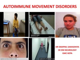
- 1. AUTOIMMUNE MOVEMENT DISORDERS DR SWAPNIL SAMADHIYA SR DM NEUROLOGY GMC KOTA
- 2. • Autoimmune syndromes mimic neurodegenerative or metabolic disorders. • Rarely occur as isolated movement disorders, accompanying clinical signs clue to diagnosis. • Detailed history and careful examination often reveal clinical red flags, help guide diagnosis • Timely identification paramount importance, treatable.
- 3. The clinical approach • Firstly 1. Recording clinical characteristics 2. Phenomenological categorization 3. Syndromic diagnosis • Secondly 1. The Movement disorder syndrome categorized into “isolated” or “combined” with other clinical symptoms Balint, B., Vincent, A., Meinck, H. M., Irani, S. R., & Bhatia, K. P. (2018). Movement disorders with neuronal antibodies: Syndromic approach, genetic parallels and pathophysiology.Brain: a journal of neurology, 141(1), 13–36
- 4. Cerebellar ataxia • Older age ,multi-system symptoms ,suspect,paraneoplastic • Subacute onset of rapidly progressive ataxia often combined with peripheral neuropathy, dementia, hearing loss and dysphagia • Anti-neuronal antibodies are detectable in up to 60% • Red flags -cognitive deficits, seizures, neuropathic pain, autonomic dysfunction and insomnia Lim, T. T. (2017). Paraneoplastic autoimmune movement disorders. Parkinsonism & Related Disorders, 44, 106– 109
- 8. Chorea and Dyskinesias • Second most common acquired cause after vascular lesions. • Examples -sydenham’s chorea ,chorea in antiphospholipid syndrome / systemic lupus erythematosus • Children -movement disorders and seizures, • Adults -behavioral changes, psychiatric disorders or cognitive impairment • Red flags -Cognitive dysfunction, seizures, prominent psychiatric disturbance, dysautonomia, optic neuritis,bulbar symptoms,weight loss and peripheral neuropathy. • m/c Ab. Collapsin response mediated protein-5 (CRMP-5) related to SCLC or Thymoma O'Toole, O., Lennon, V. A., Ahlskog, J. E., et al. (2013). Autoimmune chorea in adults.Neurology, 80(12), 1133–1144
- 11. Dystonia • Might be the only clinical finding (isolated dystonia) or can be combined with other movement disorders (combined dystonia) or the occurrence of other neurological signs • Orofacial dyskinesia, chorea,stereotypies,encephalopathy and psychiatric features, NMDAR-antibody testing is mandatory • Combination of jaw dystonia and breathing difficulties should prompt testing for Ri antibodies Duan, B. C., Weng, W. C., Lin, K. L., et al. (2016). Variations of movement disorders in anti-N-methyl-D-aspartate receptor encephalitis: A nationwide study in Taiwan.Medicine, 95(37), e4365
- 14. Myoclonus • Myoclonus -sudden, brief and involuntary jerks • Myoclonus is one of the defining clinical characteristics- opsoclonus myoclonus syndrome (OMS) • Most cases of OMS, no underlying autoantibodies can be detected • Myoclonus predominantly affects the limbs but can also present as axial myoclonus Oh, S. Y., Kim, J. S., & Dieterich, M. (2019). Update on opsoclonus-myoclonus syndrome in adults.Journal of Neurology, 266(6), 1541–1548
- 17. Parkinsonism • Autoimmune rare, m/c atypical parkinsonian syndrome, reflecting brainstem encephalitis with eye movement abnormalities or other brainstem signs, and sleep disorders. • Red flags -hypothalamic pituitary dysfunction, weight gain, prominent sleep disorders including excessive daytime sleepiness, rapid eye movement (REM) sleep behaviour disorder (RBD), narcolepsy- cataplexy, and eye movement abnormalities(vertical gaze palsy) • IgLON5 antibodies postural instability and gaze palsies, resembling PSP in up to 23% Dalmau, J., Graus, F., Villarejo, A., et al. (2004). Clinical analysis of anti-Ma2-associated encephalitis.Brain: a journal of neurology, 127(Pt 8), 1831–1844
- 20. Paroxysmal movement disorders • Paroxysmal dyskinesias and episodic ataxias -onset childhood or early adulthood ,autosomal dominant inheritance. • Antibody associated paroxysmal movement disorders -later in life • Young women, painless paroxysmal dystonic posturing should prompt consideration of NMDAR-antibody testing whereas painful tonic spasms warrant testing for NMOSD-associated antibodies (AQP4, MOG) Liu, J., Zhang, Q., Lian, Z., et al. (2017). Painful tonic spasm in neuromyelitis optica spectrum disorders: Prevalence, clinical implications and treatment options.Multiple Sclerosis and Related Disorders, 17,99–102
- 22. Stiff person spectrum disorders • Rare diseases (estimated prevalence: 1/1,000,000) • Stiffness, Spasms and Hyperekplexia • m/c antibodies against GAD65 in the serum and cerebrospinal fluid • Associated features and autoimmunity may indicate the underlying antibody (e.g. GAD antibodies with type 1 diabetes) Carvajal-Gonzalez, A., Leite, M. I., Waters, P., et al. (2014). Glycine receptor antibodies in PERM and related syndromes: Characteristics, clinical features and outcomes.Brain: a journal of neurology, 137(Pt 8), 2178–2192
- 25. Tics • Role of antibodies controversial. • Tics -paediatric autoimmune neuropsychiatric disorders associated with streptococcal infections (PANDAS) • Antibodies modulating dopamine d1 and d2 receptors have been hypothesized as being causative • No antineuronal antibody consistently shown to underlie tic disorders Morris-Berry, C. M., Pollard, M., Gao, S., Thompson, C., Tourette Syndrome Study G, & Singer, H. S. (2013). Anti- streptococcal, tubulin, and dopamine receptor 2 antibodies in children with PANDAS and Tourette syndrome:Single- point and longitudinal assessments.Journal of Neuroimmunology,264(1–2), 106–113
- 27. Tremor • Isolated tremor is highly unlikely • Isolated“whole-body tremulousness” often mistaken as generalized tremor ,actually suffer from generalized repetitive myoclonus • Beside the classic antineuronal antibodies ,paranodal Abs, such as contactin-1 (CNTN1), neurofascin-155 (NF155), nodal neurofascin (NF140/186) and contactin associated-protein-1 (CASPR1) CIDP McKeon, A., Pittock, S. J., Glass, G. A.,et al. (2007). Whole-body tremulousness:Isolated generalized polymyoclonus. Archives of Neurology, 64(9), 1318–1322
- 30. Sleep behaviour disorders • Ma2 encephalitis, affects limbic, diencephalic and brainstem structures, or in LGI1-antibody-associated limbic encephalitis (Iranzo et al., 2006) • Both RBD and non-RBD can be seen in IgLON5-antibody linked neurodegeneration (Sabateret al., 2014) • Periodic limb movement and ambiguous sleep are observed with DPPX antibodies (Tobin et al., 2014)
- 31. • Status Dissociatus (breakdown of the boundaries, which are wakefulness, REM sleep, and non-REM sleep, with motor hyperactivity) • Agrypnia Excitata (insomnia, motor and autonomic hyperactivation) hallmark Morvan syndrome (with CASPR2 antibodies, less commonly LGI1 antibodies), NMDAR antibodies or GABA-B R antibodies(can be) (Frisulloet al., 2007)
- 33. Whispering and talking (“Good morning, your ring cakes are very nice”, "Which is the difference?”), changing facial expressions, gesturing and complex movements, and stridor.
- 34. Model of B cell activation
- 35. Directed against predominantly intracellular synaptic proteins may gain access at the time of vesicle fusion (A) Antibody-dependent Cell-mediated Cytotoxicity (ADCC) (B) Direct Target Modulation Through Agonist/Antagonist Effects (C) Complement Activation (D) Antigen Internalization
- 36. Diagnosis • Clinical • Laboratory investigation • Imaging • Antibody testing
- 37. Lab.Investigations • Cerebrospinal fluid abnormalities 1. Cells >5 cells/cu mm 2. Protein >0.45 g/dl 3. Elevated IgG index or oligoclonal bands
- 42. Imaging
- 46. Antibody testing • Combination of - 1. Immunoblot 2. Enzyme-linked-immune-tests (e.G. ELISA) and/or radioimmunoassay (RIA) 3. Cell-based assays (CBA) 4. Tissue-based assays (TBA) • Complementery tests(research laboratories) 1. Live-cell cbas and non-permeabilized (live) cells 2. Primary hippocampal neurons Ganesh A, Wesley SF. Practice Current: When do you suspect autoimmune encephalitis and what is the role of antibody testing?. Neurol Clin Pract. 2018;8(1):67-73. doi:10.1212/CPJ.0000000000000423
- 47. Testing should follow five rules: (a) Always use a combination of antigen-specific tests (immunoblots, ELISA/RIA, CBAs) and tissue-based screening tests (TBA). (b) Include immunoblots for intracellular antigens,CBAs for extracellular antigens, and ELISA/RIA for GAD65, VGCC. (c) Combination testing serum and CSF improves sensitivity and specificity,prevents false positives and false-negatives (not uncommon if testing serum only). (d) Start with common antibodies. If negative and clinical suspicion high, second diagnostic step TBAs and CBAs in research laboratories utilized for less common and unknown antibodies. (e) Nevertheless, seronegativity in spite of comprehensive testing is common.
- 48. • Suspected AE cases, generally prefer to send both serum and CSF for antibody testing • Some antibodies are more sensitive when testing serum (e.g., LGI1, AQP4) whereas more sensitive when testing CSF (e.g., NMDAR , GlyR, GABA -A) Ganesh A, Wesley SF. Practice Current: When do you suspect autoimmune encephalitis and what is the role of antibody testing?. Neurol Clin Pract. 2018;8(1):67-73. doi:10.1212/CPJ.0000000000000423
- 49. The different test systems for antibody detection
- 52. MANAGEMENT
- 53. DRUG THERAPY FOR AUTOIMMUNE MOVEMENT DISORDERS
- 58. FOLLOW UP
- 59. Future Directives • Presently, treatment empirical or based on expert consensus, lack of randomized controlled trials and available evidence limited to a few observational studies (Grauset al., 2016) • In disorders where neuronal surface antibodies are the main players, overall response to immunotherapy is usually good, Although varied.
- 60. • LGI1 antibodies -response to corticosteroids, • Half patients NMDAR antibodies respond insufficiently, first line immunotherapy (corticosteroids, plasma exchange, intravenous immunoglobulins) (Irani et al., 2013; Titulaer et al., 2013) • Drawback (immunotherapies) -potentially toxic, higher risk of infections, possibly with fatal outcomes • Future-antigen-specific immunotherapy
- 61. Potential future treatment approaches with antigen-specific immunotherapy
- 62. • Small blocking molecule, or ‘AQUAPORUMAB’ as a competitive and protective antibody (Verkman et al., 2013).
- 63. • Different approach -Target the pathophysiological cascades downstream from the antibody–antigen interaction. • NMDAR antibodies disrupt the interaction between NMDAR and ephrin-B2 receptor, eventually leads to displacement of NMDAR to extrasynaptic sites before they are internalized.
- 64. CONCLUSION • Prominent and common feature in many autoantibody-mediated neurological diseases (Gable et al., 2012) • It is imperative not to miss these potentially treatable disorders (Irani et al.,2013; Titulaeret al., 2013) • Can also be an alert to an occult neoplasia. (Irani et al.,2013; Titulaeret al., 2013) • Adequate treatment (Immunosuppression or Immunomodulation, Tumour Treatment as appropriate), many patients, good recovery, lasting deficits may occur (McKeonet al., 2013;Titulaeret al., 2013; Balintet al., 2014a)
- 65. REFERENCES 1. Movement disorders with neuronal antibodies: syndromic approach, genetic parallels and pathophysiology (Bettina Balint,Angela Vincent,Hans-Michael Meinck,Sarosh R. Irani and Kailash P. Bhatia) doi:10.1093/brain/awx189, BRAIN 201 2. The Clinical Features, Underlying Immunology, and Treatment of Autoantibody-Mediated Movement Disorders,Published online 00 Month 2018 in Wiley Online Library (wileyonlinelibrary.com).DOI: 10.1002/mds.27446 3. Principles and approaches to the treatment of immune-mediated movement disorders, https://doi.org/10.1016/j.ejpn.2017.11.010 4. Autoimmune and paraneoplastic movement disorders: An update Journal of the Neurological Sciences 385 (2018) 175–184 5. Uptodate.com 6. Parkinson’s disease and movement disorders,j.jankovic,e.tolsa,6th edition
- 66. Thank you
Editor's Notes
- Model of B cell activation in neuronal surface autoantibody-associated movement disorders. Triggers of immunological activation in CNS autoimmunity may lead to exposure of antigen (red star) and its presentation in the germinal centres of the cervical lymph nodes. Interaction between naïve B cells and CD4 + T helper cells in germinal centres causes maturation of B cells into antigen-specific cells that can switch their immunoglobulin chain to express IgG. These cells can subsequently differentiate into antibody-secreting plasmablasts and become tissue-resident plasma cells. Circulating memory B cells and plasmablasts can reach the CNS through the internal carotid artery, re-encounter the antigen, and produce antibodies (intrathecal synthesis). Modified with permissions from Wilson et al. 83 IgG (immunoglobulin G), IL-2 (interleukin 2), IL-21 (interleukin 21), IgD (Immunoglobulin D), TNF alpha. CD = cluster of differentiation.
- Anti ma2 ab related encephalitis,hypothalamic involvement
- Pcd ovarian teratoma
- Crmp5,cv2 realated bg encephalitis
- Paraneoplastic limbic encephalitis
- Example of comprehensive autoantibody testing available in scientiifc research settings.aTissue-based test using sagittal rat brain sections optimized for detection of neuronal surface antibodies. Brown staining indicates specific binding of human IgG. Hippocampal and cerebellar regions are shown magnified. Shown is an example of the staining obtaining with GABA(A) receptor antibody containing patient CSF. bCell-based assay with HEK293 cells expressing human autoantigens. This example shows cells transfected with AMPA receptor subunits stained with serum of a patient with anti-AMPA receptor encephalitis. Green staining indicates human IgG, red staining indicates a commercial antibody detecting transfected cells. Merged images are shown on the right side. Yellow indicates cells transfected and detected by human IgG thus indicating presence of autoantibodies targeting the transfected antigen. In addition to the shown example of cells fixed with paraformaldehyde, for some autoantibodies non-fixed live-cell based assays, which stain cells before addition of fixatives are more sensitive for the detection of some autoantibodies (e.g. MOG, not shown).cPrimary, embryonal, rat hippocampal neurons are stained with serum from a patient with antiAMPA receptor encephalitis. Cells are alive and non-permeabilized so that only surface antigens are detected by autoantibodies. Green indicates human IgG binding to individual synapses, blue is a DAPI counterstaining of nuclei to demonstrate presence of neurons