
Recommended
More Related Content
What's hot
What's hot (20)
Similar to Rhabdomyosarcoma
Similar to Rhabdomyosarcoma (20)
Recently uploaded
Recently uploaded (20)
Rhabdomyosarcoma
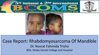
- 1. Case Report: Rhabdomyosarcoma Of Mandible Dr. Nusrat Fahmida Trisha BDS, Dhaka Dental College and Hospital
- 2. INTRODUCTON • Rhabdomyosarcoma is a tumor of skeletal muscle origin, is the most common soft tissue sarcomas in childhood and adolescence accounting for 4-8% of all malignant lesions in those younger than 15 years of age. • Genetic translocation producing new protein that interrupts skeletal muscle differentiation • Oral and maxillofacial region : 44% of all RMS, occurs mostly in the children and teenagers • 2% of RMS are present at birth • 5% occur in individuals younger than 1 year • Tumors have a peak incidence at the age 4 years and another at age 17 years • HISTOPATHOLOGICAL REPORT: The diagnosis is established by means of a deep incisional biopsy within the center of the tumor. EMBRYONAL RMS ALVOLAR RMS PLEOMORPHIC RMS • RADIOLOGY: Plain radiographs, a CT Scan, an MRI Scan is required to understand the tumor’s size, spatial anatomic relationship and extend of bony destruction. • The CT or MRI Scan should include detailed cuts of the base of the scull to assess potential invasion into this area and include the entire skull to rule out brain extension or metastasis DIAGNOSIS Treatment Protocol LOCAL EXCISIONCHEMOTHERAPY RADIOTHERAPY • Recently the treatment is done by chemotherapy followed by surgery
- 3. CASE REPORT • Baby girl Ilma, 4 years old child, hailing from New market was referred to the department of oral & maxillofacial surgery of Dhaka Dental College Hospital in 30th October 2017, with a chief complaint of painless swelling in the mandibular right posterior region of 25 days duration. • The swelling was initially small size and quickly increased ( progressive growth ). She had no past history of trauma and systemic disease. According to the statement of patient’s attendance, there was no sign of pain, sensation disorder, pus discharge and bleeding in the involved area. • Extra oral examination showed facial asymmetry and a diffuse soft tissue to firm swelling in the posterior region on the mandible with smooth surface, ill defined margin, consistency soft but near angle of mouth swelling is hard and fixed to underlying structure. • Intraoral examination showed irregular ulcerated swelling, well defined margin, consistency firm to hard, fixed to underlying structure. Measurement of lesion : 4cm X 4cm • She had no systemic diseases. • Regional Lymph node: right submandibular lymph node is palpable, size almost 1.5cm, mobile. • Incisional biopsy : soft tissue taken from right side of the body, angle and ramus of the mandible. sections showed fragmented pieces of tissue showing a neoplasm, composed of cells having irregular nuclear membrane. Background is myxoid. OPG • The paneromic xray revealed radiolucent lesion with ill defined borders. • The lesion was extending anterioposteriorly, from the retro molar area to the ramus of the mandible, • Inferiorly up to the inferior alveolar canal of the mandible • Superiorly to the alveolar ridge INVESTIGATION Fig : OPG showing bone destruction at right side of mandible
- 4. CT shows facial asymmetry cortical perforation • The computed tomography ( CT scan ) showed the large aggressive mass lesion ( 47.5 mm X 39.2 mm ) noted in right lower jaw involving right ramus and adjacent part of body of mandible • The lesion having large soft tissue component surrounding the involved part of mandible • Gross bony destruction noted in involved part of mandible • Medially the mass involved right buccal region & laterally the mass merged with muscles of mastication CT SCAN
- 5. DISCUSSION • The latest treatment protocol is Chemotherapy followed by surgical excision. • So the patient was reffered to Oncology department of Dhaka Medical College Hospital for better management on 23rd November 2017 • Radiation therapy usually begins several months after chemotherapy starts. It’s another effective way of shrinking the tumor • Often we do a PET scan after chemotherapy and radiation is done to see how the tumor responded to the treatments. Study shows that this practice can help predict the chance that the rhabdomyosarcoma will be permanently controlled by chemotherapy and radiation therapy. CONCLUSION We conclude that in children, any swelling should be carefully examined and treatment outcomes should be regularly followed up. High degree of suspicion, early diagnosis and a multidisciplinary treatment approach would be of great importance in such cases. REFERENCES • Oral and Maxillo-facial pathology Robert E. Marx,DDS Diane Stern,DDS • Oral Pathology REGEZI SCIUBBA JORDAN • The Intergroup Rhabdomyosarcoma Study Group (IRSG) • Medical Wikipedia