2. INTRODUCTION
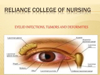
1. EYELIDS: Two movable folds with eyelashes.
Layers present are skin, aerolar tissue; muscles- orbicularis
oculi, levator palpebrae superioris & Muller’s muscle; thin
sheet of dense connective tissue, tarsal plate; thin lining of
palpebrae conjunctiva.
2. EYELID MARGIN
Covered with stratified squamous epithelium
Anterior border is round, posterior is sharp & lies closely in
contact with the eyeball.
Eyelashes originate anterior to the grey line and ducts of
meibomian are located posterior to the grey line.
3. 3. GLANDS OF EYELIDS
Zeis’s Gland: Sebaceous gland situated in close association
with cilia.
Moll’s Gland : Sweat glands & lie between the cilia.
Meibomian Gland : Enormously developed sebaceous gland
embedded in tarsal plate, secretes oily secretion that
lubricates the eye
Palpebral Fissure: Space between the two lids when the eye
is open
Outer Canthus: Outer or lateral angle of palpebrae fissure
Inner Canthus: Inner or medial angle of the palpebral fissure
Blood supply: Opthalmic & lacrimal arteries & opthalmic vein
Nerve supply: 7th , 3rd & 5th cranial nerve.
6. DEFINITION = 1. An eyelid infection is
any abnormal condition that effect eyelids.
2. Infection of an oil gland on the surface of
the upper and lower parts of eyelids
3. A bacterial infection of an oil gland, hair
follicle or sweat gland are caused drooping
, twitching (F), inflammation , itching,
burning, crustiness, redness , edema ,
tearing, irritation .
9. A. CONGENITAL ANOMALIES
I. Able-pharon: Macrostomia syndrome Extremely rare,
the lid is not developed autosomal
recessive genetic disorder .
ii. Micropharon : Rare, lids are abormally small
iii. Cryptopharon:: Rare anomaly in which a fold of skin passes
from the eyebrow over the malformed eye to the cheek.
iv. Ptosis: Common, drooping of eyelid
10. v. Epicanthus : A vertical fold of skin on either side of the nose
sometime covering the inner canthus.
(the outer or inner corner of the eye )
vi. Distichiasis: an additional row of lashers
occupies the position of meibomian glands .abnormal growth
of lashers.
vii. Coloboma: condition where normal tissue in or around the
eye is missing from birth.
11. B. EDEMA
A swelling eyelid occurs when there is inflammation or
excess fluid ( edema) in the connective tissue surrounding
the eye.
Common due to looseness of tissues
Inflammatory Edema: Found in conjunctivitis, tarsitis,( infl of
gland and lasher ) dacryo-cystitis, ( inflammation of lacrimal
sac )orbital cellulitis, ( infl of eye tissue ) drug allergy
(atropine).
Passive edema: common feature of cavernous sinus
thrombosis ( formation of blood clot with in the cavernous
sinus a cavity at the base of the brain which drains
deoxygenated blood from the brain back to the heart ),
12. C. INFLAMMATIONS
It includes
1. Blepharitis
2. Hordeolum (stye)
3. Chalazion (tarsal or meimobian cyst)
4. internal . Hordeolum
13. BLEPHARITIS
Is an inflammation of the eye lids in which
they become red, irritated and itching
dandruff like scales from on the
eyelashes…
14. 1. BLEPHARITIS
Squamous blepharitis
Ulcerative blepharitis
1. Squamous blepharitis
: it is due to abnormal metabolism (abnormal
chemical in body ) & seborrhea usually
associated with the dandruff of the scalp.
Numerous white coloured small scales
accumulate among the eyelashes.
15. 2. ULCERATIVE BLEPHARITIS
it is an ineffective condition. The yellow
crusts glue ( sticky) the lashes together. On
removing the crust there are small ulcers
seen around the bases of lashes.
Symptoms: itching, redness, soreness,
lacrimation and photophobia.
16. Treatment
Local: -removal of scales , crusts & diseased lashes is done by bathing
lid margin with 3% of NaHCO3 ( sodium bicarbonate ) lotion.
- antibiotics & ointment are applied.
General : Improvement of general health & personal hygiene.
- Dandruff of the scalp is to be adequately treated.
17. 2. Hordeolum (Stye):is a localized infection or inflammation
of the eye lid margin involving hair follicles of eyelashers
or meibomian gland (supply of meibumian, an oily substance )
Etiology: Associated with staphylococci infection, .
Common in young adults & debilitated persons( very week
person).
Symptoms: Ac. Pain & tenderness over inflamed Zeis’s gland.
Signs: Localized Pain, redness & edema near the lid margin.
Treatment: Hot fomentation, ( to apply a warm compressor eye
)Evacuation of pus,
antibiotic eye drop = tobramycin = it is killing or slow the growth of
certain type of bacteria.
& ointment & broad spectrum antibiotics is useful.
- Analgesics & anti-inflammatory drugs control pain & inflammation.
18. =
3. Chalazion: It is chronic granulomatous inflammation ( produced in
response to infection, inflammation,) or the presence of a foreign
substance. of mei-bomian gland.
Etiology: due to chronic irritation due to organism of low virulence (The
ability of bacteria to cause disease )where the glandular tissue is
replaced by granulation tissue containing giant cells.
- Occur in crops, more common in adults.
Symptoms : No pain unless secondary infected
Signs: Small non tender hard swelling slightly away from and swelling
lid margin ,swelling is red or purple, can be grey in later stages, yellow
when secondary infected with pyogenic organisms.
Treatment:
.
b) Inj. Triameinolone directly into the chalazion cause
complete resolution.
It prevents the release of substances in the body that cause
inflammation.
19. 4. Internal Hordeolum : It is an acute Supportive
inflammation ( formation of pus )of mei-bomian glands
Etiology: Occurs due to secondary infection (occurs during
or after treatment for another infection.) of chalazion.
Symptoms : More violent than stye because the gland is
larger & embedded deeply in the dense fibrous tissue.
Sign : Yellow spot (pus) seen shining through the
conjunctiva on averting ( remaining) the lid;
TREATMENT
Warm compresses an d massages of the lesions for 10
minutes 4 times per day
Tropical anti biotic ointment
Amoxicillin
Doxycycline
Erythromycin
20. D. ANOMALIES & POSITION OF EYELIDS
It includes
1.Trichiasis, abnormally positioned eyelashes
2.Entropion, the lid margin rolls inwards
3.Ectropion, lid margin rolls outwards
4. Symblepharon, the adhesions between lids
and the globe.
5. Ankyloblepharon, adhesion of the margins
of two eyelids
6. Lago-pthalamus, inability to close the
eyelids completely.
7. Ptosis. drooping of the upper lid
21. D. ANOMALIES & POSITION OF EYELIDS
It includes Trichiasis, Entropion, Ectropion, Symblepharon,
Ankyloblepharon, Blepharophimosis, Lagopthalamus, Ptosis.
1. TRICHIASIS : abnormally positioned eyelashes Few lashes or
whole lid margin involved.
Etiology: Recurrent stye, Ulcerative blepharitis, Tight bandaging,
Scars of lid following burn, injury or operation.
Symptoms
a) Foreign body sensation of photophobia due to corneal involvement
b) Irritation, pain & lacrimation
c) Treatment
d) Trichiasis treatment involves removing the eyelash, follicle or both, or
redirecting eyelash growth.
22. 2. ENTROPION: Conditions in which the lid margin rolls inwards.
Etiology
a) Spastic entropion: Due to the spasm of orbicularis oculi muscle as
may occur after tight bandaging after operation of following irritative
corneal condition
b) Cicatricial entropion :, ulcerative blepharitis, burns, operations,
diphtheritic membranous conjunctivitis.
Sign & symptoms : Same as for trichiasis
Treatment
A. Spastic
Basic cause of blepharospasm is treated
If due to prolonged & tight bandaging,
discontinue it.
Antibiotics
Anti inflammatory - corticosteroids
Botulinum toxin –using eyelid spasms
23. 3. ECTROPION : It is a condition in which lid margin rolls outwards
Symptom : Most common epiphora
excessive watering of the eyes
Signs
i) Conjunctiva become dry in appearance
ii) Chronic conjunctivitis & corneal ulcers.
iii) TREATMENT
iv) Use of lubricating ointment or mild steroid several day and
weeks to ectropion repair corneal epithelium ,,,
24. 4. SYMBLEPHARON : It is a condition of the adhesions
between lids and the globe.
Etiology : due to
- burns, ulcers, diphtheria, operation
Symptoms
1. Lagopthalamus: inability to close lids properly
2. Diplopia : double vision
3. treatment
4. Lysis and removal of subconjunctival scar tissue
25. 5. ANKYLOBLEPHARON
It is a condition of the adhesion of the margins of two eyelids. Adhesion
may be partial or complete,
Etiology
- Congenital or acquired due to chemical burn i.e. acid, alkali.
Treatment
- Separation of lid margins along with mucus membrane or conjunctival
grafting is recommended
26. 7. LAGOPTHALAMOS
It is a condition of incomplete closure of palpebral aperture when eyes
Lagophthalmos is defined as the inability to close the eyelids
completely. Blinking covers the eye with a thin layer of tear fluid,
Etiology
Loss of function of the facial nerve inhibits eyelid closure as well as the
blinking reflex .
- Congenital deformity of lids, ectropion ,proptosis (abnormal protrusion
or displacement of an eye , paralysis , absence of reflex, blinking in
extremely ill patient’s
Treatment
1. Application of antibiotic eye ointment & bandage during sleep is
recommended.
2. Levofloxacin
27. 8. PTOSIS
It is a condition in which there is drooping of the upper lid below its
normal position .
Etiology
1. Congenital Ptosis: Occurs in 80 % cases, due to maldevelopment of
levator muscle; congenital weakness of superior rectus muscle.
2. Acquired Ptosis : a). Neurogenic : partial/ complete paralysis of 3rd
nerve b) Mechanical : Due to increased weight of upper lid as a
result of edema, hypertrophy or tumor formation.
c) Myogenic : Due to trauma of levator muscle, muscular dystrophy (
increasing weakening or breakdown of muscle) & myasthenia gravis.(
neuromuscular disease weakness of skeleton muscle)
Symptoms : Visual disturbance
visible drooping of the upper eyelid
28. TRETMENT
high doses of opioid drugs such
as morphine, Morphine is a narcotic pain
reliever used to treat moderate to severe
pain. ... 0.05 mg/kg IM, IV, or subcutaneously
every 4 to 8 hours
oxycodone hydrochloride analgesic agents
heroin, or hydrocodone can cause ptosis.
Pregabalin (Lyrica), an anticonvulsant drug,
has also been known to cause mild ptosis.[6]
30. Nevus( mole on the skin red patches)
A choroidal nevus is a flat, benign pigmented
area that appears in the back of the eye
Heman-gioma
Hemangioma is a non-cancerous (benign)
tumor caused by abnormal growth of blood
vessels. cavernous hemangioma occurs in
the deeper layers of the skin or around
the eye.
31. PAPILLOMA
a benign tumor derived from
epithelium. Papillomas may arise from skin,
mucous membranes, or glandular ducts
Xanthelasma ( jenthelasma)
are yellowish plaques that occur most commonly
near the inner canthus of the eyelid, more often
on the upper lid than the lower lid. Xanthelasma
32. NEUROFIBROMA
It is a generalized disease that may involve
the lid & cause mechanical ptosis. It is
associated with unilateral infant glaucoma.
Small, multiple tumors are distributed along
the hypertrophied nerves.
35. Squamous cell carcinoma
Seen at the edge of the lid (transition zone)
where the epithelium changes. It starts as a
nodule that ulcerate. The preauricular lymph
nodes are enlarged. It spreads slowly the
surrounding structures and are painless.
Metastasis ( spread of cancer cells to new
area of the body ) common,
Basal cell carcinoma
It is most common seen in lower lid near the
inner canthus. It is locally malignant.
Epithelial growth spreads under the skin in all
direction .
36. ETIOLOGY
Environmental toxins such as exposure to
radiation
Genetics
Diet – deficiency of minerals and vitamins
Stress excessive stress cause blocking
mutation in the cells of their developing eyes
Local trauma or injury- orbital trauma and
bleeding
Inflammation or infection
37. CLINICAL MANIFESTATION
Bulging of one eye (protrusion)
Complete and partial loss of sight
Pain in or around the eye
Blurred vision
Change in the appearance of the eye
Edema
Redness
Itching
Burning
38. DIAGNOSIS
Ultrasound scans
CT SCAN
MRI
They show the size , location ,and shape of
the tumors and also show the enlarged or
affected lymph node around the eye.
39. MEDICAL MAANAGEMENT
1.Identify the cause & eliminate the cause
2. Achieve and maintain control of symptoms
3. Avoid adverse effects of medication.
4. to give antibiotics and anti inflammatory drugs .
5. Maintain normal activity level ,including
exercise .
6, Prevention foreign particles enter in eye
Use of sunglasses
To keep the eye clan ,wipe away the drainage
from around the eye ‘moisten and clan cotton
ball or wash cloth with warm water, from inner to
the outer part the eye.
40. MANAGMENT
Chemotherapy eye drops
Mitomycin C –are used to treat different
types of growths on the surface of the eye
fluorouracil
Is used treatment pre-cancerous an
cancerous cell growth
Radiation therapy uses high energy x- rays
or other types of radiation kill the cancer cell.