SlideShare a Scribd company logo
1 of 74
ENDOTRACHEAL INTUBATION IN
ORAL & MAXILLOFACIAL SURGERY
Dr. Padmasree Patowary
CONTENTS
 Introduction
 Historical prospective
 Indication and contraindication
 Armamentarium
 Procedure for Endotracheal Intubation
 Types of intubations
I. Orotracheal intubation
II. Nasoendotracheal intubation
III. Retrograde intubation
IV. Retromolar intubation
V. Submental intubation
VI. Fibroptic intubation
 Complications
 Conclusion.
Introduction
Maintaining the airway is an essential step in oral and
maxillofacial surgical cases, which are performed under
general anesthesia or maxillofacial trauma. There are various
techniques for maintaining the airway ranging from, head tilt –
chin lift to surgical airway.
Endotracheal intubation provides an artificial medium between
the atmosphere and the patient’s trachea for the purpose of
alveolar gas exchange or protection of the lungs from
inappropriate substances.
Historical prospective
Endotracheal intubation for the
purpose of providing anaesthesia
was first described by William
Mac Ewan in 1878 when he
passed a tube from the mouth into
the trachea, using fingers as a
guide in the conscious patient.
Routine
To provide anaesthesia
Emergency
Airway obstruction
Respiratory distress
Oxygenation failure (hypoxia)
Ventilation failure
Mental status alteration
Indications
Contraindication
1. Severe airway trauma or obstruction that does not permit safe
way of an endotracheal tube. Emergency cricothyrotomy is
indicated in such cases.
2. Cervical spine injury.
3. Mallampati Classification of class III / IV or other
determination of potential difficult airway.
Intubation can be achieved in following ways:
1. After induction of general anaesthesia (by IV or
inhalation method).
a. Oral—orotracheal intubation.
b. Nasal—nasotracheal intubation.
2. Awake intubation
a. Blind oral
b. Blind nasal
c. Retrograde
d. By use of fiberoptic scope.
Procedure for Endotracheal Intubation
Anatomy of airway
Upper Airway Anatomy
• Anterior Nares
• Vestibule
• Mucosal Lining
• Septum
• Nasal conchea
• Posterior Nares
• Nasopharynx
• Oropharynx
• Pharynx
• Larynx
• Epiglottis
• Vocal Folds
• Glottis
Lower Airway Anatomy
• Trachea
• C-shaped cartilaginous
rings
• Carina
• Right main bronchus
• Left main bronchus
• Bronchial Tree
• Bronchioles
• Alveolar ducts
• Alveolar sacs
• Alveoli
– ̴300,000,000
• Capillaries
Armamentarium
i. Anesthetic Mask
ii. Laryngoscope
iii. Endotracheal Tubes
iv. Airways
v. Laryngeal Mask Airway (LMA)
vi. Magill’s forceps
vii. Ambu Bag
viii. Stylet
Mask is an essential part of any anaesthetic
breathing system during the induction phase
(beginning) of anaesthesia.
A mask allows administration of gases from
the breathing system, without introducing any
invasive equipment (e.g. an endotracheal tube)
into the patient’s airway. Face-masks are
designed to fit the patient’s face
perfectly without any leaks.
• Parts:
I. Connector or mount
II. Body
III. Edge or seal
• Sizes: 1, 2, 3, 4, 5.
Anesthetic Mask
Laryngoscope : handle and blade
LARYNGOSCOPIC BLADE
• Macintosh (curved) blade (most popular)
• Miller (straight) blade (useful for neonates and small children)
Laryngoscope is designed for
doing direct laryngoscopy
(directly viewing the vocal
cords) and to pass an endo
tracheal tube into the larynx
under vision.
• Parts: Handle, blade with
light bulb.
• Sizes (for the blades) Neonate
(infant), Paediatric (child),
adult and extra large.
Endotracheal tube is a specially designed tube with one end
straight and the other being obliquely cut (beveled end).
Beveled end enters the trachea.
It is used for the surgical procedures in which it is not feasible to
administer anaesthetic gases via mask, procedures which are long-
lasting, procedures in which there are chances of having blood,
secretions, pus, vomiting etc. in the oral cavity or where patient
needs to be given muscle relaxants and controlled breathing.
Endotracheal Tubes
It is always passed with direct laryngoscopy under vision after
anaesthetizing the patient, except in cases, where patients are unable to
open mouth, e.g. T.M. joint ankylosis or when a difficult intubation is
expected.
Age Weight
(Inner) diameter
(mm)
New born 3 kg 2.5–3.0
0–6 months 3.5 kg 3.0–3.5
6–12 months 7 kg 3.5–4.0
1–3 years 10–12 kg 4.0–4.5
4–7 years 16–18 kg 5.0–5.5
8–10 years 24–30 kg 5.5–6.5
Adult female 7.0-7.5
Adult male 8.0-9.0
Types of endotracheal tube
include
1. oral (south pole) or nasal
(north pole)
2. cuffed or uncuffed
3. preformed (e.g. RAE (Ring,
Adair, and Elwyn) tube
4. reinforced tubes
5. double-lumen endobronchial
tubes.
The purpose of introducing an airway is to lift the tongue and
epiglottis away from the posterior pharyngeal wall and prevent
them from obstructing the space above the larynx.
Airways
I. Oropharyngeal (Oral) Airway (Placed from Lips to
Pharynx)
II. Nasopharyngeal (Nasal) Airway (Placed from the Nose to
the Pharynx)
Oropharyngeal airway: In basic airway
management, the oropharyngeal airway is
used to provide a right airway to make
possible for chest ventilation. In advanced
airway management, it can be inserted
following endotracheal intubation to act
as a bite-block to protect the endotracheal
tube.
• Parts: Flange, bite portion, air/suction
channel (curved portion)
• Sizes: 1, 2, 3, 4.
Nasopharyngeal airway is a tube that is designed to be inserted into
the nose to secure an open airway. When a patient becomes
unconscious, the muscles in the jaw commonly relax and can allow
the tongue to slide back and obstruct the airway. The purpose of the
flared end is to prevent the device from becoming lost inside the
patient's nose.
•Parts: Flange or a movable disc attached to a tube
• Sizes (diameter)
— 7.0/7.5 for adult males
— 6.5/7.0 for adult females
— Same/one size smaller than an appropriate endotracheal tube for
children
Laryngeal Mask Airway
(LMA)
Most popular supraglottic airway in emergency and short
procedures. Consist of a tube with an inflatable cuff that is
inserted into the pharynx . Very useful device in “can’t intubate
/ can’t ventilate” scenario.
Parts: Mask, tube at an angle of 30 degrees, black
line on tube to face upper incisors and pilot balloon.
• Sizes: 1, 1.5, 2, 2.5, 3, 4, 5.
Designed by Dr Archie Brain in UK in 1981 and released in 1988
Indications:
• When endotracheal intubation is not necessary or it is
difficult
Contraindications:
• Non-fasted patients
• Obstructive or abnormal lesions of the oropharynx
Advantages:
• Allows rapid access
• Does not require laryngoscope
• Relaxants not needed
• Provides controlled airway
• Ventilation
• Tolerated at lighter anesthetic planes
Disadvantages:
• Does not fully protect against aspiration in the non-fasted patient
• Requires re-sterilization
Magill’s forceps is an instrument, which is
most often used for guiding an endotracheal
tube, from the pharynx into the larynx during
nasal intubation. It is also used to pack the
throat with a roller gauze during oral and
pharyngeal surgery, to pick up a broken or
dislodged tooth lying in the oral cavity or to
pass a ryles (nasogastric) tube. It is L-shaped
and it has no catch. It is available in two
sizes—adult and paediatric.
Magill’s forceps
•It is a combination of a self-inflating bag, a non- rebreathing valve
and a facemask.
• As it is useful for ventilating a patient in emergency situation, it
should always be available in the operation theater as handy as
possible.
•If any problem occurs with anaesthesia machine or a breathing
system, which cannot be diagnosed or corrected quickly, this bag will
allow the user to ventilate the patient with room air (with or without
oxygen) till the problem is solved.
Ambu Bag
It is also useful and should be
available during patient transport. It
can also be used for administering
anaesthesia in the absence of an
anaesthesia machine, e.g., in rural set-
up or field situations.
A stylet can be inserted inside an
endotracheal tube to make it more rigid, or
to change the shape of the tube.
It is recommended that it can be used in all
emergency intubations.
In this way, if the shape of the tube needs to
be modified, the stylet is already in place.
The stylet should be lubricated before
insertion into the endotracheal tube, so that
it is easy to remove.
Stylet
I. Orotracheal intubation
II. Nasoendotracheal intubation
III. Retromolar intubation
IV. Submental intubation
V. Retrograde intubation
VI. Fibroptic intubation
Types of Endotracheal intubation
Orotracheal Intubation
• The oral route for intubating is preferred since it can usually be
performed more rapidly and provides for better visualization.
• In addition, the mouth can accommodate a larger tube than the
nose.
INDICATIONS
-Failure to oxygenate adequately.
-Failure to ventilate adequately.
-Failure of or need for airway maintenance or protection.
-Expected need for intubation based on progression of underlying
physiologic process.
CONTRAINDICATIONS
-Do Not Intubate (DNI) order
-Partial tracheal transection
-Difficult airway (relative)
Mallampati classification
Soft palate
Uvula
Head positioning for tracheal intubation. A . Neutral position. B. Head
elevated. C.“Sniffing” position with a flexed neck and extended head. Note that
flexing the neck while extending the head lines up the various axes and allows
direct laryngoscopy. D. Ramped position with elevation of the upper part of the
back, neck, and head.
Trachlight
• Light-guided intubation using the trachlight is a safe and
gentle technique for both oral and nasal endotracheal tube
placement and positioning .
• A well defined circumscribed glow can be seen in the
anterior neck when the endotracheal tube and light enter into
the glottic opening.
Advantages
• Minimal neck movement
• Useful for laryngoscopy
• Portable and inexpensive
• Functional in bloody airway
• Provides definitive airway
Disadvantages
• Blind technique
• May damage airway
• Expertise requires practice
Conformation
• Chest lift
• Movement of moisture column in ETT
• Auscultation
• Improvement and maintenance of SpO2
• Exhaled carbon dioxide
Nasoendotracheal intubation
• Nasal intubation is applied for dental procedures performed
in the hospital, dental office when possible.
• Although many who provide anesthesia services are familiar
and comfortable with nasal intubation techniques, some are
uncomfortable because of lack of experience or fear of
nasopharyngeal bleeding and trauma.
• It has been observed from experiences in various settings
that many approaches may be adapted to the technique of
achieving nasal intubation
• Most commonly for dental procedures and intraoral (eg, mandibular
reconstructive procedures) and oropharyngeal surgeries
• Major maxillofacial surgeries
• Securing the airway questionable cervical spine stability or severe
degenerative cervical spine disease
• Mass lesions or structural abnormalities
• Limited mouth opening (eg : trismus).
Indications
 Absolute contraindications
• Suspected inflammation of the epiglottis
• Midface instability
• Coagulopathy
• Suspected fractures in base of the skull
• Sleeping Apnea
 Relative contraindications
• Large nasal polyps
• Suspected nasal foreign bodies
• Recent nasal surgery
• Upper neck hematoma or infection
• History of frequent episodes of epistaxis
• Prosthetic heart valve (increased risk of bacteremia during the insertion)
Contraindications
Technique of Nasotracheal Intubation
Examine both nostrils to determine which is larger.
Spray the nasal passages and back of the throat with an
appropriate topical anesthetic and vasoconstrictor (neosynephrine)
in order to numb the mucosa and reduce bleeding.
 Use of xylocaine jelly and a nasal airway before intubation, it
may increase anesthesia and reduce bleeding. Remove the airway
before intubation.
Pre-oxygenate the patient at 100% and keep the patient on Oxygen
by a cannula in the mouth during the entire procedure.
Check the balloon on an appropriately sized nasotracheal tube.
 Firmly seat the 15 mm adapter in the proximal end of the tube and
lubricate the distal 4 cm with xylocaine jelly. Position the patients head
in midline neutral position if possible.
 Patient may be sitting or supine position.
Tell the patient that patient may feel like gagging or coughing when
the tube is inserted. Advise the patient to resist gagging or coughing.
Reassure and calm the patient.
 With gentle pressure, insert the tube directed towards the
occipital protuberance on the back of the skull with the bevel
turned towards the nasal septum. Do not aim the tube up towards
the vertex of the skull. If the tube will not pass on one side, try the
other. Some resistance may be encountered when the tube reaches
the posterior nasopharynx . At this point some gentle manipulation
may need for pass the resistance. Turn the tube 1/4 turn after
reaching the nasopharynx . Do not force it.
When the tube has reached the oropharynx at the back of the
mouth, pay attention at the end of the tube for air moving in and out
with each respiration. Gently but decisively push the tube further
into the nostril and tube is enter into the trachea. Do not force the
tube down as structures in the larynx. If properly placed, the tube
will easily slip into the trachea.
Confirm tube position in the same manner as for an orotracheal
procedure. Control bleeding by gently pinching the nostrils around
the tube. Secure tube with tape.
Retromolar Intubation
Patients with panfacial trauma require specific considerations for
securing airway intraoperatively. The necessity of intraoperative
restoration of dental occlusion by maxillomandibular fixation
(MMF) makes the presence of oral endotracheal tube unfeasible.
Nasotracheal intubation is contraindicated in certain conditions –
viz fracture of base of skull, fracture of naso-orbital-ethmoid
complex, etc.In such cases, retromolar intubation, a non-invasive
technique of securing airway in patients with panfacial trauma,
avoids both submento-tracheal intubation and tracheostomy.
•Orotracheal intubation is done
initially with a flexometallic
tracheal tube using standard
general anaesthesia technique. The
aim is to place the orotracheal tube
in the retromolar space (space
behind the last upper and lower
erupted molar teeth) .
• The orotracheal tube is grasped
with gloved fingers and is placed
into the retromolar space.
Technique
• The tube is then fixed by a wire
ligature to the molar/premolar
tooth.
• The wire ligature is the same that
is used for MMF. It fixes the
retromolar tracheal tube with the
tooth in a “figure of eight”
fashion.
• The retromolar tracheal tube
allows adequate dental occlusion.
Advantages
If retromolar intubation can be done with adequate dental
occlusion, then tracheostomy and submento - tracheal intubation
techniques can be avoided the.
Disadvantages
1. In some patients, the retromolar space is not adequate. After
retromolar placement of the tracheal tube dental occlusion is not
possible. Therefore, intraoperative MMF cannot be done.
2.The tracheal tube can interfere with the main surgical field that
may be partially draped.
3.The tracheal tube can also interfere with positioning and
application of dental fixation devices, especially in patients with
bilateral maxillary/ mandibular fractures.
Submental Intubation
Submental route of endotracheal intubation is a simple, safe
and useful technique in maxillofacial trauma, where oral and
nasal endotracheal intubation cannot be performed. It avoids
the need for tracheostomy.
Technique
• After a standard orotracheal intubation, a
temporary draping of the mouth and chin
was carried out.
• Then, a 2-cm skin incision was made in
the median region of the submental area,
directly adjacent to the lower border of
the mandible.
• The muscular layers (platisma and mylohyoid muscles) were
traversed by blunt dissection using a Kelly forceps that was
always in contact with the lingual cortex of the mandible.
• The mucosal layer on the floor of the mouth was incised over
the distal end of the forceps, and the forceps were then opened,
creating a tunnel. During the dissection it is important that the
width of the submental access should be sufficient to pass the
tube without any interference.
•It is important to make sure that the
tube has not been displaced during its
passage through the submental
tunnel.
•After the surgery, the submental
intubation was converted to an
orotracheal intubation by replacing
the tube in the mouth and carrying
out extubation in the classical
manner.
Minimal scar postoperative.
•If the patient needed long-period postoperative ventilation, the
orotracheal intubation was maintained until extubation was
complete.
•The suture was done with 5-0 nylon in the skin and 4-0 vycril in
the internal layers.
Retrograde Intubation
Ankylosing spondylitis (AS) patients are most challenging.
Sound clinical judgment is critical for timing and selecting the
method for airway intervention. The retrograde intubation
technique is an important option when fiberoptic bronchoscope is
not available, and other method is not applicable for gaining
airway access for surgery in supaine position.
Indications
1. Facial Anomalies:
a. Maxillary hypoplasia
b. Mandibular hypoplasia
c. Mandibular hyperplasia
2. Temporomandibular joint pathology:
a. Ankylosis or reduced movment
3. Anomalies of the mouth and tongue:
a. Microstomia (burns, trauma scarring)
b. Diseases of the tongue (burns, trauma, Ludwig angina)
all lead to tongue swelling
c. Tumors of the mouth and tongue (hemangioma,
lymphangioma)
d. Macroglossia (Down syndrome, hypothyroidism)
4. Problem with teeth (missing left upper incisors, protruding
upper incisors)
Contraindication
• Adequate mouth opening and easily performed orotracheal
intubation
• Relative contraindication: Systemic coagulopathy, infection in
the skin overlying the cricothyroid membrane.
Fiberoptically guided tracheal intubation represents one of the
most important advances in airway management to occur in
the past thirty years. Perhaps its most important role is in
management of the anticipated difficult airway. This is a
situation in which the dangers of encountering the life-
threatening "can't intubate, can't ventilate" situation can be
avoided by placement of an endotracheal tube while the patient
is awake.
Fiberoptic Intubation
Indications:
• Predictable difficult intubation (upper airway abnormality)
• Cases where neck extension is not desirable
• Nasal fiberoptic intubation in Oral Submucous Fibrosis
Contraindications:
• Inability to oxygenate
• Major bleeding
1. Eye piece
2. Diopter ring for focusing
3. Control lever
4. Working channel port
5. Body
6. Insertion cord
7. Light source
8. Suction valve and port
Fiberoptic Bronchoscope
Technique
• Oral or nasal route is preferable
• Jaw thrust maneuver to improve visualization
• After the broncoscope is lubricated and loaded with
endotracheal tube it is introduced strictly in the midline
following the base of the tongue, pass the uvula, behind
epiglottis and between the vocal cords.
• Once the carina is visualized endotracheal tube is introduced
by rotating movement over the broncoscope.
Complications
1) During intubation
2) During remained intubation
3) During extubation
4) After extubation
During intubation
• Trauma to lip, tongue or teeth
• Hypertension and tachycardia
• Pulmonary aspiration.
• Laryngospasm
• Bronchospasm
• Laryngeal edema
• Arytenoid dislocation
• Increased intracranial pressure
• Spinal cord trauma in cervical spine injury
During remained intubation
• Accidental extubation or endobronchial intubation.
• Disconnection from breathing circuit Pulmonary aspiration.
• Lip or nasal ulcer in case with prolong period of intubation.
• Sinusitis in case with prolong nasoendotracheal intubation.
• Esophageal intubation
• Epistaxis
• Laryngospasm
• Pulmonary aspiration
• Edema of upper airway
During extubation
After extubation
• Sore throat
• Tracheal infection (Prolong intubation)
• Laryngeal granuloma
Conclusion
Airway management is the most important skill required of an
anaesthesiologist and also for oral and maxillofacial surgeons.
Endotracheal intubation is a skill based on the knowledge of airway
anatomy, equipments for airway manipulation, also the use of
pharmacological agents and experience.
REFERENCES
• Miller’s Anesthesia, 7th Edition by Ronald D. Miller.
• Clinical Anesthesiology, 4th Edition by G. Edward Morgan, Jr.,
Maged S. Mikhail, Michael J. Murray.
• Bailey & Love’s Short Practice of Surgery, 25th Edition.
• M Polansky. Airway Management: The Basics Of Endotracheal
Intubation. The Internet Journal of Academic Physician Assistants
1996;(1):1.
• Christopher Kabrhel, Todd W. Thomsen, Gary S. Setnik, Ron M.
Walls. Orotracheal Intubation. N Engl J Med 2007;356:e15
• Endotracheal intubation: MedlinePlus Medical Encyclopedia
• Textbook of OMFS , 3rd edition by Neelima malik
• Malhotra N. Bhardwaj Chari P. :SUBMENTAL ENDOTRACHEAL
INTUBATION:A USEFUL ALTERNATIVE TO TRACHEOSTOMYINDIAN
JOURNAL OF ANAESTHESIA, OCTOBER 2002 2002; 46 (5) : 400-402
THANK YOU

More Related Content

What's hot

Surgical airway techniques
Surgical airway techniquesSurgical airway techniques
Surgical airway techniquesNida fatima
 
Hypotensive anesthesia
Hypotensive anesthesiaHypotensive anesthesia
Hypotensive anesthesiaDr Kumar
 
ENDOTRACHEAL TUBE INTUBATION II Parts II Details II Clinical Discussion
ENDOTRACHEAL TUBE INTUBATION II Parts II Details II Clinical DiscussionENDOTRACHEAL TUBE INTUBATION II Parts II Details II Clinical Discussion
ENDOTRACHEAL TUBE INTUBATION II Parts II Details II Clinical DiscussionSwatilekha Das
 
Anesthetic management of facio maxillary trauma
Anesthetic management of facio maxillary traumaAnesthetic management of facio maxillary trauma
Anesthetic management of facio maxillary traumaMadhan Chandramohan
 
Video & fibreoptic laryngoscope
Video & fibreoptic laryngoscopeVideo & fibreoptic laryngoscope
Video & fibreoptic laryngoscopeAji Kumar
 
Tracheostomy tubes by Dr.Ashwin Menon
Tracheostomy tubes by Dr.Ashwin MenonTracheostomy tubes by Dr.Ashwin Menon
Tracheostomy tubes by Dr.Ashwin MenonDr.Ashwin Menon
 
Fiberoptic intubation
Fiberoptic  intubationFiberoptic  intubation
Fiberoptic intubationWesam Mousa
 
Endotracheal intubation nursing procedure
Endotracheal intubation nursing procedureEndotracheal intubation nursing procedure
Endotracheal intubation nursing procedureABHIJIT BHOYAR
 
Endotracheal tubes
Endotracheal tubesEndotracheal tubes
Endotracheal tubesPratik Kumar
 
LAYNGEAL MASK AIRWAY
LAYNGEAL MASK AIRWAYLAYNGEAL MASK AIRWAY
LAYNGEAL MASK AIRWAYNiresh Raja
 

What's hot (20)

Laryngeal mask-airway
Laryngeal mask-airwayLaryngeal mask-airway
Laryngeal mask-airway
 
Surgical airway techniques
Surgical airway techniquesSurgical airway techniques
Surgical airway techniques
 
Hypotensive anesthesia
Hypotensive anesthesiaHypotensive anesthesia
Hypotensive anesthesia
 
Airway Management
Airway ManagementAirway Management
Airway Management
 
Airway Management
Airway ManagementAirway Management
Airway Management
 
ENDOTRACHEAL TUBE INTUBATION II Parts II Details II Clinical Discussion
ENDOTRACHEAL TUBE INTUBATION II Parts II Details II Clinical DiscussionENDOTRACHEAL TUBE INTUBATION II Parts II Details II Clinical Discussion
ENDOTRACHEAL TUBE INTUBATION II Parts II Details II Clinical Discussion
 
Endotracheal tube
Endotracheal tubeEndotracheal tube
Endotracheal tube
 
Airway Management
Airway ManagementAirway Management
Airway Management
 
Laryngoscopy
LaryngoscopyLaryngoscopy
Laryngoscopy
 
Tracheostomy
Tracheostomy Tracheostomy
Tracheostomy
 
premedication
 premedication premedication
premedication
 
Oropharyngeal Airway.pptx
Oropharyngeal Airway.pptxOropharyngeal Airway.pptx
Oropharyngeal Airway.pptx
 
Anesthetic management of facio maxillary trauma
Anesthetic management of facio maxillary traumaAnesthetic management of facio maxillary trauma
Anesthetic management of facio maxillary trauma
 
Video & fibreoptic laryngoscope
Video & fibreoptic laryngoscopeVideo & fibreoptic laryngoscope
Video & fibreoptic laryngoscope
 
Tracheostomy tubes by Dr.Ashwin Menon
Tracheostomy tubes by Dr.Ashwin MenonTracheostomy tubes by Dr.Ashwin Menon
Tracheostomy tubes by Dr.Ashwin Menon
 
Fiberoptic intubation
Fiberoptic  intubationFiberoptic  intubation
Fiberoptic intubation
 
Endotracheal intubation nursing procedure
Endotracheal intubation nursing procedureEndotracheal intubation nursing procedure
Endotracheal intubation nursing procedure
 
Endotracheal tubes
Endotracheal tubesEndotracheal tubes
Endotracheal tubes
 
LAYNGEAL MASK AIRWAY
LAYNGEAL MASK AIRWAYLAYNGEAL MASK AIRWAY
LAYNGEAL MASK AIRWAY
 
Tracheostomy decanulation
Tracheostomy   decanulationTracheostomy   decanulation
Tracheostomy decanulation
 

Similar to Endotracheal intubation in oral & maxillofacial surgery

endotrachial intubation
endotrachial intubationendotrachial intubation
endotrachial intubationAasma Poudel
 
INTUBATION AND EXTUBATION in medicine.pptx
INTUBATION AND EXTUBATION in medicine.pptxINTUBATION AND EXTUBATION in medicine.pptx
INTUBATION AND EXTUBATION in medicine.pptxJuma675663
 
Seminar presentation 14october 2020 (1)
Seminar presentation 14october 2020 (1)Seminar presentation 14october 2020 (1)
Seminar presentation 14october 2020 (1)SumaiyaShams
 
Restoring airway patency
Restoring airway patencyRestoring airway patency
Restoring airway patencyUmesh Kumar
 
Airway management
Airway managementAirway management
Airway managementNatsu Amir
 
AIRWAY MANAGEMENT in the medical field.pptx
AIRWAY MANAGEMENT in the medical field.pptxAIRWAY MANAGEMENT in the medical field.pptx
AIRWAY MANAGEMENT in the medical field.pptxJuma675663
 
Difficults airway
Difficults airwayDifficults airway
Difficults airwayisakakinada
 
Intubation_Presentation_20150119
Intubation_Presentation_20150119Intubation_Presentation_20150119
Intubation_Presentation_20150119LariRN
 
Clinica Cirurgica - Manejo de la via aérea
Clinica Cirurgica - Manejo de la via aéreaClinica Cirurgica - Manejo de la via aérea
Clinica Cirurgica - Manejo de la via aéreaBrunaCares
 

Similar to Endotracheal intubation in oral & maxillofacial surgery (20)

endotrachial intubation
endotrachial intubationendotrachial intubation
endotrachial intubation
 
Airway management final
Airway management finalAirway management final
Airway management final
 
INTUBATION AND EXTUBATION in medicine.pptx
INTUBATION AND EXTUBATION in medicine.pptxINTUBATION AND EXTUBATION in medicine.pptx
INTUBATION AND EXTUBATION in medicine.pptx
 
Seminar presentation 14october 2020 (1)
Seminar presentation 14october 2020 (1)Seminar presentation 14october 2020 (1)
Seminar presentation 14october 2020 (1)
 
AIRWAY MANAGEMENT-INTUBATION (2).ppt
AIRWAY MANAGEMENT-INTUBATION (2).pptAIRWAY MANAGEMENT-INTUBATION (2).ppt
AIRWAY MANAGEMENT-INTUBATION (2).ppt
 
Restoring airway patency
Restoring airway patencyRestoring airway patency
Restoring airway patency
 
Airway management
Airway managementAirway management
Airway management
 
AIRWAY ADJUNCT
AIRWAY ADJUNCTAIRWAY ADJUNCT
AIRWAY ADJUNCT
 
Anesthesia 5th year, 2nd, 3rd, & 4th lectures (Dr. Gona)
Anesthesia 5th year, 2nd, 3rd, & 4th lectures (Dr. Gona)Anesthesia 5th year, 2nd, 3rd, & 4th lectures (Dr. Gona)
Anesthesia 5th year, 2nd, 3rd, & 4th lectures (Dr. Gona)
 
Surgical airway procedures
Surgical airway proceduresSurgical airway procedures
Surgical airway procedures
 
AIRWAY MANAGEMENT in the medical field.pptx
AIRWAY MANAGEMENT in the medical field.pptxAIRWAY MANAGEMENT in the medical field.pptx
AIRWAY MANAGEMENT in the medical field.pptx
 
Airway management
Airway managementAirway management
Airway management
 
Difficult airway management
Difficult airway managementDifficult airway management
Difficult airway management
 
Difficults airway
Difficults airwayDifficults airway
Difficults airway
 
Endotracheal tubes.pptx
Endotracheal tubes.pptxEndotracheal tubes.pptx
Endotracheal tubes.pptx
 
airway management
airway managementairway management
airway management
 
Difficult airway
Difficult airwayDifficult airway
Difficult airway
 
Intubation_Presentation_20150119
Intubation_Presentation_20150119Intubation_Presentation_20150119
Intubation_Presentation_20150119
 
Face mask, airways,et tubes and laryngoscopes
Face mask, airways,et tubes and laryngoscopesFace mask, airways,et tubes and laryngoscopes
Face mask, airways,et tubes and laryngoscopes
 
Clinica Cirurgica - Manejo de la via aérea
Clinica Cirurgica - Manejo de la via aéreaClinica Cirurgica - Manejo de la via aérea
Clinica Cirurgica - Manejo de la via aérea
 

More from Padmasree Patowary

More from Padmasree Patowary (8)

Genioplasty
 Genioplasty Genioplasty
Genioplasty
 
neck dissection part 2
neck dissection part 2neck dissection part 2
neck dissection part 2
 
Neck dissection part 1
Neck dissection part 1 Neck dissection part 1
Neck dissection part 1
 
local reconstruction flaps in maxillofacial surgery
local reconstruction flaps in maxillofacial surgerylocal reconstruction flaps in maxillofacial surgery
local reconstruction flaps in maxillofacial surgery
 
THIRD MOLAR IMPACTION (BASIC)
THIRD MOLAR IMPACTION (BASIC)THIRD MOLAR IMPACTION (BASIC)
THIRD MOLAR IMPACTION (BASIC)
 
Nasal fracture
Nasal fractureNasal fracture
Nasal fracture
 
Muscles of facial expression
Muscles of facial expressionMuscles of facial expression
Muscles of facial expression
 
1.osteology of zmc and nose
1.osteology of zmc and nose1.osteology of zmc and nose
1.osteology of zmc and nose
 

Recently uploaded

ECONOMIC CONTEXT - PAPER 1 Q3: NEWSPAPERS.pptx
ECONOMIC CONTEXT - PAPER 1 Q3: NEWSPAPERS.pptxECONOMIC CONTEXT - PAPER 1 Q3: NEWSPAPERS.pptx
ECONOMIC CONTEXT - PAPER 1 Q3: NEWSPAPERS.pptxiammrhaywood
 
Keynote by Prof. Wurzer at Nordex about IP-design
Keynote by Prof. Wurzer at Nordex about IP-designKeynote by Prof. Wurzer at Nordex about IP-design
Keynote by Prof. Wurzer at Nordex about IP-designMIPLM
 
Food processing presentation for bsc agriculture hons
Food processing presentation for bsc agriculture honsFood processing presentation for bsc agriculture hons
Food processing presentation for bsc agriculture honsManeerUddin
 
Barangay Council for the Protection of Children (BCPC) Orientation.pptx
Barangay Council for the Protection of Children (BCPC) Orientation.pptxBarangay Council for the Protection of Children (BCPC) Orientation.pptx
Barangay Council for the Protection of Children (BCPC) Orientation.pptxCarlos105
 
ANG SEKTOR NG agrikultura.pptx QUARTER 4
ANG SEKTOR NG agrikultura.pptx QUARTER 4ANG SEKTOR NG agrikultura.pptx QUARTER 4
ANG SEKTOR NG agrikultura.pptx QUARTER 4MiaBumagat1
 
Activity 2-unit 2-update 2024. English translation
Activity 2-unit 2-update 2024. English translationActivity 2-unit 2-update 2024. English translation
Activity 2-unit 2-update 2024. English translationRosabel UA
 
ROLES IN A STAGE PRODUCTION in arts.pptx
ROLES IN A STAGE PRODUCTION in arts.pptxROLES IN A STAGE PRODUCTION in arts.pptx
ROLES IN A STAGE PRODUCTION in arts.pptxVanesaIglesias10
 
Difference Between Search & Browse Methods in Odoo 17
Difference Between Search & Browse Methods in Odoo 17Difference Between Search & Browse Methods in Odoo 17
Difference Between Search & Browse Methods in Odoo 17Celine George
 
4.16.24 Poverty and Precarity--Desmond.pptx
4.16.24 Poverty and Precarity--Desmond.pptx4.16.24 Poverty and Precarity--Desmond.pptx
4.16.24 Poverty and Precarity--Desmond.pptxmary850239
 
Global Lehigh Strategic Initiatives (without descriptions)
Global Lehigh Strategic Initiatives (without descriptions)Global Lehigh Strategic Initiatives (without descriptions)
Global Lehigh Strategic Initiatives (without descriptions)cama23
 
Incoming and Outgoing Shipments in 3 STEPS Using Odoo 17
Incoming and Outgoing Shipments in 3 STEPS Using Odoo 17Incoming and Outgoing Shipments in 3 STEPS Using Odoo 17
Incoming and Outgoing Shipments in 3 STEPS Using Odoo 17Celine George
 
HỌC TỐT TIẾNG ANH 11 THEO CHƯƠNG TRÌNH GLOBAL SUCCESS ĐÁP ÁN CHI TIẾT - CẢ NĂ...
HỌC TỐT TIẾNG ANH 11 THEO CHƯƠNG TRÌNH GLOBAL SUCCESS ĐÁP ÁN CHI TIẾT - CẢ NĂ...HỌC TỐT TIẾNG ANH 11 THEO CHƯƠNG TRÌNH GLOBAL SUCCESS ĐÁP ÁN CHI TIẾT - CẢ NĂ...
HỌC TỐT TIẾNG ANH 11 THEO CHƯƠNG TRÌNH GLOBAL SUCCESS ĐÁP ÁN CHI TIẾT - CẢ NĂ...Nguyen Thanh Tu Collection
 
ISYU TUNGKOL SA SEKSWLADIDA (ISSUE ABOUT SEXUALITY
ISYU TUNGKOL SA SEKSWLADIDA (ISSUE ABOUT SEXUALITYISYU TUNGKOL SA SEKSWLADIDA (ISSUE ABOUT SEXUALITY
ISYU TUNGKOL SA SEKSWLADIDA (ISSUE ABOUT SEXUALITYKayeClaireEstoconing
 
Music 9 - 4th quarter - Vocal Music of the Romantic Period.pptx
Music 9 - 4th quarter - Vocal Music of the Romantic Period.pptxMusic 9 - 4th quarter - Vocal Music of the Romantic Period.pptx
Music 9 - 4th quarter - Vocal Music of the Romantic Period.pptxleah joy valeriano
 
Student Profile Sample - We help schools to connect the data they have, with ...
Student Profile Sample - We help schools to connect the data they have, with ...Student Profile Sample - We help schools to connect the data they have, with ...
Student Profile Sample - We help schools to connect the data they have, with ...Seán Kennedy
 
Visit to a blind student's school🧑‍🦯🧑‍🦯(community medicine)
Visit to a blind student's school🧑‍🦯🧑‍🦯(community medicine)Visit to a blind student's school🧑‍🦯🧑‍🦯(community medicine)
Visit to a blind student's school🧑‍🦯🧑‍🦯(community medicine)lakshayb543
 
Daily Lesson Plan in Mathematics Quarter 4
Daily Lesson Plan in Mathematics Quarter 4Daily Lesson Plan in Mathematics Quarter 4
Daily Lesson Plan in Mathematics Quarter 4JOYLYNSAMANIEGO
 

Recently uploaded (20)

ECONOMIC CONTEXT - PAPER 1 Q3: NEWSPAPERS.pptx
ECONOMIC CONTEXT - PAPER 1 Q3: NEWSPAPERS.pptxECONOMIC CONTEXT - PAPER 1 Q3: NEWSPAPERS.pptx
ECONOMIC CONTEXT - PAPER 1 Q3: NEWSPAPERS.pptx
 
Keynote by Prof. Wurzer at Nordex about IP-design
Keynote by Prof. Wurzer at Nordex about IP-designKeynote by Prof. Wurzer at Nordex about IP-design
Keynote by Prof. Wurzer at Nordex about IP-design
 
Food processing presentation for bsc agriculture hons
Food processing presentation for bsc agriculture honsFood processing presentation for bsc agriculture hons
Food processing presentation for bsc agriculture hons
 
Barangay Council for the Protection of Children (BCPC) Orientation.pptx
Barangay Council for the Protection of Children (BCPC) Orientation.pptxBarangay Council for the Protection of Children (BCPC) Orientation.pptx
Barangay Council for the Protection of Children (BCPC) Orientation.pptx
 
ANG SEKTOR NG agrikultura.pptx QUARTER 4
ANG SEKTOR NG agrikultura.pptx QUARTER 4ANG SEKTOR NG agrikultura.pptx QUARTER 4
ANG SEKTOR NG agrikultura.pptx QUARTER 4
 
Activity 2-unit 2-update 2024. English translation
Activity 2-unit 2-update 2024. English translationActivity 2-unit 2-update 2024. English translation
Activity 2-unit 2-update 2024. English translation
 
ROLES IN A STAGE PRODUCTION in arts.pptx
ROLES IN A STAGE PRODUCTION in arts.pptxROLES IN A STAGE PRODUCTION in arts.pptx
ROLES IN A STAGE PRODUCTION in arts.pptx
 
Difference Between Search & Browse Methods in Odoo 17
Difference Between Search & Browse Methods in Odoo 17Difference Between Search & Browse Methods in Odoo 17
Difference Between Search & Browse Methods in Odoo 17
 
FINALS_OF_LEFT_ON_C'N_EL_DORADO_2024.pptx
FINALS_OF_LEFT_ON_C'N_EL_DORADO_2024.pptxFINALS_OF_LEFT_ON_C'N_EL_DORADO_2024.pptx
FINALS_OF_LEFT_ON_C'N_EL_DORADO_2024.pptx
 
4.16.24 Poverty and Precarity--Desmond.pptx
4.16.24 Poverty and Precarity--Desmond.pptx4.16.24 Poverty and Precarity--Desmond.pptx
4.16.24 Poverty and Precarity--Desmond.pptx
 
Global Lehigh Strategic Initiatives (without descriptions)
Global Lehigh Strategic Initiatives (without descriptions)Global Lehigh Strategic Initiatives (without descriptions)
Global Lehigh Strategic Initiatives (without descriptions)
 
Raw materials used in Herbal Cosmetics.pptx
Raw materials used in Herbal Cosmetics.pptxRaw materials used in Herbal Cosmetics.pptx
Raw materials used in Herbal Cosmetics.pptx
 
Incoming and Outgoing Shipments in 3 STEPS Using Odoo 17
Incoming and Outgoing Shipments in 3 STEPS Using Odoo 17Incoming and Outgoing Shipments in 3 STEPS Using Odoo 17
Incoming and Outgoing Shipments in 3 STEPS Using Odoo 17
 
HỌC TỐT TIẾNG ANH 11 THEO CHƯƠNG TRÌNH GLOBAL SUCCESS ĐÁP ÁN CHI TIẾT - CẢ NĂ...
HỌC TỐT TIẾNG ANH 11 THEO CHƯƠNG TRÌNH GLOBAL SUCCESS ĐÁP ÁN CHI TIẾT - CẢ NĂ...HỌC TỐT TIẾNG ANH 11 THEO CHƯƠNG TRÌNH GLOBAL SUCCESS ĐÁP ÁN CHI TIẾT - CẢ NĂ...
HỌC TỐT TIẾNG ANH 11 THEO CHƯƠNG TRÌNH GLOBAL SUCCESS ĐÁP ÁN CHI TIẾT - CẢ NĂ...
 
ISYU TUNGKOL SA SEKSWLADIDA (ISSUE ABOUT SEXUALITY
ISYU TUNGKOL SA SEKSWLADIDA (ISSUE ABOUT SEXUALITYISYU TUNGKOL SA SEKSWLADIDA (ISSUE ABOUT SEXUALITY
ISYU TUNGKOL SA SEKSWLADIDA (ISSUE ABOUT SEXUALITY
 
Music 9 - 4th quarter - Vocal Music of the Romantic Period.pptx
Music 9 - 4th quarter - Vocal Music of the Romantic Period.pptxMusic 9 - 4th quarter - Vocal Music of the Romantic Period.pptx
Music 9 - 4th quarter - Vocal Music of the Romantic Period.pptx
 
Student Profile Sample - We help schools to connect the data they have, with ...
Student Profile Sample - We help schools to connect the data they have, with ...Student Profile Sample - We help schools to connect the data they have, with ...
Student Profile Sample - We help schools to connect the data they have, with ...
 
LEFT_ON_C'N_ PRELIMS_EL_DORADO_2024.pptx
LEFT_ON_C'N_ PRELIMS_EL_DORADO_2024.pptxLEFT_ON_C'N_ PRELIMS_EL_DORADO_2024.pptx
LEFT_ON_C'N_ PRELIMS_EL_DORADO_2024.pptx
 
Visit to a blind student's school🧑‍🦯🧑‍🦯(community medicine)
Visit to a blind student's school🧑‍🦯🧑‍🦯(community medicine)Visit to a blind student's school🧑‍🦯🧑‍🦯(community medicine)
Visit to a blind student's school🧑‍🦯🧑‍🦯(community medicine)
 
Daily Lesson Plan in Mathematics Quarter 4
Daily Lesson Plan in Mathematics Quarter 4Daily Lesson Plan in Mathematics Quarter 4
Daily Lesson Plan in Mathematics Quarter 4
 

Endotracheal intubation in oral & maxillofacial surgery

  • 1. ENDOTRACHEAL INTUBATION IN ORAL & MAXILLOFACIAL SURGERY Dr. Padmasree Patowary
  • 2. CONTENTS  Introduction  Historical prospective  Indication and contraindication  Armamentarium  Procedure for Endotracheal Intubation  Types of intubations I. Orotracheal intubation II. Nasoendotracheal intubation III. Retrograde intubation IV. Retromolar intubation V. Submental intubation VI. Fibroptic intubation  Complications  Conclusion.
  • 3. Introduction Maintaining the airway is an essential step in oral and maxillofacial surgical cases, which are performed under general anesthesia or maxillofacial trauma. There are various techniques for maintaining the airway ranging from, head tilt – chin lift to surgical airway. Endotracheal intubation provides an artificial medium between the atmosphere and the patient’s trachea for the purpose of alveolar gas exchange or protection of the lungs from inappropriate substances.
  • 4. Historical prospective Endotracheal intubation for the purpose of providing anaesthesia was first described by William Mac Ewan in 1878 when he passed a tube from the mouth into the trachea, using fingers as a guide in the conscious patient.
  • 5. Routine To provide anaesthesia Emergency Airway obstruction Respiratory distress Oxygenation failure (hypoxia) Ventilation failure Mental status alteration Indications
  • 6. Contraindication 1. Severe airway trauma or obstruction that does not permit safe way of an endotracheal tube. Emergency cricothyrotomy is indicated in such cases. 2. Cervical spine injury. 3. Mallampati Classification of class III / IV or other determination of potential difficult airway.
  • 7. Intubation can be achieved in following ways: 1. After induction of general anaesthesia (by IV or inhalation method). a. Oral—orotracheal intubation. b. Nasal—nasotracheal intubation. 2. Awake intubation a. Blind oral b. Blind nasal c. Retrograde d. By use of fiberoptic scope. Procedure for Endotracheal Intubation
  • 9. Upper Airway Anatomy • Anterior Nares • Vestibule • Mucosal Lining • Septum • Nasal conchea • Posterior Nares • Nasopharynx • Oropharynx • Pharynx • Larynx • Epiglottis • Vocal Folds • Glottis
  • 10. Lower Airway Anatomy • Trachea • C-shaped cartilaginous rings • Carina • Right main bronchus • Left main bronchus • Bronchial Tree • Bronchioles • Alveolar ducts • Alveolar sacs • Alveoli – ̴300,000,000 • Capillaries
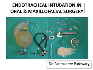
  • 11. Armamentarium i. Anesthetic Mask ii. Laryngoscope iii. Endotracheal Tubes iv. Airways v. Laryngeal Mask Airway (LMA) vi. Magill’s forceps vii. Ambu Bag viii. Stylet
  • 12. Mask is an essential part of any anaesthetic breathing system during the induction phase (beginning) of anaesthesia. A mask allows administration of gases from the breathing system, without introducing any invasive equipment (e.g. an endotracheal tube) into the patient’s airway. Face-masks are designed to fit the patient’s face perfectly without any leaks. • Parts: I. Connector or mount II. Body III. Edge or seal • Sizes: 1, 2, 3, 4, 5. Anesthetic Mask
  • 14. LARYNGOSCOPIC BLADE • Macintosh (curved) blade (most popular) • Miller (straight) blade (useful for neonates and small children)
  • 15. Laryngoscope is designed for doing direct laryngoscopy (directly viewing the vocal cords) and to pass an endo tracheal tube into the larynx under vision. • Parts: Handle, blade with light bulb. • Sizes (for the blades) Neonate (infant), Paediatric (child), adult and extra large.
  • 16. Endotracheal tube is a specially designed tube with one end straight and the other being obliquely cut (beveled end). Beveled end enters the trachea. It is used for the surgical procedures in which it is not feasible to administer anaesthetic gases via mask, procedures which are long- lasting, procedures in which there are chances of having blood, secretions, pus, vomiting etc. in the oral cavity or where patient needs to be given muscle relaxants and controlled breathing. Endotracheal Tubes
  • 17. It is always passed with direct laryngoscopy under vision after anaesthetizing the patient, except in cases, where patients are unable to open mouth, e.g. T.M. joint ankylosis or when a difficult intubation is expected. Age Weight (Inner) diameter (mm) New born 3 kg 2.5–3.0 0–6 months 3.5 kg 3.0–3.5 6–12 months 7 kg 3.5–4.0 1–3 years 10–12 kg 4.0–4.5 4–7 years 16–18 kg 5.0–5.5 8–10 years 24–30 kg 5.5–6.5 Adult female 7.0-7.5 Adult male 8.0-9.0
  • 18. Types of endotracheal tube include 1. oral (south pole) or nasal (north pole) 2. cuffed or uncuffed 3. preformed (e.g. RAE (Ring, Adair, and Elwyn) tube 4. reinforced tubes 5. double-lumen endobronchial tubes.
  • 19. The purpose of introducing an airway is to lift the tongue and epiglottis away from the posterior pharyngeal wall and prevent them from obstructing the space above the larynx. Airways I. Oropharyngeal (Oral) Airway (Placed from Lips to Pharynx) II. Nasopharyngeal (Nasal) Airway (Placed from the Nose to the Pharynx)
  • 20. Oropharyngeal airway: In basic airway management, the oropharyngeal airway is used to provide a right airway to make possible for chest ventilation. In advanced airway management, it can be inserted following endotracheal intubation to act as a bite-block to protect the endotracheal tube. • Parts: Flange, bite portion, air/suction channel (curved portion) • Sizes: 1, 2, 3, 4.
  • 21. Nasopharyngeal airway is a tube that is designed to be inserted into the nose to secure an open airway. When a patient becomes unconscious, the muscles in the jaw commonly relax and can allow the tongue to slide back and obstruct the airway. The purpose of the flared end is to prevent the device from becoming lost inside the patient's nose. •Parts: Flange or a movable disc attached to a tube • Sizes (diameter) — 7.0/7.5 for adult males — 6.5/7.0 for adult females — Same/one size smaller than an appropriate endotracheal tube for children
  • 22. Laryngeal Mask Airway (LMA) Most popular supraglottic airway in emergency and short procedures. Consist of a tube with an inflatable cuff that is inserted into the pharynx . Very useful device in “can’t intubate / can’t ventilate” scenario. Parts: Mask, tube at an angle of 30 degrees, black line on tube to face upper incisors and pilot balloon. • Sizes: 1, 1.5, 2, 2.5, 3, 4, 5.
  • 23. Designed by Dr Archie Brain in UK in 1981 and released in 1988
  • 24. Indications: • When endotracheal intubation is not necessary or it is difficult Contraindications: • Non-fasted patients • Obstructive or abnormal lesions of the oropharynx
  • 25. Advantages: • Allows rapid access • Does not require laryngoscope • Relaxants not needed • Provides controlled airway • Ventilation • Tolerated at lighter anesthetic planes Disadvantages: • Does not fully protect against aspiration in the non-fasted patient • Requires re-sterilization
  • 26. Magill’s forceps is an instrument, which is most often used for guiding an endotracheal tube, from the pharynx into the larynx during nasal intubation. It is also used to pack the throat with a roller gauze during oral and pharyngeal surgery, to pick up a broken or dislodged tooth lying in the oral cavity or to pass a ryles (nasogastric) tube. It is L-shaped and it has no catch. It is available in two sizes—adult and paediatric. Magill’s forceps
  • 27. •It is a combination of a self-inflating bag, a non- rebreathing valve and a facemask. • As it is useful for ventilating a patient in emergency situation, it should always be available in the operation theater as handy as possible. •If any problem occurs with anaesthesia machine or a breathing system, which cannot be diagnosed or corrected quickly, this bag will allow the user to ventilate the patient with room air (with or without oxygen) till the problem is solved. Ambu Bag
  • 28. It is also useful and should be available during patient transport. It can also be used for administering anaesthesia in the absence of an anaesthesia machine, e.g., in rural set- up or field situations.
  • 29. A stylet can be inserted inside an endotracheal tube to make it more rigid, or to change the shape of the tube. It is recommended that it can be used in all emergency intubations. In this way, if the shape of the tube needs to be modified, the stylet is already in place. The stylet should be lubricated before insertion into the endotracheal tube, so that it is easy to remove. Stylet
  • 30. I. Orotracheal intubation II. Nasoendotracheal intubation III. Retromolar intubation IV. Submental intubation V. Retrograde intubation VI. Fibroptic intubation Types of Endotracheal intubation
  • 31. Orotracheal Intubation • The oral route for intubating is preferred since it can usually be performed more rapidly and provides for better visualization. • In addition, the mouth can accommodate a larger tube than the nose.
  • 32. INDICATIONS -Failure to oxygenate adequately. -Failure to ventilate adequately. -Failure of or need for airway maintenance or protection. -Expected need for intubation based on progression of underlying physiologic process. CONTRAINDICATIONS -Do Not Intubate (DNI) order -Partial tracheal transection -Difficult airway (relative)
  • 34. Head positioning for tracheal intubation. A . Neutral position. B. Head elevated. C.“Sniffing” position with a flexed neck and extended head. Note that flexing the neck while extending the head lines up the various axes and allows direct laryngoscopy. D. Ramped position with elevation of the upper part of the back, neck, and head.
  • 35.
  • 36. Trachlight • Light-guided intubation using the trachlight is a safe and gentle technique for both oral and nasal endotracheal tube placement and positioning . • A well defined circumscribed glow can be seen in the anterior neck when the endotracheal tube and light enter into the glottic opening.
  • 37.
  • 38. Advantages • Minimal neck movement • Useful for laryngoscopy • Portable and inexpensive • Functional in bloody airway • Provides definitive airway Disadvantages • Blind technique • May damage airway • Expertise requires practice
  • 39. Conformation • Chest lift • Movement of moisture column in ETT • Auscultation • Improvement and maintenance of SpO2 • Exhaled carbon dioxide
  • 40. Nasoendotracheal intubation • Nasal intubation is applied for dental procedures performed in the hospital, dental office when possible. • Although many who provide anesthesia services are familiar and comfortable with nasal intubation techniques, some are uncomfortable because of lack of experience or fear of nasopharyngeal bleeding and trauma. • It has been observed from experiences in various settings that many approaches may be adapted to the technique of achieving nasal intubation
  • 41. • Most commonly for dental procedures and intraoral (eg, mandibular reconstructive procedures) and oropharyngeal surgeries • Major maxillofacial surgeries • Securing the airway questionable cervical spine stability or severe degenerative cervical spine disease • Mass lesions or structural abnormalities • Limited mouth opening (eg : trismus). Indications
  • 42.  Absolute contraindications • Suspected inflammation of the epiglottis • Midface instability • Coagulopathy • Suspected fractures in base of the skull • Sleeping Apnea  Relative contraindications • Large nasal polyps • Suspected nasal foreign bodies • Recent nasal surgery • Upper neck hematoma or infection • History of frequent episodes of epistaxis • Prosthetic heart valve (increased risk of bacteremia during the insertion) Contraindications
  • 43. Technique of Nasotracheal Intubation Examine both nostrils to determine which is larger. Spray the nasal passages and back of the throat with an appropriate topical anesthetic and vasoconstrictor (neosynephrine) in order to numb the mucosa and reduce bleeding.  Use of xylocaine jelly and a nasal airway before intubation, it may increase anesthesia and reduce bleeding. Remove the airway before intubation.
  • 44. Pre-oxygenate the patient at 100% and keep the patient on Oxygen by a cannula in the mouth during the entire procedure. Check the balloon on an appropriately sized nasotracheal tube.  Firmly seat the 15 mm adapter in the proximal end of the tube and lubricate the distal 4 cm with xylocaine jelly. Position the patients head in midline neutral position if possible.  Patient may be sitting or supine position. Tell the patient that patient may feel like gagging or coughing when the tube is inserted. Advise the patient to resist gagging or coughing. Reassure and calm the patient.
  • 45.  With gentle pressure, insert the tube directed towards the occipital protuberance on the back of the skull with the bevel turned towards the nasal septum. Do not aim the tube up towards the vertex of the skull. If the tube will not pass on one side, try the other. Some resistance may be encountered when the tube reaches the posterior nasopharynx . At this point some gentle manipulation may need for pass the resistance. Turn the tube 1/4 turn after reaching the nasopharynx . Do not force it.
  • 46. When the tube has reached the oropharynx at the back of the mouth, pay attention at the end of the tube for air moving in and out with each respiration. Gently but decisively push the tube further into the nostril and tube is enter into the trachea. Do not force the tube down as structures in the larynx. If properly placed, the tube will easily slip into the trachea. Confirm tube position in the same manner as for an orotracheal procedure. Control bleeding by gently pinching the nostrils around the tube. Secure tube with tape.
  • 47. Retromolar Intubation Patients with panfacial trauma require specific considerations for securing airway intraoperatively. The necessity of intraoperative restoration of dental occlusion by maxillomandibular fixation (MMF) makes the presence of oral endotracheal tube unfeasible. Nasotracheal intubation is contraindicated in certain conditions – viz fracture of base of skull, fracture of naso-orbital-ethmoid complex, etc.In such cases, retromolar intubation, a non-invasive technique of securing airway in patients with panfacial trauma, avoids both submento-tracheal intubation and tracheostomy.
  • 48. •Orotracheal intubation is done initially with a flexometallic tracheal tube using standard general anaesthesia technique. The aim is to place the orotracheal tube in the retromolar space (space behind the last upper and lower erupted molar teeth) . • The orotracheal tube is grasped with gloved fingers and is placed into the retromolar space. Technique
  • 49. • The tube is then fixed by a wire ligature to the molar/premolar tooth. • The wire ligature is the same that is used for MMF. It fixes the retromolar tracheal tube with the tooth in a “figure of eight” fashion. • The retromolar tracheal tube allows adequate dental occlusion.
  • 50. Advantages If retromolar intubation can be done with adequate dental occlusion, then tracheostomy and submento - tracheal intubation techniques can be avoided the. Disadvantages 1. In some patients, the retromolar space is not adequate. After retromolar placement of the tracheal tube dental occlusion is not possible. Therefore, intraoperative MMF cannot be done.
  • 51. 2.The tracheal tube can interfere with the main surgical field that may be partially draped. 3.The tracheal tube can also interfere with positioning and application of dental fixation devices, especially in patients with bilateral maxillary/ mandibular fractures.
  • 52. Submental Intubation Submental route of endotracheal intubation is a simple, safe and useful technique in maxillofacial trauma, where oral and nasal endotracheal intubation cannot be performed. It avoids the need for tracheostomy.
  • 53. Technique • After a standard orotracheal intubation, a temporary draping of the mouth and chin was carried out. • Then, a 2-cm skin incision was made in the median region of the submental area, directly adjacent to the lower border of the mandible.
  • 54. • The muscular layers (platisma and mylohyoid muscles) were traversed by blunt dissection using a Kelly forceps that was always in contact with the lingual cortex of the mandible. • The mucosal layer on the floor of the mouth was incised over the distal end of the forceps, and the forceps were then opened, creating a tunnel. During the dissection it is important that the width of the submental access should be sufficient to pass the tube without any interference.
  • 55. •It is important to make sure that the tube has not been displaced during its passage through the submental tunnel. •After the surgery, the submental intubation was converted to an orotracheal intubation by replacing the tube in the mouth and carrying out extubation in the classical manner. Minimal scar postoperative.
  • 56. •If the patient needed long-period postoperative ventilation, the orotracheal intubation was maintained until extubation was complete. •The suture was done with 5-0 nylon in the skin and 4-0 vycril in the internal layers.
  • 57. Retrograde Intubation Ankylosing spondylitis (AS) patients are most challenging. Sound clinical judgment is critical for timing and selecting the method for airway intervention. The retrograde intubation technique is an important option when fiberoptic bronchoscope is not available, and other method is not applicable for gaining airway access for surgery in supaine position.
  • 58. Indications 1. Facial Anomalies: a. Maxillary hypoplasia b. Mandibular hypoplasia c. Mandibular hyperplasia 2. Temporomandibular joint pathology: a. Ankylosis or reduced movment
  • 59. 3. Anomalies of the mouth and tongue: a. Microstomia (burns, trauma scarring) b. Diseases of the tongue (burns, trauma, Ludwig angina) all lead to tongue swelling c. Tumors of the mouth and tongue (hemangioma, lymphangioma) d. Macroglossia (Down syndrome, hypothyroidism) 4. Problem with teeth (missing left upper incisors, protruding upper incisors)
  • 60. Contraindication • Adequate mouth opening and easily performed orotracheal intubation • Relative contraindication: Systemic coagulopathy, infection in the skin overlying the cricothyroid membrane.
  • 61.
  • 62. Fiberoptically guided tracheal intubation represents one of the most important advances in airway management to occur in the past thirty years. Perhaps its most important role is in management of the anticipated difficult airway. This is a situation in which the dangers of encountering the life- threatening "can't intubate, can't ventilate" situation can be avoided by placement of an endotracheal tube while the patient is awake. Fiberoptic Intubation
  • 63. Indications: • Predictable difficult intubation (upper airway abnormality) • Cases where neck extension is not desirable • Nasal fiberoptic intubation in Oral Submucous Fibrosis Contraindications: • Inability to oxygenate • Major bleeding
  • 64. 1. Eye piece 2. Diopter ring for focusing 3. Control lever 4. Working channel port 5. Body 6. Insertion cord 7. Light source 8. Suction valve and port Fiberoptic Bronchoscope
  • 65. Technique • Oral or nasal route is preferable • Jaw thrust maneuver to improve visualization • After the broncoscope is lubricated and loaded with endotracheal tube it is introduced strictly in the midline following the base of the tongue, pass the uvula, behind epiglottis and between the vocal cords. • Once the carina is visualized endotracheal tube is introduced by rotating movement over the broncoscope.
  • 66.
  • 68. 1) During intubation 2) During remained intubation 3) During extubation 4) After extubation
  • 69. During intubation • Trauma to lip, tongue or teeth • Hypertension and tachycardia • Pulmonary aspiration. • Laryngospasm • Bronchospasm • Laryngeal edema • Arytenoid dislocation • Increased intracranial pressure • Spinal cord trauma in cervical spine injury
  • 70. During remained intubation • Accidental extubation or endobronchial intubation. • Disconnection from breathing circuit Pulmonary aspiration. • Lip or nasal ulcer in case with prolong period of intubation. • Sinusitis in case with prolong nasoendotracheal intubation. • Esophageal intubation • Epistaxis
  • 71. • Laryngospasm • Pulmonary aspiration • Edema of upper airway During extubation After extubation • Sore throat • Tracheal infection (Prolong intubation) • Laryngeal granuloma
  • 72. Conclusion Airway management is the most important skill required of an anaesthesiologist and also for oral and maxillofacial surgeons. Endotracheal intubation is a skill based on the knowledge of airway anatomy, equipments for airway manipulation, also the use of pharmacological agents and experience.
  • 73. REFERENCES • Miller’s Anesthesia, 7th Edition by Ronald D. Miller. • Clinical Anesthesiology, 4th Edition by G. Edward Morgan, Jr., Maged S. Mikhail, Michael J. Murray. • Bailey & Love’s Short Practice of Surgery, 25th Edition. • M Polansky. Airway Management: The Basics Of Endotracheal Intubation. The Internet Journal of Academic Physician Assistants 1996;(1):1. • Christopher Kabrhel, Todd W. Thomsen, Gary S. Setnik, Ron M. Walls. Orotracheal Intubation. N Engl J Med 2007;356:e15 • Endotracheal intubation: MedlinePlus Medical Encyclopedia • Textbook of OMFS , 3rd edition by Neelima malik • Malhotra N. Bhardwaj Chari P. :SUBMENTAL ENDOTRACHEAL INTUBATION:A USEFUL ALTERNATIVE TO TRACHEOSTOMYINDIAN JOURNAL OF ANAESTHESIA, OCTOBER 2002 2002; 46 (5) : 400-402

Editor's Notes

  1. airway
  2. Armamentarium
  3. Class I: Pharyngeal pillars, entire palate, and uvula are visible. Class II: Pharyngeal pillars and soft palate are visible, with visualization of the uvula obstructed by the tongue. Class III: Soft palate is visible, but pharyngeal pillars and uvula are not visualized. Class IV: Only the hard palate is visible; the soft palate, pillars, and uvula are not visualized.
  4. Proper positioning of the clinician, patient, and assistant for tracheal intubation. The following points are demonstrated: the difficult airway cart is adjacent to the patient, the suction device is at the head of the bed, the patient is in the “sniffing position” with the occiput elevated and the clinician’s right hand ready for additional adjustment if necessary, the bed is elevated and the clinician is at the appropriate distance from the patient, and the laryngoscope handle is angled at 45 degrees.
  5. Eye piece: Can be attached to a camera for display on screen. fiberoptic scopes have an eye piece; video scopes do not. Diopter ring for focusing Control lever: Controls the tip. Only permits movement in a vertical plane. Two wires extend from the handle to the tip in the insertion cord. Moving the lever down, moves the tip up and moving the lever up, points the tip down. Side to side movement is accomplished by rotation of the body of the bronchoscope with the operator's wrist and shoulder. Working channel port: For suction, instillation of local anesthetic, oxygen delivery. Body: Incorporates the eye piece, diopter ring, control level and working channel. Grasped by the operators non-dominant hand. Insertion cord: Contains fiberoptic bundle for light and image transmission, tip bending control wires and working channel. Average length 600mm (range 500 – 650mm). Light source: Can be a portable battery powered source or via a cable. Light source may be halogen, incandescent or LED. Suction valve and port