SlideShare a Scribd company logo
1 of 32
Download to read offline
Metabolic Syndrome
and associated pathologies
TECOmedical Clinical and Technical Review
March 2013
Authors:
Andreas Pfützner, M.D., Ph.D.,1, 2
and Peter Haima, Ph.D.,3
1.	IKFE – Institute for Clinical Research and Development, Mainz/Germany
Concept: BEVAIR (Beta-cell dysfunction, Visceral Adipogenesis  Insulin Resistance) Patent pending
2.	 University of Applied Sciences, Rheinbach/Germany
3.	 Life-Force biomedical communication, Netherlands
2
Contents
1 Introduction 	 .  .  .  .  .  .  .  .  .  .  .  .  .  .  .  .  .  .  .  .  .  .  .  .  .  .  .  .  .  .  .  .  .  .  .  .  .  .  .  .  .  .  .  .  .  .  .  .  .  .  .  .  .  .  .  .  .  .  .  .  .  .  .  .  .  .  .  .  .  .  .  .  .  .  .  .  .  .  . 	3
2 Metabolic syndrome .  .  .  .  .  .  .  .  .  .  .  .  .  .  .  .  .  .  .  .  .  .  .  .  .  .  .  .  .  .  .  .  .  .  .  .  .  .  .  .  .  .  .  .  .  .  .  .  .  .  .  .  .  .  .  .  .  .  .  .  .  .  .  .  .  .  .  .  .  .  .  .  . 	4
	 a Definition and pathophysiology of metabolic syndrome . . . . . . . . . . . . . . . . . . . . . . . . . . . . . . . . . . . . . . . . . 	4
	 b The role of adipokines. . . . . . . . . . . . . . . . . . . . . . . . . . . . . . . . . . . . . . . . . . . . . . . . . . . . . . . . . . . . . . . . . . . 	5
		I	 Adiponectin . . . . . . . . . . . . . . . . . . . . . . . . . . . . . . . . . . . . . . . . . . . . . . . . . . . . . . . . . . . . . . . . . . . . . 	5
		II	Leptin. . . . . . . . . . . . . . . . . . . . . . . . . . . . . . . . . . . . . . . . . . . . . . . . . . . . . . . . . . . . . . . . . . . . . . . . . . 	6
	 c Development into diabetes. . . . . . . . . . . . . . . . . . . . . . . . . . . . . . . . . . . . . . . . . . . . . . . . . . . . . . . . . . . . . . . . 	6
3 Insulin resistance and diabetes mellitus type 2 .  .  .  .  .  .  .  .  .  .  .  .  .  .  .  .  .  .  .  .  .  .  .  .  .  .  .  .  .  .  .  .  .  .  .  .  .  .  .  .  .  .  .  .  .  .  .  .  .  .  . 	6
	 a Pathophysiology . . . . . . . . . . . . . . . . . . . . . . . . . . . . . . . . . . . . . . . . . . . . . . . . . . . . . . . . . . . . . . . . . . . . . . . 	7
	 b Development stages of β-cell dysfunction. . . . . . . . . . . . . . . . . . . . . . . . . . . . . . . . . . . . . . . . . . . . . . . . . . . . 	7
	 c Diagnosis of insulin resistance and diabetes type 2. . . . . . . . . . . . . . . . . . . . . . . . . . . . . . . . . . . . . . . . . . . . . 	8
		I	 Glucose testing . . . . . . . . . . . . . . . . . . . . . . . . . . . . . . . . . . . . . . . . . . . . . . . . . . . . . . . . . . . . . . . . . . 	8
		II	 HOMA score to assess early stages of β-cell dysfunction . . . . . . . . . . . . . . . . . . . . . . . . . . . . . . . . . 	8
		III	Intact proinsulin testing to assess late stage β-cell dysfunction. . . . . . . . . . . . . . . . . . . . . . . . . . . . . 	8
		IV	Intact proinsulin combined with oral glucose tolerance testing to identify prediabetes patients. . . . 	9
4 Diabetes related cardiovascular disease .  .  .  .  .  .  .  .  .  .  .  .  .  .  .  .  .  .  .  .  .  .  .  .  .  .  .  .  .  .  .  .  .  .  .  .  .  .  .  .  .  .  .  .  .  .  .  .  .  .  .  .  .  .  .  . 	10
	 a pathophysiology . . . . . . . . . . . . . . . . . . . . . . . . . . . . . . . . . . . . . . . . . . . . . . . . . . . . . . . . . . . . . . . . . . . . . . . . 	10
	 b Biochemical markers for diagnosis and therapy selection. . . . . . . . . . . . . . . . . . . . . . . . . . . . . . . . . . . . . . . . 	12
		I	 Proinsulin – a blood glucose independent marker to assess insulin resistance and β-cell dysfunction	12
		II	 Adiponectin – a visceral adipose tissue activity marker to predict cardiovascular risk. . . . . . . . . . . . 	12
		III	hsCRP – a chronic inflammation marker to predict cardiovascular risk . . . . . . . . . . . . . . . . . . . . . . . 	13
5 Other metabolic syndrome-associated diseases. .  .  .  .  .  .  .  .  .  .  .  .  .  .  .  .  .  .  .  .  .  .  .  .  .  .  .  .  .  .  .  .  .  .  .  .  .  .  .  .  .  .  .  .  .  .  .  .  . 	13
	 a Fatty liver disease . . . . . . . . . . . . . . . . . . . . . . . . . . . . . . . . . . . . . . . . . . . . . . . . . . . . . . . . . . . . . . . . . . . . . . 	13
	 b PCOS: polycystic ovary syndrome . . . . . . . . . . . . . . . . . . . . . . . . . . . . . . . . . . . . . . . . . . . . . . . . . . . . . . . . . 	14
6 Risk assessment and therapy of metabolic syndrome-associated pathologies .  .  .  .  .  .  .  .  .  .  .  .  .  .  .  .  .  .  .  .  .  .  .  . 	15
	 a Diagnosis and risk assessment . . . . . . . . . . . . . . . . . . . . . . . . . . . . . . . . . . . . . . . . . . . . . . . . . . . . . . . . . . . . 	15
	 b Therapy and monitoring . . . . . . . . . . . . . . . . . . . . . . . . . . . . . . . . . . . . . . . . . . . . . . . . . . . . . . . . . . . . . . . . . . 	15
7 References 	 .  .  .  .  .  .  .  .  .  .  .  .  .  .  .  .  .  .  .  .  .  .  .  .  .  .  .  .  .  .  .  .  .  .  .  .  .  .  .  .  .  .  .  .  .  .  .  .  .  .  .  .  .  .  .  .  .  .  .  .  .  .  .  .  .  .  .  .  .  .  .  .  .  .  .  .  .  .  . 	16
3
1 Introduction
Metabolic syndrome is a combination of risk factors — abdominal obesity, hypertension, high blood sugar levels, insulin
resistance (IR), abnormal cholesterol levels, hyperlipidemia and inflammatory states — that increase the risk of heart
disease, stroke, diabetes type 2, fatty liver disease and PCOS (in young women). Having any of these conditions increase
the risk of serious disease. If more than one of these conditions occur in combination, the risk is even greater.
Metabolic syndrome is now present in up to 40 % of the United States adult population (prevalence on average 25 % of
the population and 40+ % from age 60 and higher) [2] and is associated with a nearly a two fold increase in cardiovascular
events, independent of the presence of diabetes mellitus. Obesity is the dominant key feature of metabolic syndrome
[1, 2], although patients of normal weight may also suffer from IR and metabolic syndrome, and obesity and metabolic
syndrome do not always occur in concordance as there is some evidence for conditions of benign obesity [3–7]. The
epidemic of obesity in in adults and children in both industrialized and third world countries is regarded as one of the most
serious public health problems of the 21st
century.
Many (especially overweight) people with IR are able to compensate for the increasing insulin requirement, so that the
blood sugar does not rise at first. Late stage β-cell dysfunction then develops into clinically manifest type 2 diabe-
tes in approximately one third of these patients [31]. Development of macrovascular damages may, therefore, already
have developed before type 2 diabetes is clinically manifest (normal or impaired glucose tolerance test result). These
macrovascular damages are, in part, irreversible, need to be treated lifelong and still are the actual cause of death in
75 % of diabetes type 2 patients.
Up to now, the underlying pathomechanisms of metabolic syndrome are not detected at all or only very late and
inadequately. In this review we discuss the latest clinical insights into Metabolic syndrome and related disease like
diabetes type 2, fatty liver disease, coronary disease and PCOS. Conventional diagnosis is discussed alongside the use
of new biomarkers. Three new biomarkers appear to be particularly suitable for early diagnosis and therapy selection due
to their stability and studies that are presently available: intact Proinsulin, Adiponectin and hsCRP [29]. In addition, the
effect of the therapy used on pathophysiological basic components can be checked by means of these new markers.
4
(1)	 TG ≥ 1.695 mmol/L and HDL ≤ 0.9 mmol/L (male), ≤1.0 mmol/L (female).
(2)	 Waist/hip ratio  0.90 (male) 0.85 (female), or body mass index  30 kg/m2.
(3)	 Urinary albumin excretion ratio ≥ 20 μg/min or albumin/creatinine ratio ≥ 30 mg/g.
(4)	 Waist circumference ≥ 94 cm (male), ≥80 cm (female).
(5)	 TG ≥ 2.0 mmol/L and/or HDL  1.0 mmol/L or treated for dyslipidemia.
(6)	 Men, greater than 40 inches (102 cm) and women, greater than 35 inches (88 cm).
(7)	 Equal to or greater than 150 mg/dL (1.7 mmol/L).
(8)	 Men, Less than 40 mg/dL (1.03 mmol/L) and women, Less than 50 mg/dL (1.29 mmol/L).
(9)	 Equal to or greater than 130/85 mm Hg or use of medication for hypertension.
(10)	 Equal to or greater than 100 mg/dL (5.6 mmol/L) or use of medication for hyperglycemia.
(11)	Defined as waist circumference with ethnicity specific values (If BMI is 30 kg/m², central obesity can be assumed and waist
circumference does not need to be measured).
(12)	 TG  150 mg/dL (1.7 mmol/L), or specific treatment for this lipid abnormality.
(13)	 HDL  40 mg/dL (1.03 mmol/L) in males, 50 mg/dL (1.29 mmol/L) in females, or specific treatment for this lipid abnormality.
(14)	 Systolic BP  130 or diastolic BP  85 mm Hg, or treatment of previously diagnosed hypertension.
(15)	 FPG  100 mg/dL (5.6 mmol/L), or previously diagnosed type 2 diabetes.
2 Metabolic syndrome
a. Definition and pathophysiology of metabolic syndrome
Metabolic syndrome is a cluster of risk factors — abdominal obesity, hypertension, high blood sugar levels, IR, abnormal
cholesterol levels, hyperlipidemia and inflammatory states — that increase the risk of heart disease, stroke, diabetes
type 2, fatty liver disease and PCOS (in young women). It has also been variously termed X syndrome, insulin resistance
syndrome, metabolic syndrome X, cardiometabolic syndrome, syndrome X, Reaven's syndrome, and CHAOS (in Australia).
The diagnostic criteria for metabolic syndrome have been set out by different organizations with slight variations in these
criteria as shown in Table 1 [8].
WHO
World Health Organization
EGIR
European Group for the Study
of Insulin Resistance
NCEP
US National Cholesterol
Education Program
IDF
International Diabetes
Federation
Presence of one of
following:
Insulin resistance AND two
or more of following:
Presence of three of
following (2004):
Central obesity (11)
AND
any two of following:
DM / IGT / IFG / Insulin
resistance
Central obesity (4)
Elevated waist
circumference (6)
Raised TG (12)
AND two of the following: Dyslipidemia (5) Elevated TG (7) ▼ HDL (13)
BP ≥ 140/90 BP ≥ 140/90
Reduced HDL (8) ▲ BP (16)
Dyslipidemia (1) FBG ≥ 6.1 mmol/L (110
mg/dL)
Elevated BP (9) ▲ FBG (17)
Central obesity (2) Elevated fasting glucose (10)
Microalbuminuria (3)
BP: blood pressure, DM: diabetes mellitus, FBG: fasting blood glucose, HDL: high density lipoproteins, IFG: impaired fasting glucose, IGT: impaired glucose
tolerance, TG: triglycerides.
Table 1: Comparison of definitions of the metabolic syndrome.
5
Figure 1: the link between obesity, inflammation, β-cell dysfunction, insulin
resistance and cardiovascular risk [28, 29]
Oxidative stress, which is defined as imbalance between
the production and inactivation of reactive oxygen
species, has a major pathophysiological role in all the
components of metabolic syndrome [20–24]. Oxidative
stress and consequent inflammation induce insulin
resistance (IR) which likely initiates metabolic syndrome
and massive damage of pancreatic β-cell dysfunction.
The association between the metabolic syndrome and
inflammation is well documented [9]. Welsh et al. [10]
demonstrated that adiposity leads to higher levels of
the “acute phase” inflammatory protein CRP (C-reactive
protein) and accumulating evidence demonstrates a close
link among metabolic syndrome, chronic inflammation and
oxidativestress[11].Infact,theoxidativestress-inflammation
pathway has important roles in all the individual components of Metabolic syndrome including vascular alterations
[11-15]. Figure 1 shows the link between obesity, inflammation, insulin resistance, β-cell dysfunction, and cardiovascular
risk. [28, 29]. Adipo(cyto)kines (e.g. Adiponectin) and other factors produced by fat tissue and antiinsulinemic hormones
play a key role in the process.
b. The role of adipokines
Multiple adipokines can be held responsible for the negative consequences of abdominal adipogenesis on insulin
resistance. The growth of lipid tissue is induced by the differentiation of mesenchymal stem cells to become
preadopocytes and finally mature lipid cells. In this stage peripheral monocytes/macrophages migrate into the lipid tissue
and are kept at a constantly increased level of activation by the secretion of a whole pattern of proinflammatory cytokines
from the pre-adipocyte. In consequence many adipokines have been identified which have been previously described
to be associated with inflammatory conditions in other parts of the body, and which have a known negative influence on
insulin sensitivity, e. g. IL-6 and TNFα. A list of some recently described prominent adipokines is provided in Table 2 [31].
Adiponectin and leptin have been studied most extensively
andplayamajorroleinlipidmetabolismandthedevelopment
of obesity. Further adipokines are resistin and visfatin that
also seem to be linked to insulin resistance and metabolic
syndrome. They are currently under evaluation regarding
their clinical value.
I. Adiponectin
Adiponectin owns an exceptional place in this listing. It is secreted by the mature adipocytes (and the connective tissue)
and not by the pre-adipocytes and has a synergistic action to insulin. High plasma adiponectin concentrations result
in an improvement of insulin sensitivity. An increase in body weight with differentiation of stem cells to pre-adipocytes
is associated with a suppression of adiponectin concentrations in the circulation [38]. Other disease conditions that
have been described to be correlated with a suppression of adiponectin levels include, but are not limited to metabolic
syndrome, atherosclerosis and any kind of obesity. Adiponectin may, therefore, be regarded as an indicator of the
activity of pre-adipocytes. Female patients have higher reference values than male patients. Several plasma sub-fractions
have been described that are differentiated by the agglomeration of different numbers of single adiponectin molecules.
However, they have as of yet not shown any difference in their changing behaviour following therapeutic interventions.
For practical use it does, therefore, not really matter, whether “High Molecular Weight” or “Low Molecular Weight”
adiponectin is determined for diagnostic purposes, as long as the same sub-fraction is used to draw any diagnostic or
clinical conclusions [40]. Adiponectin levels respond quickly to changes in insulin resistance and the metabolic situation
in the lipid tissue. It is, therefore, suitable to track slight changes in insulin resistance, e.g. the metabolic deterioration
induced by the hormonal changes in women with polycystic ovary syndrome (PCOS) [41, 42].
Leptin Free Fatty acids
Interleukine-6 PAI-I and tPA
Retinol Binding protein 4 Angiotensin II
Adipsin (Complementation factor D) TNF-alpha
Adiponectin Visfatin
Resistin Vaspin
Table 2: Recently described adipokines
Anti-insulinemic hormones
Insulin Resistance
β-Cell
Dysfunction
β-Cell
Dysfunction
IL-6, TNFα etc.
Free
Fatty Acids
Angiotensin
Insulin-
requirement
Insulin
Proinsulin
Insulin
Proinsulin
Adiponectin
Adipogenesis
Lipid Cell
Pre-Adipocyte
Stem Cell
Stem Cell
Stem Cell
Leptin
Anti-insulinemic hormones
Insulin Resistance
β-Cell
Dysfunction
IL-6, TNFα etc.
Free
Fatty Acids
Angiotensin
Insulin-
requirement
Insulin
Proinsulin
Adiponectin
Adipogenesis
OBESITY
Lipid Cell
Pre-Adipocyte
Leptin AdiponectinC
B
Arteriosclerosis HypertensionDyslipidemia
Hyperglycemia
Intact
Proinsulin
hsCRP
6
II. Leptin
Leptin is a 16kDa non-glycosylated protein that is predo-
minantly secreted by mature lipid cells, but can also de-
rive in minor amounts from the stomach, intestine tract,
muscle and breast tissue. The plasma leptin levels reflect
the actual amount of lipid tissue, the size of the adipocytes
and their triglyceride content. In consequence, plasma leptin
concentrations are elevated in case of obesity and decre-
ase with a loss in body weight [43]. These changes are in-
fluenced by the actual insulin and glucose concentrations
and by inflammatory cytokines. In addition, leptin plays a
role in the control of energy consumption, in angiogenesis,
fertility, bone formation and many other endocrine body
functions [44]. Leptin levels are higher in female patients,
most probably because of the larger amount of subcuta-
neous lipid tissue and a higher stimulation by estrogens in
women. Leptin concentrations decrease in a cold environment and during adrenergic stimulation. The brain uses leptin as
an important control variable for appetite regulation. It carries the information, whether “sufficient” amounts of lipid tissue
are prevalent and, therefore, leptin, like insulin, belongs to the lipostatic molecules. The lipostatic factors orchestrate to-
gether with the short-acting incretines (e. g. GLP-1, ghrelin, GIP, cholecystokinin (CCK), obestatin, PYY etc.) the nutritional
behaviour of the human organism (see Figure 2, [45]).
c. Development into diabetes
Oxidative stress and consequent inflammation induce IR, which likely initiates metabolic syndrome. Afterwards, there are
2 principal pathways of metabolic syndrome development [16]:
A. With preserved pancreatic beta cells function and
insulin hypersecretion which can compensate for
insulin resistance. This pathway leads mainly to the
macrovascular complications of metabolic syndrome.
B. With massive damage of pancreatic beta cells leading
to progressively decrease of insulin secretion and to
hyperglycemia (e.g. overt type 2 diabetes).
This pathway leads to both microvascular and
macrovascular complications.
An insulin-resistant state – as the key phase of metabolic syndrome – constitutes the major risk factor for the development
of diabetes mellitus. Hyperinsulinemia appears to be a compensatory mechanism that responds to increased levels of
circulating glucose. Fasting glucose is presumed to remain normal as long as insulin hypersecretion can compensate for
insulin resistance. The fall in insulin secretion leading to hyperglicemia occurs as a late phenomenon.
3 Insulin resistance and diabetes mellitus type 2
In type 2 diabetes, fat, liver, and muscle cells do not respond correctly to insulin anymore. Due to this insulin resistance
(IR), blood sugar does not enter these cells and consequently high levels of sugar build up in the blood. This is called
hyperglycemia. Type 2 diabetes usually occurs slowly over time. Most people with the disease are overweight when they
are diagnosed. Type 2 diabetes can also develop in people who are thin. This is more common in the elderly. Family
history and genes play a large role in type 2 diabetes. Low activity level, poor diet, and excess body weight around the
waist increase the risk.
Figure 3: The relationship between metabolic syndrome, insulin resistance,
hyper-insulinemia and hyperglycemia (overt type 2 diabetes), adapted from [16]
M e t a b o l i c S y n d r o m e
insulin resistance
Life time
Diabetes type 2
Pancreatic beta cells
stress  damage
Hyperinsulinemia
Macrovascular
+ Microvascular
complications
Macrovascular
complications
Food intake
excess
Genetic
background
Physical
inactivity
Adipogenesis
Overweight
A
Obesity
Hyperinsulinemia HyperglycemiaB
Figure 2: Factor controlling appetite and food uptake
7
Diabetes type 2 is one possible outcome of Metabolic syndrome. Diabetes type 2 and obesity are two diseases with
continuously growing prevalence over the past decades that have both reached pandemic dimensions in their distribution.
Approximately 4–5 % of the world population are currently affected and the annual incidence in Western Europe is
approximately 10 %. Type 2 diabetes is conventionally diagnosed via elevated blood glucose and glycosylated
hemoglobin (HbA1c) levels.
a. Pathophysiology
The major underlying mechanisms of diabetes type 2 are
the development of systemic insulin resistance and a col-
lapsed insulin secretion by pancreatic β-cells. IR is cha-
racterized by a general decrease of the insulin sensitivity
of the peripheral cells, which on a receptor level is asso-
ciated with a genetically determined change in the insulin
receptor molecule and a reduction of the overall number of
insulin receptors on the cells (post-receptor defects have
also been described in literature). Figure 4, shows the rela-
tion between IR, decreased insulin secretion and adiposity.
b. Development stages of β-cell dysfunction
If a patient shows hereditary or acquired insulin resistance, this will initially be compensated for by an appropriate additional
secretion of insulin. Insulin, however, is the only (known) physiological hormone that stimulates adipogenesis. As a
consequence, this leads to a strong tendency to develop adipose tissue, especially with increased intake of calories. In
advanced stage, the processing of the insulin precursor molecule proinsulin becomes insufficient and increasing amounts
of intact proinsulin are being secreted. Proinsulin has only a fraction of the blood sugar reducing effect of insulin, but has
the same adipogenic potency [3–5].
In consequence, elevated plasma intact Proinsulin levels
are a highly specific direct indicator for advanced β-cell
dysfunction and a highly specific indirect indicator for cli-
nically relevant insulin resistance [21]. Using the fasting
intact proinsulin concentrations and under consideration
of the level of insulin resistance (e. g. by means of the
HOMA score [22]), it is possible to introduce a clinically
useful staging of β-cell dysfunction that allows for a dif-
ferential diagnosis and selection of a pathophysiologically
oriented differential therapy of type 2 diabetes mellitus [23].
This staging is presented in Figure 5 and further detailed
in Table 3.
Figure 5: Staging of β-cell dysfunction by means of insulin resistance and
composition of the β-cell secretion product [23]
Stage Description Insulin Proinsulin Glucose
I Insulin sensitive Normal Normal Normal
II
Insulin resistance without
qualitative secretion disorder
Elevated Normal
Normal or
elevated
III a
Insulin resistance with major
β-cell secretion disorder
Normal/Elevated Elevated
Normal or
elevated
III b Collapsed β-cell secretion Low Elevated to normal (in end stage) Elevated
Table 3: Staging of β-cell dysfunction by means of insulin resistance and composition of the β-cell secretion product [23]
1. Cause of Disease:
Reduced Insulin Effectiveness
(Insulin resistance)
Increased
Insulin
requirements
Visceral Adipogenesis
Weight gain
Hormones and cytokines
from the lipid tissue reduce
insulin sensitivity and induce
dyslipidemia, hypertension
and atherosclerosis
With sufficient food supply new adipose
tissue is built up supported by insulin
but even more by proinsulin
2. Cause of Disease:
Deteriorated Insulin Secretion
(β-Cell dysfunction)
Increased Insulin- and Proinsulin-
Secretion
D
Y
S
L
I
P
I
D
E
M
I
A
A
T
H
E
R
O
S
C
L
E
R
O
S
I
S
H
Y
P
E
R
T
E
N
S
I
O
N
Figure 4: Relation between insulin resistance, β-cell dysfunction, obesity
and the resulting complications [19, 20]
8
II. HOMA score to assess early stages of β-cell dysfunction
The HOMA (Homeostasis model assessment) score is an easy method to estimate the degree of insulin resistance
non-diabetic or early stage diabetic patients [24,25]. It can be applied only if intact proinsulin levels are in the normal range,
as the total secretion activity of the β-cell is under this condition represented by the fasting insulin levels. The HOMA score
is based on the assumption that under normal conditions, a normal blood glucose value is associated with a matching
normal insulin level, which may vary individually from patient to patient. Insulin resistance is indicated, if at this normal
insulin level, an elevated blood glucose is observed, or if more insulin is required to maintain blood glucose at its normal
level.
After determining the fasting serum insulin and fasting plasma glucose levels the HOMA-IR score is calculated as follows:
HOMA = Insulin [μU/ml] x Glucose [mmol/l] /22.5.
Insulin resistance is assumed if the score value exceeds 2 [25]. Of particular interest are changes in the HOMA score
during therapeutic interventions, as a score reduction represents an improvement in insulin sensitivity. Again, the HOMA
score should only be used in patients with stage I and II of β-cell dysfunction (see Figure 3), because intact Proinsulin is
an additional marker for β-cell activity that is not considered in the HOMA-IR score equation.
III. Intact proinsulin testing to assess late stage β-cell dysfunction
Intact proinsulin is produced in the pancreatic β-cells and is normally further processed to insulin and C-peptide. It is
only seen in low concentrations in the plasma of healthy subjects, as it is rapidly degraded (T 1/2
is 15 minutes). Proinsulin
cleavage products (like des32,33) are stable for several hours. As these fragments are inactive and can make up to 50 %
of total Proinsulin, only assays specifically measuring intact proinsulin are suitable to indicate advanced β-cell dysfunction
and IR.
An increase in the insulin demand, as provided by insulin resistance in later stages of type 2 diabetes mellitus, can result
in increased expression of proinsulin into the blood (see Figure 3). When intact proinsulin is secreted together with or
instead of insulin in the fasting state (stage III of β-cells dysfunction, Figure 3) the HOMA score cannot be used to assess
the level of β-cells dysfunction [26, 27]. Intact proinsulin is also able to lower glucose values but is not considered in the
HOMA equations. Measurement of fasting intact proinsulin has been shown to be a very specific indicator and clinically
significant IR.
In clinical practice, fasting morning intact proinsulin can be used as highly specific indicator of of late stage β-cell
dysfunction and clinically relevant IR. It can also to serve as the basis for the selection of an insulin resistance therapy,
and to monitor the therapeutic effect on β-cell dysfunction.
c. Diagnosis of insulin resistance and diabetes type 2
I. Glucose testing
Today, diagnosis and therapy of the type 2 diabetes mellitus is still based on blood sugar values and the associated
values for glycosylated hemoglobin (HbA1c). WHO 2011 guidelines describe that fasting glucose should be 127 mg/dl
and HbA1c  48 mmol/mol. In addition an oral glucose tolerance test (OGTT) is often performed. During an OGGT glucose
is given and blood samples taken afterward to determine how quickly it is cleared from the blood. WHO recommends for
a 75g oral dose in all adults: the dose is adjusted for weight only in children.
Glucose levels  11.1 mmol/L ( 200 mg/dL) at 2 hours confirms the diagnosis of diabetes (see Table 4).
Glucose
levels
Normal
Impaired fasting
glycaemia
Impaired glucose
tolerance
Diabetes
mellitus
Venous Plasma Fasting 2hrs Fasting 2hrs Fasting 2hrs Fasting 2hrs
(mmol/L) 6.1 7.8  6.1  7.0 7.8 7.0 7.8 7.0 11.1
(mg/dL) 110 140 110  126 140 126 140 126 200
Table 4: 1999 WHO Diabetes criteria - Interpretation of Oral Glucose Tolerance Test
9
IV. Intact proinsulin combined with oral glucose tolerance testing to identify prediabetes
patients.
Because of the blood sugar reducing effect of Proinsulin, some patients may already be suffering from β-cell dysfunction
years before clinical manifestation of elevated blood sugar levels. It has been shown that elevation of fasting intact
proinsulin is an indicator of insulin resistance and severe β-cell dysfunction. Earlier detection of prediabetic patients is
important to prevent irreversible cardiovascular damages.
In a recent pilot study [57] it was investigated whether elevation of intact proinsulin after 1 and 2 hours in the course of
an oral glucose tolerance test (OGTT) may be an indicator of the development of type 2 diabetes. Patients were enrolled
based on previous results of OGTT: 11 healthy subjects (7 female, 4 male, age: 59 ± 20 yrs.), 10 patients with Impaired
Glucose Tolerance (IGT; 6 female, 6 male, age: 62 ± 10 yrs.), and 10 patients with overt type 2 diabetes (6 female, 4 male,
age: 53 ± 11 yrs.). Another OGTT was performed with measurement of glucose and intact proinsulin levels after 0h, 1 h and
2 hours. Five years later. The diabetes status of the patients was confirmed and correlated with the earlier OGTT results.
Patients with diabetes (Fig. 6) had elevated fasting glucose levels after 1 and 2 h (diabetes: 0/1/2 h:
121 ± 20/230 ± 51/213 ± 24 mg/dL; prediabetes: 102 ± 9/168 ± 57/149 ± 34 mg/dL; normals: 94 ± 8/140 ± 29/90 ± 24 mg/dL).
Proinsulin values after 2 hours were elevated in diabetes and prediabetes vs. control 27 ± 10 pmol/L and 28 ± 6 pmol/L,
respectively, vs. 10 ± 5 pmol/L (p0.05 vs. both other groups).
Five years later, all patients with IGT and three normal subjects had developed overt type 2 diabetes. All manifesting
patients had elevated intact proinsulin levels ( 11 pmol/L) after 1 and 2 h. A value  20 pmol/L after 2 h was always
indicative for β-cell dysfunction and progressive disease development. However, because of the multiple existing pheno-
types of type 2 diabetes, a value  20 pmol/L, however, does not automatically exclude diabetes in an individual patient.
Two out of 10 patients with initial IGT were in fact normal according to the WHO diabetes criteria during the second
OGTT. However, proinsulin values were 33 and 36 pmol/L after 2 hours, confirming β-cell dysfunction and progressive
disease development.
In conclusion: Five year after an initial oral glucose
tolerance test, all patients with elevated intact proinsulin
levels after 1 h and 2 h had developed overt Type 2 diabetes,
irrespective of the observed blood glucose values in the
OGTT. Increasing intact proinsulin levels after 1 h and 2 h in
the OGTT indicate stress-related β-cell dysfunction and may
be an effective predictor for type 2 diabetes development
in a mid-term future.
This first study is very promising, as early detection of pre-
diabetes using intact proinsulin as biomarker, may allow
life style and other interventions to prevent irreversible
cardiovascular damages in diabetes patients that are often
the actual cause of death.
60
time [min]
glucose[mg/dl]
glucose
0
0
50
200
150
100
250
120
Normal
Prediabetes
Diabetes
Normal
Prediabetes
Diabetes
60
time [min]
intactproinsulin[pmd/L]
intact proinsulin
0
0
5
20
15
10
25
30
35
120
Figure 6: Glucose and intact Proinsulin levels during oral glucose tolerance
testing of healthy, Prediabetes and clinically manifest diabetes type 2
subjects [30]
10
4 Diabetes related cardiovascular disease
Today, diagnosis and therapy of the type 2 diabetes mellitus is still based on blood sugar values and the associated
values for glycosylated hemoglobin (HbA1c). But even with good blood sugar adjustment, patients still have an increased
cardiovascular risk. Still, 75 % of patients with type 2 diabetes die of cardiovascular events, whereas this is only true for
35 % of patients with type 1 diabetes, although these patients also have an increased blood sugar level. Today, differences
in mortality can be explained by the underlying pathologic developments in type 2 diabetes on a metabolic and vascular
level.
a. Pathophysiology
Pathophysiologically, type 2 diabetes is characterized by insulin resistance and defective secretion of the pancreas. In
particular, IR is closely associated with macrovascular complications, since insulin receptors exist on the endothelial cells
of large vessels, whose function is not to absorb glucose, but to activate NO synthesis in the cells. NO is the mediator
of numerous vasoprotective mechanisms and finally protects the organism against the development and progression of
atherosclerosis [29]. In case of IR not only metabolic but also vascular receptors will be affected and, consequently,
not only the insulin requirement or the blood sugar will rise, but usually there is a parallel decrease of the protection of
the vessel cells against the deposition of foam cells, a key step in the development of atherosclerosis. Many (especially
overweight) people with insulin resistance are able to compensate for the increasing insulin requirement, so that the
blood sugar does not rise at first. An additional malfunction of the insulin-producing β-cells of the pancreas then leads
to clinically manifest type 2 diabetes in approximately one third of these patients [31]. Development of macrovascular
damages may, therefore, already commence in a stage of the disease in which type 2 diabetes is not yet clinically manifest,
but where, for example, “only” a disturbed glucose tolerance is present. For this reason, many type 2 diabetes patients
already show cardiovascular damages during the first clinical diagnosis of their disease and these damages are, in part,
not reversible any more. They need to be treated lifelong and cardiovascular damages are often the actual cause of death.
IR-induced hyperinsulinemia leads to a strong tendency to develop adipose tissue (insulin is the only physiological
hormone that stimulates adipogenesis), especially with increased intake of calories. If there is a simultaneous β-cell
dysfunction, proinsulin is increasingly present in the secretion product, which has only a fraction of the blood sugar
reducing effect of insulin, but has the same adipogenic potency [32-34] (Fig. 7A).
Both hormones also increase adipogenesis and lead to intensified differentiation of mesenchymal stem cells into pre-
adipocytes and finally into adipocytes [35]. At this stage, adipose tissue, a highly active endocrine organ, secretes
hormones directed against insulin, e. g. estrogenes, which in turn enhances insulin resistance [36]. At the same time, the
amount of circulating adiponectin is suppressed, a hormone of the white adipose tissue and the connective tissue, which
has strong vessel-protective and anti-atherosclerotic properties [37,38]. A vicious circle is created within which insulin
resistance, β-cell dysfunction and adipogenesis mutually affect each other negatively. The differentiated pre-adipocytes
in turn secrete further molecules, which in their totality can maintain or even enhance the metabolic syndrome, e. g.
angiotensin, IL-6, TNFα, free fatty acids, RBP4 or PAI-1 Fig. 7B). The consequence is the development or enhancement
of hypertension, dyslipidemia and increased macrophage activation which ultimately contributes to atherosclerosis [39].
These pathophysiological associations result in a higher atherosclerosis risk, especially if the insulin requirement rises
further due to hyperglycemia and toxic concentrations of glucose occurring in the plasma, and, at the same time, the
present insulin resistance seriously interferes with the vessel-protective NO-production in the endothelium (Fig. 7C).
11
Anti-insulinemic hormones
Insulin Resistance
Insulin Resistance
β-Cell
Dysfunction
β-Cell
Dysfunction
IL-6, TNFα etc.
Free
Fatty Acids
Angiotensin
Insulin-
requirement
Insulin
Proinsulin
Insulin
Proinsulin
Insulin-
requirement
Adiponectin
Anti-insulinemic hormones
Adiponectin
Adipogenesis
Lipid Cell
Pre-Adipocyte
Adipogenesis
Lipid Cell
Stem Cell
Stem Cell
Stem Cell
Pre-Adipocyte
Leptin
Anti-insulinemic hormones
Insulin Resistance
β-Cell
Dysfunction
IL-6, TNFα etc.
Free
Fatty Acids
Angiotensin
Insulin-
requirement
Insulin
Proinsulin
Adiponectin
Adipogenesis
OBESITY
Lipid Cell
Pre-Adipocyte
Leptin AdiponectinC
B
A
Arteriosclerosis HypertensionDyslipidemia
Hyperglycemia
Intact
Proinsulin
hsCRP
Figure 7: The relation between insulin resistance, obesity and the development of cardiovascular risk
12
b. Biochemical markers for diagnosis and therapy selection
Routine diagnostics of high blood pressure involves measurement of lipids, HbA1c and glucose. However, the underlying
pathomechanisms described above are not detected at all or only very late and inadequately. Therefore, numerous new
laboratory markers for classification of metabolic and vascular risk have been investigated and described in recent years.
Three of these markers appear to be particularly suitable for diagnosis and therapy selection due to their stability and
studies that are presently available: intact Proinsulin, Adiponectin and hsCRP [29].
I. Proinsulin – a blood glucose independent marker to assess insulin resistance and β-cell
dysfunction
The significance of intact proinsulin as β-cell function marker has already been described in chapter 3. Intact proinsulin
occurs in plasma in increased fasting levels only if a clinically significant IR exists already [27]. Thus, intact Proinsulin is
an indirect, but highly specific marker for late stage β-cell dysfunction.
Many (especially overweight) people with insulin resistance are able to compensate for the increasing insulin requirement,
so that the blood sugar does not rise at first. Late stage β-cell dysfunction then develops into clinically manifest type 2
diabetes in approximately one third of these patients [31]. Development of macrovascular damages may, therefore, already
have developed before type 2 diabetes is clinically manifest (normal or impaired glucose tolerance test result). These
macrovascular damages are, in part, irreversible and need to be treated lifelong (and often are the actual cause of death).
Measurement of Proinsulin allows for early detection and treatment of these prediabetic patients to prevent irreversible
cardiovascular damages.
In view of this it appears to be reasonable to adapt the therapy to the β-cell dysfunction. Prospective studies have
demonstrated already that intervention by mobility, Metformin, glitazones or insulin will protect the β-cell and lead to a
decrease of the proinsulin level, an effect that could not be observed with sulfonylurea [46-48].
Only assays specifically measuring intact Proinsulin are suitable. Fasting values  11 pmol/L are considered as normal.
Fasting values  11 pmol/L are indicative of β-cell dysfunction, insulin resistance and cardiovascular risk.
Increasing intact proinsulin levels after 1 and 2 hours in an oral glucose tolerance test are highly indicative for stressrelated
β-cell dysfunction and may be a strong predictor for type 2 diabetes development and cardiovascular damages in a
mid-term future (see chapter 3 [57]). A proinsulin value  20 pmol/L at any time point (1 or 2 hours) is indicative for β-cell
dysfunction and progressive disease development.
II. Adiponectin – a visceral adipose tissue activity marker to predict cardiovascular risk
The fat and connective tissue hormone adiponectin is a reverse indicator of visceral adipose tissue activity. As such it is
regarded as a good marker of insulin resistance and metabolic syndrome. High plasma adiponectin concentrations result
in an improvement of insulin sensitivity. Even though adiponectin appears to be less suitable for the initial diagnosis of
insulin resistance than intact proinsulin [49], it is an excellent indicator of the metabolic overall situation, which responds
very sensitively to successful interventional therapeutic approaches. An increase of adiponectin under therapy shows an
improvement of the risk profile.
Clinical studies showed that values below 7 mg/l were associated with an increased risk of cardiovascular events [37, 38].
Values between 7 and 10 mg/L are regarded as grey zone, values  10 mg/L are considered as normal.
13
III. hsCRP – a chronic inflammation marker to predict cardiovascular risk
The increasing amount of abdominal lipid tissue exposes the patient to an increased macrovascular risk. The
proinflammatory adipokines deriving from the pre-adipocyte activate the immune system not only locally but also
systematically, i.e. mononuclear cells in the circulation are also alerted. Especially in the postprandial state, these
monocytes/macrophages may be loaded with LDL particles. At the same time, these cells play a key role in the
pathophysiology of atherosclerosis, as they penetrate into the vessel wall by means of further inflammatory proteins and
enzymes, which finally leads to cholesterol deposit and plaque formation. A known, “acute phase” inflammatory protein
involved in this process is C-reactive protein (CRP), which is produced in the liver.
Whereas the application of intact proinsulin and adiponectin for therapy selection and therapy control is just beginning
to assert itself now in type 2 diabetes, the use of highly sensitive C-reactive protein (hsCRP) as inflammatory marker of
cardiovascular risk especially in cardiology has already reached a high level of general acceptance. While CRP has been
considered to be an unspecific indicator of inflammation of any origin in the past, it could be shown tha t h sCRP-values,
stratified into three risk groups, have their own predictive value for cardiovascular risk in the low measurement range
( 10 mg/l) [50]. Values in this range, when determined with a highly sensitive test method (therefore: “high sensitivity CRP”
or “hsCRP”), describe a stepwise increased cardiovascular risk in patients with and without diabetes mellitus (Table 5
[50, 52]). This staging has been confirmed in numerous studies and meta analyses and has been included in the official
diagnosis criteria of the American Heart Association [51].
A reduction of the hsCRP in the course of the observation shows an improvement of the cardiovascular risk profile [53].
5 Other metabolic syndrome-associated diseases
a. Fatty liver disease
With the increasing prevalence of obesity and Metabolic syndrome an increase of nonalcoholic fatty liver disease (NAFLD)
is obvious. Patients with insulin resistance and other symptoms of metabolic syndrome should therefore be screened for
NAFLD and its progressive and chronic form NASH (non-alcoholic steatohepatitis) [56]. While most patients with steatosis
tend to have a benign clinical course, a significant proportion of those with NASH have a progressive disease with a risk of
developing liver cirrhosis and hepatocellular carcinoma [12]. In the USA, 7 % of all liver transplants are based on diagnosis
of NASH [23]. The diagnostic challenge is to predict NAFLD patients that are likely to progress into liver disease, initiate
therapy and life style changes and to monitor the efficacy of the measures.
Conventional markers of liver damage, liver transaminases (AST/ALT), frequently provide incorrect information about
liver damage. For example, it could be shown that up to 25–30 % of the patient with fibrosis liver damage have normal
transaminase levels [59, 60]. Hepatocyte cell death, specifically hepatocyte apoptosis, is considered to play a crucial role
in the formation of liver fibrosis or liver cirrhosis. Numerous studies have demonstrated that hepatocyte apoptosis can
be specifically assessed by means of caspases cleaved fragments of Keratin 18 (ccK18), a major intermediate filament
protein, expressed by hepatocytes. The biomarker CcK18 as determined by the M30 Apoptosense®
ELISA allows prediction
of the level of fibrosis (staging), steatosis and NASH and can improve decisions on therapeutic regiments for patients.
Canbay et al. concluded that serological investigation, including the biomarker ccK18 can predict progression of NAFLD
into NASH in obese patients [55].
hsCRP fasting value Cardiovascular risk
 10 mg/L No assessment possible
 3-10 mg/L High
 1-3 mg/L Average
	 0-1 mg/L Low Table 5: hsCRP cardiovascular risk groups
14
Liver damage biomarker Cardiovascular risk
Caspases cleaved Keratin 18
(ccK18: M30 Elisa)
Hepatocyte apoptosis
Keratin 18 (cleaved and
uncleaved: M65 Elisa)
Hepatocyte apoptosis
and necrosis
Alpha Glutathione
STransferase (α GST, serum)
Hepatocyte damage
Pi Glutathione
S-Transferase (π GST, serum)
Bile duct damage
Collagen IV (serum) Increased collagen
deposition
Table 6: Various liver damage biomarkers
For each diabetic patient, liver biomarker levels were expressed as a percentage of the upper limit of the normal range for the specific marker (upper limit for
ccK18: 186 U/L; K18: 183 U/L; α GST: 12 µg/L).
Liver damage biomarkers in diabetic patients
%ofupperlimitnormalrange
Patients
600 %
500 %
300 %
200 %
100 %
0 %
400 %
CCK18 (M30)
K18 (M65)
alpha GST
Figure 8: Liver damage biomarkers in diabetic patients
In a recent study by Pfützner et al [58], liver transaminases (ALT/AST) and liver damage biomarkers ccK18, K18 and α GST
were measured in 32 diabetic patients and 36 healthy subjects. ALT/AST levels were elevated in only 22/13 % of diabetic
patients. In contrast, all biomarkers were highly elevated in up to 65 % of the cases (above reference range, ccK18: 50 %;
K18: 65 %; α GST: 58 %) and showed similar behavior in most patients (Figure 8), indicating ongoing liver damage. This
pilot study supports the use of liver damage biomarkers in diabetic patients to diagnose fatty liver disease and predict
possible progression to NASH.
b. PCOS: polycystic ovary syndrome
The polycystic ovary syndrome (PCOS) is induced by a deterioration of the hormonal regulation in female patients and is
characterized by chronic anovulation and hyperandrogenism. It is one of the most frequent endocrine disorders in young
female patients (prevalence 6 %). PCOS is often associated with obesity and IR. During the PCOS development increased
LH levels induce an increased synthesis of steroids in the ovaries, which in turn leads to an increased modification of
androgens into estrogens in the lipid tissue. The acyclic production of these estrogens results in increased secretion of
LH from the pituary gland. Another source of increased androgen concentrations in patients with PCOS is a suppression
of the production of sex-hormone-binding globulin (SHBG) in the liver, which leads to increased formation of biologically
active androgens. The increased formation of all these “anti-insulinemic” hormones may frequently result in development
of a metabolic insulin resistance and an increased insulin secretion to compensate for the higher needs.
Insulin resistance does not represent the only cause for PCOS development, but the accompanying hyperinsulinemia
supports the development by the acceleration of ovarian and adrenal androgen production. This understanding of the
pathophysiological disease background has led to the use of insulin sensitizing drugs in the affected patients. Therapy
with metformin resulted in a significant decrease in circulating androgen levels, an increase in SHBG concentrations,
a normalization of the menstrual cycle and an improvement in the fertility [54]. Similar effects have been reported for
intervention with glitazones (rosiglitazone, pioglitazone). The key parameters for diagnosing insulin resistance in PCOS
patients appear to be the HOMA score and adiponectin or intact proinsulin.
Canbay et al. demonstrated that patients with PCOS also have increased risk for developing non-alcoholic steatohepatitis
(NASH). Due to this association they advise to investigate female NASH patients for PCOS and PCOS patients for NASH
[55].
Liver damage can also be assessed using other biomarkers. A list of some recently described liver damage biomarkers
is shown in Table 6.
15
6 Risk assessment and therapy of metabolic syndrome-associated pathologies
a. Diagnosis and risk assessment
Up to now, the underlying pathomechanisms described above are not detected at all or only very late and inadequately.
As discussed in chapter 3, intact Proinsulin, Adiponectin and hsCRP appear to be particularly suitable for diagnosis and
therapy selection due to their stability and studies that are presently available [29]. In addition, the effect of the therapy
used on pathophysiological basic components can be checked by means of these new risk markers.
The values for intact proinsulin, adiponectin and hsCRP can be used to assess
•	 β-cell function,
• 	 insulin sensitivity
• 	 patient's individual cardiovascular risk
Increased proinsulin and hsCRP levels and low adiponec-
tin values indicate insulin resistance with β-cell dysfunction
and impending macrovascular complications. Adiponectin
increase, on the other hand, is accompanied by significant
improvement of metabolic status and cardiovascular pro-
gnosis. In addition biomarkers for liver damage and pro-
gression to NASH can be included like ccK18, K18, α GST
and collagen IV (see chapter 4a).
b. Therapy and monitoring
Proinsulin, adiponectin and hsCRP are independent risk factors for type 2 diabetes as well as for macrovascular
complications. According to present knowledge, all three risk factors will be improved in particular through changes in
lifestyle (weight reduction and more physical activity) and through pathophysiologically oriented medicinal treatment.
Improvements have been observed especially with pioglitazone, GLP-1 analogs, SGLT-2 inhibitors and insulin (Table 7,
Pfützner et al [58] ). Such positive evidence is not available for other oral antidiabetic drugs, for example, sulfonylurea [11,
13, 14, 19].
Intact Proinsulin: 11 pmol/l = low risk
≥11 pmol/l = high risk
≥10 mg/l = low risk
≥1–3 mg/l = average risk
≥3–10 mg/l = high risk
≥10 mg/l = unspecific
0–1 mg/l = low risk
7–10 mg/l = grey zone
≤ 7 mg/l = high risk
Adiponectin:
hsCRP:
Figure 9: Assessment of biomarkers for risk assessment of metabolic
syndrome associated pathologies
Intervention intact Proinsulin Adiponectin hsCRP
Diet  Exercise
Sulphonylurea/Glinides
Metformin
Pioglitazone
DPPIV-Inhibitors
GLP-1 Analogs
SGLT-2 Inhibitors
Insulin (early)
Insulin (late)
β-Cell Dysfunction Visceral tissue
activity
Chronic systemic
inflammation
( (
((
Table 7: effect of various therapies on biomarkers
16
7 References
[1]	 Fujita T.
Insulin resistance and salt-sensitive
hypertension in metabolic syndrome.
Nephrology Dialysis Transplantation.
2007;22(11):3102–3107.
[2]	 Fujita T.
Aldosterone in salt-sensitive hypertension and
metabolic syndrome.
Journal of Molecular Medicine. 2008;86(6):729–734.
[3]	 Uretsky S, Messerli FH, Bangalore S, et al.
Obesity paradox in patients with hpertension
and coronary artery disease.
American Journal of Medicine.
2007;120(10):863–870.
[4]	 Aguilar-Salinas CA, García E, Robles L, et al.
High adiponectin concentrations are associated
with the metabolically healthy obese phenotype.
Journal of Clinical Endocrinology and Metabolism.
2008;93(10):4075–4079.
[5]	 Wildman RP, Muntner P, Reynolds K, et al.
The obese without cardiometabolic risk factor
clustering and the normal weight with
cardiometabolic risk factor clustering:
prevalence and correlates of 2 phenotypes
among the US population
(NHANES 1999–2004) Archives of Internal Medicine.
2008;168(15):1617–1624.
[6]	 Stefan N, Kantartzis K, Machann J, et al.
Identification and characterization of
metabolically benign obesity in humans.
Archives of Internal Medicine. 2008;168(15):1609–
1616.
[7]	 Wildman RP. Healthy obesity.
Current Opinion in Clinical Nutrition  Metabolic
Care.
2009;12:438–443.]
[8]	 Saeid Golbidi, 1 Azam Mesdaghinia, 2 and Ismail
Laher.
Exercise in the Metabolic syndrome
Oxid Med Cell Longev. 2012; 2012: 349710.
[9]	 Fernández-Real JM, Ricart W.
Insulin resistance and chronic cardiovascular
inflammatory syndrome.
Endocrine Reviews. 2003;24(3):278–301. [PubMed]
[10]	 Welsh P, Polisecki E, Robertson M, et al.
Unraveling the directional link between
adiposity and inflammation: a bidirectional
mendelian randomization approach.
Journal of Clinical Endocrinology and Metabolism.
2010;95(1):93– 99.
[11]	 Wellen KE, Hotamisligil GS.
Inflammation, stress, and diabetes.
Journal of Clinical Investigation.
2005;115(5):1111–1119.
[12]	 Stocker R, Keaney JF.
Role of oxidative modifications in
atherosclerosis.
Physiological Reviews. 2004;84(4):1381–1478.
[13]	 Furukawa S, Fujita T, Shimabukuro M, et al.
Increased oxidative stress in obesity and its
impact on metabolic syndrome.
Journal of Clinical Investigation.
2004;114(12):1752–1761.
[14]	 Keaney JF, Jr., Larson MG, Vasan RS, et al.
Obesity and systemic oxidative stress: clinical
correlates of oxidative stress in the Framingham
study. Arteriosclerosis, Thrombosis, and Vascular
Biology. 2003;23(3):434–439.
[15]	 Pinzani M, Marra F, Carloni V.
Signal transduction in hepatic stellate cells.
Liver. 1998;18(1):2–13.
[16]	 Alexander Tenenbaum, Enrique Z Fisman and
Michael Motro.
Metabolic syndrome and type 2 diabetes +
mellitus: focus on peroxisome proliferator
activated receptors (PPAR).
Cardiovascular Diabetology 2003,
2:4 doi:10.1186/1475-2840-2-4
[17]	 Wolf G Nutr Rev.
Energy regulation by the skeleton.
2008 Apr;66(4):229-33
[18]	 Fukushima N, Hanada R, Teranishi H, Fukue Y,
Tachibana T, Ishikawa H, Takeda S, Takeuchi Y,
Fukumoto S, Kangawa K, Nagata K, Kojima M.
Ghrelin directly regulates bone formation.
J Bone Miner Res. 2005 May;20(5):790-8
17
[19]	 Benetos A, Zervoudaki A, Kearney-Schwartz A,
Perret-Guillaume C, Pascal-Vigneron V, Lacolley P,
Labat C, Weryha
Effects of lean and fat mass on bone mineral
density and arterial stiffness in elderly men.
G. Osteoporos Int. 2009 Aug;20(8):1385-91.
Epub 2008 Dec 4.
[20]	 Lee NK, Sowa H, Hinoi E, Ferron M, Ahn JD,
Confavreux C, Dacquin R, Mee PJ, McKee MD,
Jung DY, Zhang Z, Kim JK, Mauvais-Jarvis F, Ducy
P, Karsenty
Endocrine regulation of energy metabolism by
the skeleton.
G. Cell. 2007 Aug 10;130(3):456-69
[21]	 Janghorbani M, Van Dam RM, Willett WC, Hu FB.
Systematic review of type 1 and type 2 diabetes
mellitus and risk of fracture.
Am J Epidemiol. 2007 Sep 1;166(5):495-505.
Epub 2007 Jun 16. Review.
[22]	 Hofbauer LC, Brueck CC, Singh SK, Dobnig H.
Osteoporosis in patients with diabetes mellitus.
J Bone Miner Res. 2007 Sep;22(9):1317-28. Review.
[23]	 Benetos A, Zervoudaki A, Kearney-Schwartz A,
Perret-Guillaume C, Pascal-Vigneron V, Lacolley P,
Labat C, Weryha G.
Effects of lean and fat mass on bone mineral
density and arterial stiffness in elderly men.
Osteoporos Int. 2009 Aug;20(8):1385-91.
Epub 2008 Dec 4.
[24]	 Matthews DR, Hosker JP, Rudenski AS, Naylor BA,
Treacher DF, Turner RC.
Homeostasis model assessment: insulin
resistance and beta-cell function from fasting
plasma glucose and insulin concentrations in
man.
Diabetologia 28:412-419, 1985
[25]	 Hedblad B, Nilsson P, Janzon L, Berglund G.
Relation between insulin resistance and carotid
in- tima-media thickness and stenosis in non-
diabetic subjects. Results from a cross-sectional
study in Malmö, Sweden.
Diabet Med. 17:299-307, 2000
[26]	 Pfützner A, Pfu¨ tzner AH, Larbig M, Forst T:
Role of intact proinsulin in diagnosis and
treatment of type 2 diabetes mellitus.
Diabetes Technol Ther 2004;6:405–412.
[27]	 Pfützner A, Kunt T, Mondok A, Pahler S, Konrad T,
Luebben, G, Forst T:
Fasting intact proinsulin is a highly specific
predictor of insulin resistance in type 2 diabetes.
Diabetes Care 2004;27:682–687.
[28]	 Andreas Pfützner †, Christian A Schneider and
Thomas Forst
Pioglitazone: an antidiabetic drug with
cardiovascular therapeutic effects.
Expert Review of Cardiovascular Therapy July 2006,
Vol. 4, No. 4, Pages 445-459
[29]	 Kintscher U, Marx N, Koenig W, Pfützner A, Forst T,
Schnell O.
Kardiodiabetologie: Der aktuelle Stand –
Epidemiologie, Pathophysiologie, Therapie,
Klinik und Praxis.
Diabetes Stoffwechsel und Herz 15 (2006) 31-45
[30]	 Pfützner A.1, Kann P., Weber M., Stute R., Forst T.
Erhöhte Spiegel von intaktem Proinsulin nach
einer oralen Glukosebelastung sind unabhängig
vom Diabetesstatus ein Indikator der
progressiven β-Zelldysfunktion.
Diabetologie und Stoffwechsel 2012; 7 - P_45
[31]	 Buchanan T. Pancreatic Beta-Cell
Loss and Preservation in type 2 diabetes.
Clin. Ther. 23 (Suppl. B) (2003) B32-B46
[32]	 Pfützner A, Forst T.
Intaktes Proinsulin als kardiovaskulärer Risiko-
marker und prädiktiver diagnostischer Marker
für die Insulinresistenz bei Patienten mit
Typ-2-Diabetes.
Diabetes Stoffwechsel 14 (2004) 193-200
[33]	 Galloway JA, Hooper SA, Spradlin CT, Howey DC,
Frank BH, Bowsher RR, Anderson JH.
Biosynthetic human proinsulin. Review of
chemistry, in vitro and in vivo receptor binding,
animal and human phar- macology studies, and
clinical trial experience.
Diabetes Care 15 (1992) 666-692
[34]	 Pfützner A, Pansky A, Maiworm A, Matthey M,
Lückerath K, Roitzheim B, Forst T, Tobiasch E.
Mesen- chymal stem cell differentiation into
adipocytes is equally induced by insulin and
proinsulin in vitro.
Diabetologia, 49 (Suppl.1) (2006) P-707
18
[35]	 Smith SA.
Central role of the adipocyte in the insulin-
sensitising and cardiovascular risk modifying
actions of the thiazolidinediones.
Biochemie. 85 (2003) 1219-1230
[36]	 Feher T, Bodrogi L, Vallent K, Ribai Z.
Role of human adipose tissue in the production
and meta- bolism of steroid hormones.
Endokrinologie. 80 (1982) 173-80
[37]	 Trujillo ME, Scherer PE.
Adiponectin – journey from an adipocyte
secretory protein to biomarker of the metabolic
syndrome.
J. Intern. Med. 257 (2005) 167-175
[38]	 Schöndorf T, Maiworm A, Emission N, Forst T,
Pfützner A.
Biological Background and Role of Adiponectin
as Marker for Insulin Resistance and
Cardiovascular Risk.
Clin Lab. 51 (2005) 489-94
[39]	 Boyle JJ.
Macrophage activation in atheroscle- rosis:
pathogenesis and pharmacology of plaque
rupture.
Curr. Vasc. Pharmacol. 3 (2005) 3:63-8
[40]	 Blüher M, Brennan AM, Kelesidis T, Kratzsch J,
Fasshauer M, Kralisch S, Williams CJ, Mantzoros CS.
Total and highmolecular weight adiponectin in
relation to metabolic variables at baseline and
in response to an exercise treatment program:
comparative evaluation of three assays.
Diabetes Care. 30:280-285, 2007
[41]	 Bik W, Baranowska-Bik A, Wolinska-Witort E,
Chmielowska M, Martynska L, Baranowska B.
The relationship between metabolic status and
levels of adiponectin and ghrelin in lean women
with poly- cystic ovary syndrome.
Gynecol Endocrinol. 23:325-31, 2007
[42]	 Xita N, Papassotiriou I, Georgiou I, Vounatsou M,
Margeli A, Tsatsoulis A.
The adiponectin-to-leptin ratio in women with
polycystic ovary syndrome: relation to insulin
resistance and pro-inflammatory markers.
Metab. 56:766-771, 2007
[43]	 Ahima RS, Saper CB, Flier JS, Elmquist JK.
Leptin regulation of neuroendocrine systems.
Front Neuroendocrinol 21:263-307, 2000
[44]	 Lam QLK, Lu L.
Role of Leptin in Immunity.
Cellular  Molecular Immunology. 4:1-13, 2007
[45]	 Konturek SJ, Konturek JW, Pawlik T, Brzozowki T.
Brain-Gut Axis and its role in the control of food
intake.
J. Physiol. Pharmacol., 55:137-154, 2004
[46]	 Pfützner A, Hohberg C, Lübben G, Pahler S,
Pfützner AH, Kann P, Forst T.
Pioneer Study: PPAR - Activation Results in an
Overall Improvement of Clinical and Metabolic
Markers associated with Insulin Resistance
independent from Long-Term Glucose Control.
Horm. Metab. Res. 37 (2005) 510-515
[47]	 Pfützner A, Schöndorf T, Seidel D, Winkler K,
Matthaei S, Hamann A, Forst T.
Impact of Rosiglitazone on β-Cell Function,
Insulin Resistance and Adipo- nektin
Concentrations – Results from a Double Blind
Oral Combination Study with Glimepiride.
Metabolism 55 (2006) 20-25
[48]	 Pfützner A, Lorra B, Abdollhania M, Kann PH,
Ma- thieu D, Pehnert C, Oligschleger C, Kaiser M,
Forst T.
Preprandial Short-acting Insulin Analogue
Substitu- tion has an Immediate and
Comprehensive β-Cell Protective Effect in
Patients with Type-2-Diabetes Mellitus –
Results from a Randomized Comparator Study
vs. Glimepiride.
Diab. Technol. Ther. 8 (2006) 375-384
[49]	 Langenfeld M, Forst T, Standl E, Strotmann HJ,
Luebben G, Pahler S, Kann P, Pfuetzner A. IRIS II
Study:
Sensitivity and Specificity of Intact Proinsulin,
Adiponectin and the Proinsulin/Adiponectin
Ratio as Markers for Insulin Resistance.
Diab. Technol. Ther. 6 (2004) 836-843
[50]	 Ridker PM, Wilson PF, Grundy SM.
Should C- reactive protein be added to
metabolic syndrome and to assessment of
global cardiovascular risk.
Circulation 109 (2004) 2818-2815
19
[51]	 Pearson TA, Mensah GA, Alexander RW, Anderson
JL, Cannon RO 3rd, Criqui M, Fadl YY, Fortmann
SP, Hong Y, Myers GL, Rifai N, Smith SC Jr, Taubert
K, Tracy RP, Vinicor F; Centers for Disease Control
and Prevention; American Heart Association.
Markers of inflammation and cardiovascular
disease: application to clinical and public
health practice. A statement for health care
professionals from the Centers for Disease
Control and Prevention and the American Heart
Association.
Circulation 107 (2003) 499-511
[52]	 Pfützner A, Forst T.
High-sensitivity C-reactive protein as
cardiovascular risk marker in patients with
diabetes mellitus.
Diab. Technol. Ther. 8:28-36, 2006
[53]	 Pfützner A, Marx N, Lübben G, Langenfeld M,
Walcher D, Konrad T, Forst T.
Improvement of Cardiovascular Risk Markers
by Pioglitazone is Independent from Glycemic
Control – Results from the Pioneer Study.
J. Am. Coll. Card. 45 (2005) 1925-1931
[54]	 Moghetti P, Castello R, Negri C, Tosi F, Perrone F,
Caputo M, Zanolin E, Muggeo M.
Metformin effects on clinical features, endocrine
and metabolic profi-les, and insulin sensitivity
in polycystic ovary syn- drome: a randomized,
double-blind, placebo controlled 6-month trial,
followed by open, long-term clinical evaluation.
J Clin Endocrinol Metab. 85:139-146, 2000
[55]	 Julia Kalsch, Lars P. Bechmann, Hagen K¨alsch,
Martin Schlattjan, Jochen Erhard, Guido Gerken,
and Ali Canbay.
Evaluation of Biomarkers of NAFLD in a Cohort
of Morbidly Obese Patients.
Journal of Nutrition and Metabolism Volume 2011,
doi:10.1155/2011/369168.
[56]	 Bernsmeier et al.
Nicht-alkoholische Fettleber und Steatohepatitis.
Hepatische Manifestationen des metabolischen
Syndroms.
Schweiz Med Forum 2011; 11: 43-57
[57]	 Pfützner A., Hengesbach C., Ramljak S., Forst T.,
IKFE, Mainz, Germany, paper in preparation.
[58]	 Pfützner A,
IKFE, Mainz, Germany, paper in preparation.
[59]	 Marcellin et al.
Therapy of hepatitis C: patients with normal
aminotransferase levels.
Hepatology 1997; 26: 133–136.
[60]	 Kronenberger et al.
Hepatocellular proliferation in patients with
chronic hepatitis C and persistently normal or
abnormal aminotransferase levels.
J Hepatol 2000 ; 33(4):640-7.
20
Intact Proinsulin (TECO®
)
					
Cat. No.: 	 TE1012
Tests: 	 96
Method: 	 ELISA
Range: 	 ~ 3 – 100 pmol/l
Sensitivity: 	 0.3 pmol/l
Incubation time: 	 2.5 hours
Sample volume: 	 50 µl
Sample type: 	 Serum, EDTA / Heparin plasma, cell culture
Sample preparation: 	Fasting blood sample collection.
	Due to higher stability, EDTA or heparin plasma samples are preferred to serum samples.
Plasma: the sample collection can take place in HbA1C-tubes.
These samples are stable at room temperature and should be centrifuged within 48 hours.
Plasma should be used in the assay or can be stored in aliquots, stable  2 years at -20 °C.
	Serum: centrifuge whole blood within 4 hours. Proteases degrade intact proinsulin in serum,
do not store longer than 1 day at 2–8 °C.
Serum should be used in the assay or can be stored in aliquots at -20 °C.
Avoid repeated freeze/thaw cycles.
Reference values: 	 After fasting:	 mean 	3.99 pmol/l +/- 1.58 SD
			 ≤ 11 pmol/l (normal secretion)
			  11 pmol/l (dysfunction of secretion)
Species:	Human
Specificity:	 No cross-reactivity has been observed:
	 *not present in Serum and Plasma samples
Intended use:
Proinsulin is produced in the pancreatic β-cells and is normally further processed to insulin and C-peptide. It is only
seen in low concentrations in the plasma of healthy subjects. An increase in the insulin demand, as provided by insulin
resistance in later stages of type 2 diabetes mellitus, can result in increased expression of proinsulin into the blood. Intact
proinsulin is rapidly degraded, but is considered to be an independent cardiovascular risk factor. The intact molecule and
its degradation products are known to block fibrinolysis because of plasminogen-activator inhibitor (PAI-1) stimulation.
In clinical practice, fasting morning intact proinsulin can be used as highly specific indicator of clinically relevant insulin
resistance, to serve as the basis for the selection of an insulin resistance therapy, and to monitor the therapeutic effect
on β-cell dysfunction.
Patients with type 2 diabetes mellitus and with elevated fasting intact proinsulin levels should be regarded and treated
as insulin resistant, in order to reduce the risk for further cardiovascular damage. Elevated fasting intact proinsulin levels
may also be seen in patients with insulinoma, a benign insulin producing tumor of the pancreas.
•  Diabetes II
•  Staging of insulin resistance and ß-cell dysfunction
•  Therapy selection
•  Therapy monitoring
•  Identification of high risk patients for CAD
•  Polycystic ovary Syndrome (PCOS)
•  Insulinoma
Human Insulin  10 000 pmol/l
Human C-Peptide 50 000 pmol/l
Des (31,32) - Proinsulin  200 pmol/l
Split (32,33) - Proinsulin 5000 pmol/l
Des (64,65) - Proinsulin* 200 pmol/l
1000 pmol/l Split (65,66) - Proinsulin
21
Adiponectin high sensitive (TECO®
)
Total Human Adiponectin
					
Cat. No.: 	 TE1013
Tests: 	 96
Method: 	 ELISA
Range: 	 1 – 100 ng/ml native Adiponectin
Sensitivity: 	  0.6 ng/ml
Incubation time: 	 2 hours
Sample volume: 	5 µl (dilute 1:300 serum and plasma).
For other biological fluids see protocol for dilutions
Sample type: 	 Serum, heparin plasma, breast milk, urine, saliva, CSF, cell culture
Sample preparation: 	Blood collection - fasting is recommended.
Samples are stable for maximum 2 days at room temperature.
Long-term storage stable for maximum 2 years at -20 °C.
Max. 5 freeze and thaw cycles.
Reference values:
	 Comprehensive clinical reference data related to age and gender are available for this test.
Species:	Human
Intended use:
Adiponectin is a 30kDa protein and its percentage in serum proteins is 0.01 %. In vivo, it appears with different oligomers
and it is mainly synthesized by adipocytes. Until now, IGF-1 is the only known natural inductor of synthesis.
Low Adiponectin levels are closely associated with insulin resistance and metabolic syndrome as well as an increased risk
of type 2 diabetes mellitus and cardiovascular disease.
Today, Adiponectin is thought to act as an endogenic insulin sensitizer by decreasing excessive glucose levels without
increasing insulin concentrations and by stimulating the burning of adipose tissue in muscle and liver.
Adiponectin is associated with glucose and lipid metabolism and is assumed to have direct antiatherogenic characteristics.
Furthermore, it is involved in inflammatory processes.
Clinical significance:
•  Obesity
•  Arteriosclerosis
•  Energy metabolism
•  Coronary diseases
•  Metabolic syndrome
•  Polycystic ovary syndrome (PCOS)
mg/l
Men Adult 8-10
Female Adult 10-12
Cut-off 10
22
Adiponectin, Mouse
Total Adiponectin
Cat. No.: 	 E091M
Tests: 	 96
Method: 	 ELISA
Range: 	 0.025 - 1 ng/ml native Adiponectin
Sensitivity: 	 ~ 0.01 ng/ml
Incubation time: 	 3 hours
Sample volume: 	100 µl (after dilution 1:10’000)
Sample type: 	Serum and plasma
Sample preparation: 	Generally, samples should be refrigerated as soon as possible following collection. Samples
are stable maximum 2 days at room temperature. Long-term storage up to 2 years at -20 °C
or below. Maximun 5 freeze and thaw cycles.
Species:	Mouse
Adiponectin, Rat
Total Adiponectin
Cat. No.: 	 E091R
Tests: 	 96
Method: 	 ELISA
Range: 	 0.25 – 10 ng/ml native Adiponectin
Sensitivity: 	 ~ 0.01 ng/ml
Incubation time: 	 3 hours
Sample volume: 	100 µl (after dilution 1:1’500)
Sample type: 	Serum and plasma
Sample preparation: 	Samples are stable for maximal 2 days at room temperature.
Long-term storage up to 2 years at -20 °C or below. Maximum 5 freeze/thaw cycles.
Species:	Rat
23
hsCRP
					
Cat. No.: 	 7033
Tests: 	 96
Method: 	 Sandwich ELISA
Range: 	 0.005 – 0.1 mg/L (0.5 – 10 mg/L), standardisation NIBSC 85/506
Sensitivity: 	 0.1 mg/L
Incubation time: 	 65 minutes
Sample volume: 	 5 uL (1 :100 diluted)
Sample type: 	 Serum
Sample preparation: 	Centrifuge collected blood within 60 minutes.
	Specimen which cannot be assayed within 24 hours, should be frozen at minus 20°C or lower,
and will be stable for up to 6 months. Specimen should not be repeatedly frozen and thawed
before testing. Avoid grossly hemolytic, lipemic or turbid samples.
Species: 	 Human, Monkey
Intended use:
CRP is synthesized in the liver and is a well established indicator for inflammatory processes. CRP assays provide useful
information for the diagnosis, therapy and monitoring of inflammatory processes and associated diseases. Measurement
of low level CRP by using hsCRP assays is useful in the risk assessment of coronary heart diseases, diabetes mellitus
type 2 and metabolic syndrome (American Heart association).
24
Leptin, human (TECO®
)		
					
Cat. No.: 	 TE1015
Tests: 	 96
Method: 	 ELISA
Range: 	 1 – 100 ng/ml, recombinant Leptin WHO NIBSC 97/594
Sensitivity: 	 0.2 ng/ml
Incubation time: 	 2 hours
Sample volume: 	 20 µl
Sample type: 	 Serum, heparin and EDTA plasma, urine, saliva, cell culture.
Sample preparation: 	Normal food intake rhythm provided, samples should be collected till 2 p.m. Leptin shows a
moderate circadian variation with a peak at 2 a.m., the leptin values at that time are about 30
to 100 % higher.
	This variation together with the influence of food intake needs to be taken into account when
blood samples are collected. Whole blood should be refrigerated as soon as possible
following collection.
	 Samples are stable for maximal 2 days at room temperature.
	 Long-term storage stable for maximal 2 years at -20 °C.
	 Max. 5 freeze and thaw cycles.
Reference values:	Leptin levels depend on age and gender and must be referred to the percentage body fat
(such as BMI). Comprehensive clinical reference data are available for this test.
Species:	Human
Intended use:
Leptin, the product of the ob gene, is a recently discovered proteohormone. It is almost exclusively produced by
differentiated adipocytes and is thought to play a key role in the regulation of body weight. Leptin has an influence on the
central nervous system, mainly on the hypothalamus, by suppressing food ingestion and increasing energy consumption.
Beside its influence on food intake, leptin has been shown to have a strong effect on reproduction and a number of
metabolic and endocrine axes.
As leptin is of great importance for reproductive functions, infertility may be due to inadequate leptin production. The
most important variable determining the circulating leptin concentration is the body fat mass as leptin level and fat mass
increase exponentially. Due to its pleiotropic effects, leptin is a valuable parameter with regard to:
• Metabolic syndrome
• Obesity
• Cachexia and other metabolic disorders
• Eating disorders
25
Leptin, Mouse/Rat
		
Cat. No.: 	 E06
Tests: 	 96
Method: 	 ELISA
Range: 	 25 – 1600 pg/ml
Sensitivity: 	 10 pg/ml
Incubation time: 	 3.5 hours
Calibration: 	 WHO NIBSC 97/626 (39, 40)
Sample volume: 	 100 µl (rat: after 1:5 – 1:10 dilution; mouse: after 1:20 dilution)
Sample type: 	 Serum, plasma, cell culture
Sample preparation: 	Serum samples could be stored at -20 °C.
	 Avoid repeated freezing/thawing of specimens.
Species:	 Mouse, rat
Intended use:
This mouse/rat-Leptin EIA provides a tool for investigation of leptin effects on energy metabolism. Beside energy metabolism
leptin influences several further endocrine axes.
In male mice leptin reduces the effect of starvation on testosterone, ACTH and corticosterone. In female mice leptin delays
starvation induced ovulation.
Leptin, the product of the ob gene, is a recently discovered proteohormone. It is almost exclusively produced by
differentiated adipocytes and is thought to play a key role in the regulation of body weight. Leptin has an influence on the
central nervous system, mainly on the hypothalamus, by suppressing food ingestion and increasing energy consumption.
Beside its influence on food intake, leptin has been shown to have a strong effect on reproduction and a number of
metabolic and endocrine axes. As leptin is of great importance for reproductive functions, infertility may be due to
inadequate leptin production. The most important variable determining the circulating leptin concentration is the body fat
mass as leptin level and fat mass increase exponentially.
26
GLP-1, Total
Total Glucagon-like peptide 1
		
Cat. No.: 	 KT-876
Tests: 	 96
Method: 	 ELISA
Range: 	 1.8 – 55 pmol/l
Sensitivity: 	 0.91 pmol/l
Incubation time: 	 20 – 24 hours
Sample volume: 	 100 µl
Sample type: 	 EDTA plasma, serum, cell culture
Sample preparation: 	Fasting sample collection by using a Vacutainer EDTA plasma tube. Separation of plasma
within 1 hour after blood collection.	The use of a protease inhibitor cocktail is required. DPP-4
inhibitor should be added right after blood collection. Recommended is the BDTM P700 Blood
Collection and Preservation System containing DPP-4 protease inhibitor.
	Extraction of the samples is strongly recommended by using Oasis® HLB 3 cc Cartridge,
Extraction Kit KT-910, or ethanol protein precipitation. Store maximum 3 hours at 2 – 8 °C.
For longer storage at -70 °C.
	 Maximum 3 freeze and thaw cycles.
Reference values: 	 Depending on blood collection fasting or none fasting the values are different.
Species:	 Human, rat, mouse, goat
Specificity:	 GLP-1 (7-36)		 100 %
	 GLP-1 (9-36)		 100 %
	 GLP-1 (9-37)		  0.1 %
	 GLP-1 (7-37)		  0.1 %
	 GLP-1 (1-36)		  0.1 %
	 GLP-2		  0.1 %
	 Glucagon		  0.1 %
27
GLP-1 (7-36), Active Human
Active Glucagon-like peptide 1 (7-36)
		
Cat. No.: 	 KT-871
Tests: 	 96
Method: 	 ELISA
Range: 	 2.11 – 158.3 pg/ml = 0.64 - 48 pmol/l
Sensitivity: 	 1 pg/ml = 3.298 pmol/l
Incubation time: 	 20 – 24 hours
Sample volume: 	 100 µl
Sample type: 	 Plasma
Sample preparation: 	Fasting sample collection by using a Vacutainer EDTA plasma tube. Separation of plasma
within 1 hour after blood collection. The use of a protease inhibitor cocktail is required. DPP-4
inhibitor should be added right after blood collection. Recommended is the BDTM P700 Blood
Collection and Preservation System containing DPP-4 protease inhibitor.
	Extraction of the samples is strongly recommended by using Oasis®
HLB 3 cc Cartridge,
Extraction Kit KT-910, or ethanol protein precipitation. Store maximum at 2 – 8 °C for 3 hours.
For longer storage at -70 °C. Avoid freeze and thaw cycles.
Reference values: 	 Depending on blood collection fasting or none fasting the values are different.
Species:	Human
Specificity:	 GLP-1 (7-36)		 100 %
	 GLP-1 (9-36)		  0.1 %
	 GLP-1 (9-37)		  0.1 %
	 GLP-1 (7-37)		  0.1 %
	 GLP-1 (1-36)		  0.1 %
	 GLP-2		  0.1 %
	 Glucagon		  0.1 %
28
Resistin
					
Cat. No.: 	 E50
Tests: 	 96
Method: 	 ELISA
Range: 	 20 – 1000 pg/ml
Sensitivity: 	 12 pg/ml
Incubation time: 	 4 hours
Sample volume: 	 15 µl (dilute 1:21; recommended)
Sample type: 	 Serum, plasma, cell culture medium, salvia, breast milk and urine.
Sample preparation: 	Haemolytic samples appear to show falsely high Resistin levels. Whole blood should be chilled
as soon as possible following collection. Serum and plasma samples are stable for maximal
2 days at room temperature. Long-term storage at -20 °C, stable for maximal 2 years.
Maximum 3 freeze/thaw cycles.
Reference values: 	 Women: 	 7.0 ng/ml +/- 2.5 SD (referred to BMI ~ 25 kg/m²)
	 Men: 	 6.0 ng/ml +/- 2.5 SD (referred to BMI ~ 25 kg/m²)
Species:	Human
Intended use:
Resistin (FIZZ3) is a hormone influencing fat metabolism and inflammation processes. In humans, it is expressed in bone
marrow and transported by macrophages into adipose tissue. Resistin stimulates pre-adipocyte proliferation and lipolysis
of mature adipocytes probably by influencing MAPK signaling. With regard to the importance of Resistin in disorders of
energy metabolism, a significant reduction could be shown in patients with anorexia nervosa. It has been demonstrated
that Resistin enhances the expression of specific cell markers such as VACM-1 and ICAM-1 and thus may influence
endothelial inflammatory processes, and thereby arteriosclerosis. Moreover, due to its association with Endothelin-1,
Resistin also plays a role in cardiovascular diseases.
Resistin is relevant to medical conditions such as:
•  Obesity
•  Insulin resistance, diabetes
•  Arteriosclerosis
•  Inflammation
•  Lipolysis
29
Fetuin-A, Human
					
Cat. No.: 	 KT-800
Tests: 	 96
Method: 	 ELISA
Range: 	 12.5 – 370 ng/ml
Sensitivity: 	 5.0 ng/ml
Incubation time: 	 3 hours
Sample volume: 	10 µl (dilute 1:10.000)
For other biological fluids see protocol for dilutions
Sample type: 	 Serum
Sample preparation: 	Serum should be separated within 3 hours after blood collection,
	 measure or store at -20 °C. Max. 3 freeze and thaw cycles.
Reference values:
Species:	Human
Intended use:
Fetuin-A synthesized in the liver is secreted into the blood stream and it is deposited, accumulated as a non-collagenous
protein in mineralized bones and teeth.
Fetuin-A acts as an important circulating inhibitor of ectopic calcification, a frequently seen complication in degenerative
diseases. Low Fetuin-A level may be associated with higher cardiovascular mortality in chronic renal failure, liver cancer
and liver cirrhosis patients on long-term dialysis.
Human Fetuin-A represents a natural inhibitor of tyrosine kinase activity of the insulin receptor. Fetuin-A may play a
significant role in regulating post-prandial glucose disposition, insulin sensitivity, weight gain, and fat accumulation and
may be a novel therapeutic target in the treatment of type 2 diabetes, obesity, and other insulin-resistant conditions.
•  Fetuin-A level ( 0.35 g/l) indicates a higher risk of cardiovascular calcification and increase mortality in ESRD-patients.
•  Fetuin-A level ( 1.00 g/l) in elderly population, an independent risk factor of type II diabetes.
•  Fetuin-A is an important predictor of death in acute myocardial infarction.
•  Involved with the regulation of calcium metabolism and osteogenesis.
mg/l Mean (g/l) SD (g/l)
0.35 – 0.95 0.57 0.13
30
Notes
31
Notes
© 10/2013 | TECOmedical Group, Switzerland
Headquarters / Switzerland
TECO medical AG
Gewerbestrasse 10
4450 Sissach
Phone	 +41 (0) 61 985 81 00
Fax 	 +41 (0) 61 985 81 09
Mail	 info@tecomedical.com
Germany
TECO medical GmbH
Wasserbreite 57
32257 Bünde
Phone 	+49 (0) 52 23 985 99 99
Fax 	 +49 (0) 52 23 985 99 98
Mail	 info@tecomedical.com
France
TECO medical SARL
20 rue du Bois Chaland
91090 Lisses
Phone 	0800 100 437
Fax 	 0800 100 480
Mail	 chdu@tecomedical.com
Germany
TECO development GmbH
GTZ Gebäude A1
Marie-Curie-Str. 1
53359 Rheinbach
Phone 	+49 (0)222 687 2450
Mail	 info@tecodevelopment.com
Benelux
TECO medical NL
´t Hazeveld 34
3862 XB Nijkerk
Phone 	+31 (0) 33 49 51 473
Fax 	 +31 (0) 33 49 51 635
Mail	 sbk@tecomedical.com
Germany
TECO biosciences GmbH
Habichtstrasse 21
84036 Landshut
Phone 	+49 (0)871 97 473 505
Fax 	 +49 (0)871 97 473 506
Mail	 prang@tecobiosciences.com
www.tecomedical.com
Supported by:

More Related Content

Viewers also liked

Valderrobles
ValderroblesValderrobles
Valderroblesnatalianc
 
Combinar correspondensia cbtis 26 1R
Combinar correspondensia cbtis 26 1RCombinar correspondensia cbtis 26 1R
Combinar correspondensia cbtis 26 1RDamaris Gaytan
 
CD Manual PHSt
CD Manual PHStCD Manual PHSt
CD Manual PHStheiko.vogl
 
Social Media Creativo: Del Branding al Engagement
Social Media Creativo: Del Branding al EngagementSocial Media Creativo: Del Branding al Engagement
Social Media Creativo: Del Branding al EngagementMónica De Salazar
 
TUTORIAL Intel® Learning Series Software Suite Classroom Management Tutorial
TUTORIAL Intel® Learning Series Software Suite Classroom Management TutorialTUTORIAL Intel® Learning Series Software Suite Classroom Management Tutorial
TUTORIAL Intel® Learning Series Software Suite Classroom Management Tutorialnorali100
 
The Art Gallery Excursion as a Bridge to Idea Generation
The Art Gallery Excursion as a Bridge to Idea GenerationThe Art Gallery Excursion as a Bridge to Idea Generation
The Art Gallery Excursion as a Bridge to Idea GenerationRozekRuzaini Tajuddin
 

Viewers also liked (13)

Valderrobles
ValderroblesValderrobles
Valderrobles
 
CV
CVCV
CV
 
Combinar correspondensia cbtis 26 1R
Combinar correspondensia cbtis 26 1RCombinar correspondensia cbtis 26 1R
Combinar correspondensia cbtis 26 1R
 
Promo cursos octubre diciembre gigante modificado
Promo cursos octubre diciembre gigante modificadoPromo cursos octubre diciembre gigante modificado
Promo cursos octubre diciembre gigante modificado
 
2017 Welcome Day Campus Gandia Library UPV
2017 Welcome Day Campus Gandia Library UPV2017 Welcome Day Campus Gandia Library UPV
2017 Welcome Day Campus Gandia Library UPV
 
CD Manual PHSt
CD Manual PHStCD Manual PHSt
CD Manual PHSt
 
Social Media Creativo: Del Branding al Engagement
Social Media Creativo: Del Branding al EngagementSocial Media Creativo: Del Branding al Engagement
Social Media Creativo: Del Branding al Engagement
 
TUTORIAL Intel® Learning Series Software Suite Classroom Management Tutorial
TUTORIAL Intel® Learning Series Software Suite Classroom Management TutorialTUTORIAL Intel® Learning Series Software Suite Classroom Management Tutorial
TUTORIAL Intel® Learning Series Software Suite Classroom Management Tutorial
 
Geolodia 12.pdf
Geolodia 12.pdfGeolodia 12.pdf
Geolodia 12.pdf
 
Qué tengo que preparar?
Qué tengo que preparar?Qué tengo que preparar?
Qué tengo que preparar?
 
The Art Gallery Excursion as a Bridge to Idea Generation
The Art Gallery Excursion as a Bridge to Idea GenerationThe Art Gallery Excursion as a Bridge to Idea Generation
The Art Gallery Excursion as a Bridge to Idea Generation
 
GESTION Y OBRAS ENERO 2015
GESTION Y OBRAS ENERO 2015GESTION Y OBRAS ENERO 2015
GESTION Y OBRAS ENERO 2015
 
Horno axati
Horno axatiHorno axati
Horno axati
 

Similar to TECO_Katalog_Diabetes_GB_0113_F

Teza de doctorat dr goel-semnat (1)
Teza de doctorat  dr  goel-semnat (1)Teza de doctorat  dr  goel-semnat (1)
Teza de doctorat dr goel-semnat (1)PopescuAnca8
 
2013 - The impact of coffee on health.pdf
2013 - The impact of coffee on health.pdf2013 - The impact of coffee on health.pdf
2013 - The impact of coffee on health.pdfJonh85
 
4_5956284027894760655.pdf
4_5956284027894760655.pdf4_5956284027894760655.pdf
4_5956284027894760655.pdfjamal sh
 
2017 eacts guidelines on perioperative medication in adult cardiac surgery
2017 eacts guidelines on perioperative medication in adult cardiac surgery2017 eacts guidelines on perioperative medication in adult cardiac surgery
2017 eacts guidelines on perioperative medication in adult cardiac surgeryJimmy Wea
 
Diabetes covid - Dr. Freddy Flores Malpartida
Diabetes covid - Dr. Freddy Flores MalpartidaDiabetes covid - Dr. Freddy Flores Malpartida
Diabetes covid - Dr. Freddy Flores MalpartidaFreddy Flores Malpartida
 
OCDE_Health at a glance 2013
OCDE_Health at a glance 2013OCDE_Health at a glance 2013
OCDE_Health at a glance 2013FIAB
 
2018 esc guidelines for the management of cardiovascular disease during pregn...
2018 esc guidelines for the management of cardiovascular disease during pregn...2018 esc guidelines for the management of cardiovascular disease during pregn...
2018 esc guidelines for the management of cardiovascular disease during pregn...Vinh Pham Nguyen
 
Nutrition for the ageing brain: Towards evidence for an optimal diet
Nutrition for the ageing brain: Towards evidence for an optimal dietNutrition for the ageing brain: Towards evidence for an optimal diet
Nutrition for the ageing brain: Towards evidence for an optimal dietNutricia
 
Mathur A Biochimie Review Paper 2016
Mathur A Biochimie Review Paper 2016Mathur A Biochimie Review Paper 2016
Mathur A Biochimie Review Paper 2016Aditi Mathur
 
Eur heart j 2014 esc guidelines on diagnosis and management of hypertrophic c...
Eur heart j 2014 esc guidelines on diagnosis and management of hypertrophic c...Eur heart j 2014 esc guidelines on diagnosis and management of hypertrophic c...
Eur heart j 2014 esc guidelines on diagnosis and management of hypertrophic c...Alexandria University, Egypt
 
American Dietetic Association._ Elliott, Laura_ McCallum, Paula Davis_ Molsee...
American Dietetic Association._ Elliott, Laura_ McCallum, Paula Davis_ Molsee...American Dietetic Association._ Elliott, Laura_ McCallum, Paula Davis_ Molsee...
American Dietetic Association._ Elliott, Laura_ McCallum, Paula Davis_ Molsee...MarthyRavello1
 
Angiotensin converting enzyme inhibitors and angiotensin
Angiotensin converting enzyme inhibitors and angiotensinAngiotensin converting enzyme inhibitors and angiotensin
Angiotensin converting enzyme inhibitors and angiotensinBrayan Romero Pajaro
 
patterson-ghrelinthedefenderoffatinthefaceofstressimplications
patterson-ghrelinthedefenderoffatinthefaceofstressimplicationspatterson-ghrelinthedefenderoffatinthefaceofstressimplications
patterson-ghrelinthedefenderoffatinthefaceofstressimplicationsZach Patterson, PhD
 
Philippine health advisories- DOH
Philippine health advisories- DOHPhilippine health advisories- DOH
Philippine health advisories- DOHReynel Dan
 

Similar to TECO_Katalog_Diabetes_GB_0113_F (20)

Teza de doctorat dr goel-semnat (1)
Teza de doctorat  dr  goel-semnat (1)Teza de doctorat  dr  goel-semnat (1)
Teza de doctorat dr goel-semnat (1)
 
2013 - The impact of coffee on health.pdf
2013 - The impact of coffee on health.pdf2013 - The impact of coffee on health.pdf
2013 - The impact of coffee on health.pdf
 
4_5956284027894760655.pdf
4_5956284027894760655.pdf4_5956284027894760655.pdf
4_5956284027894760655.pdf
 
2017 eacts guidelines on perioperative medication in adult cardiac surgery
2017 eacts guidelines on perioperative medication in adult cardiac surgery2017 eacts guidelines on perioperative medication in adult cardiac surgery
2017 eacts guidelines on perioperative medication in adult cardiac surgery
 
Malignant hypertermia slides
Malignant hypertermia slidesMalignant hypertermia slides
Malignant hypertermia slides
 
Diabetes covid - Dr. Freddy Flores Malpartida
Diabetes covid - Dr. Freddy Flores MalpartidaDiabetes covid - Dr. Freddy Flores Malpartida
Diabetes covid - Dr. Freddy Flores Malpartida
 
OCDE_Health at a glance 2013
OCDE_Health at a glance 2013OCDE_Health at a glance 2013
OCDE_Health at a glance 2013
 
2018 esc guidelines for the management of cardiovascular disease during pregn...
2018 esc guidelines for the management of cardiovascular disease during pregn...2018 esc guidelines for the management of cardiovascular disease during pregn...
2018 esc guidelines for the management of cardiovascular disease during pregn...
 
Nutrition for the ageing brain: Towards evidence for an optimal diet
Nutrition for the ageing brain: Towards evidence for an optimal dietNutrition for the ageing brain: Towards evidence for an optimal diet
Nutrition for the ageing brain: Towards evidence for an optimal diet
 
Mathur A Biochimie Review Paper 2016
Mathur A Biochimie Review Paper 2016Mathur A Biochimie Review Paper 2016
Mathur A Biochimie Review Paper 2016
 
Eur heart j 2014 esc guidelines on diagnosis and management of hypertrophic c...
Eur heart j 2014 esc guidelines on diagnosis and management of hypertrophic c...Eur heart j 2014 esc guidelines on diagnosis and management of hypertrophic c...
Eur heart j 2014 esc guidelines on diagnosis and management of hypertrophic c...
 
American Dietetic Association._ Elliott, Laura_ McCallum, Paula Davis_ Molsee...
American Dietetic Association._ Elliott, Laura_ McCallum, Paula Davis_ Molsee...American Dietetic Association._ Elliott, Laura_ McCallum, Paula Davis_ Molsee...
American Dietetic Association._ Elliott, Laura_ McCallum, Paula Davis_ Molsee...
 
Sunlight robbery - Vitamin D
Sunlight robbery - Vitamin DSunlight robbery - Vitamin D
Sunlight robbery - Vitamin D
 
Angiotensin converting enzyme inhibitors and angiotensin
Angiotensin converting enzyme inhibitors and angiotensinAngiotensin converting enzyme inhibitors and angiotensin
Angiotensin converting enzyme inhibitors and angiotensin
 
cescon2013phd
cescon2013phdcescon2013phd
cescon2013phd
 
Case Study_Pancreatic Cancer patient with TPN
Case Study_Pancreatic Cancer patient with TPNCase Study_Pancreatic Cancer patient with TPN
Case Study_Pancreatic Cancer patient with TPN
 
Hyperprolactinemia endocrin society
Hyperprolactinemia endocrin societyHyperprolactinemia endocrin society
Hyperprolactinemia endocrin society
 
patterson-ghrelinthedefenderoffatinthefaceofstressimplications
patterson-ghrelinthedefenderoffatinthefaceofstressimplicationspatterson-ghrelinthedefenderoffatinthefaceofstressimplications
patterson-ghrelinthedefenderoffatinthefaceofstressimplications
 
Std 2011
Std 2011Std 2011
Std 2011
 
Philippine health advisories- DOH
Philippine health advisories- DOHPhilippine health advisories- DOH
Philippine health advisories- DOH
 

More from Peter Haima, Ph.D.

Herstel-Yoga bij kanker en de rol in de NL gezondheidszorg.
Herstel-Yoga bij kanker en de rol in de NL gezondheidszorg.Herstel-Yoga bij kanker en de rol in de NL gezondheidszorg.
Herstel-Yoga bij kanker en de rol in de NL gezondheidszorg.Peter Haima, Ph.D.
 
Hoe herstelyoga werkt 20191013
Hoe herstelyoga werkt 20191013Hoe herstelyoga werkt 20191013
Hoe herstelyoga werkt 20191013Peter Haima, Ph.D.
 
Voeding & Kanker, gezonde voedingskeuzes voor preventie en bestrijding van ka...
Voeding & Kanker, gezonde voedingskeuzes voor preventie en bestrijding van ka...Voeding & Kanker, gezonde voedingskeuzes voor preventie en bestrijding van ka...
Voeding & Kanker, gezonde voedingskeuzes voor preventie en bestrijding van ka...Peter Haima, Ph.D.
 
STRESS UIT, HERSTEL AAN. Neem de regie over je eigen gezondheid
STRESS UIT, HERSTEL AAN. Neem de regie over je eigen gezondheidSTRESS UIT, HERSTEL AAN. Neem de regie over je eigen gezondheid
STRESS UIT, HERSTEL AAN. Neem de regie over je eigen gezondheidPeter Haima, Ph.D.
 
GEZONDE VOEDING VOOR LICHAAM & GEEST- Dr. Peter Haima – Food Passionistas, 25...
GEZONDE VOEDING VOOR LICHAAM & GEEST- Dr. Peter Haima – Food Passionistas, 25...GEZONDE VOEDING VOOR LICHAAM & GEEST- Dr. Peter Haima – Food Passionistas, 25...
GEZONDE VOEDING VOOR LICHAAM & GEEST- Dr. Peter Haima – Food Passionistas, 25...Peter Haima, Ph.D.
 
Bonepresentationgeneral.compressed
Bonepresentationgeneral.compressedBonepresentationgeneral.compressed
Bonepresentationgeneral.compressedPeter Haima, Ph.D.
 
Braamhuislezing voedingenkanker20141002alleslideszichtbaar
Braamhuislezing voedingenkanker20141002alleslideszichtbaarBraamhuislezing voedingenkanker20141002alleslideszichtbaar
Braamhuislezing voedingenkanker20141002alleslideszichtbaarPeter Haima, Ph.D.
 

More from Peter Haima, Ph.D. (8)

Herstel-Yoga bij kanker en de rol in de NL gezondheidszorg.
Herstel-Yoga bij kanker en de rol in de NL gezondheidszorg.Herstel-Yoga bij kanker en de rol in de NL gezondheidszorg.
Herstel-Yoga bij kanker en de rol in de NL gezondheidszorg.
 
Hoe herstelyoga werkt 20191013
Hoe herstelyoga werkt 20191013Hoe herstelyoga werkt 20191013
Hoe herstelyoga werkt 20191013
 
Voeding & Kanker, gezonde voedingskeuzes voor preventie en bestrijding van ka...
Voeding & Kanker, gezonde voedingskeuzes voor preventie en bestrijding van ka...Voeding & Kanker, gezonde voedingskeuzes voor preventie en bestrijding van ka...
Voeding & Kanker, gezonde voedingskeuzes voor preventie en bestrijding van ka...
 
STRESS UIT, HERSTEL AAN. Neem de regie over je eigen gezondheid
STRESS UIT, HERSTEL AAN. Neem de regie over je eigen gezondheidSTRESS UIT, HERSTEL AAN. Neem de regie over je eigen gezondheid
STRESS UIT, HERSTEL AAN. Neem de regie over je eigen gezondheid
 
GEZONDE VOEDING VOOR LICHAAM & GEEST- Dr. Peter Haima – Food Passionistas, 25...
GEZONDE VOEDING VOOR LICHAAM & GEEST- Dr. Peter Haima – Food Passionistas, 25...GEZONDE VOEDING VOOR LICHAAM & GEEST- Dr. Peter Haima – Food Passionistas, 25...
GEZONDE VOEDING VOOR LICHAAM & GEEST- Dr. Peter Haima – Food Passionistas, 25...
 
Hemo_reviewDEF6
Hemo_reviewDEF6Hemo_reviewDEF6
Hemo_reviewDEF6
 
Bonepresentationgeneral.compressed
Bonepresentationgeneral.compressedBonepresentationgeneral.compressed
Bonepresentationgeneral.compressed
 
Braamhuislezing voedingenkanker20141002alleslideszichtbaar
Braamhuislezing voedingenkanker20141002alleslideszichtbaarBraamhuislezing voedingenkanker20141002alleslideszichtbaar
Braamhuislezing voedingenkanker20141002alleslideszichtbaar
 

TECO_Katalog_Diabetes_GB_0113_F

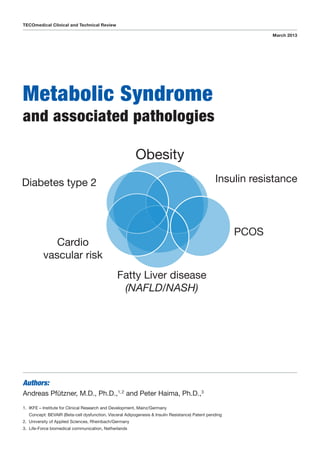
  • 1. Metabolic Syndrome and associated pathologies TECOmedical Clinical and Technical Review March 2013 Authors: Andreas Pfützner, M.D., Ph.D.,1, 2 and Peter Haima, Ph.D.,3 1. IKFE – Institute for Clinical Research and Development, Mainz/Germany Concept: BEVAIR (Beta-cell dysfunction, Visceral Adipogenesis Insulin Resistance) Patent pending 2. University of Applied Sciences, Rheinbach/Germany 3. Life-Force biomedical communication, Netherlands
  • 2. 2 Contents 1 Introduction . . . . . . . . . . . . . . . . . . . . . . . . . . . . . . . . . . . . . . . . . . . . . . . . . . . . . . . . . . . . . . . . . . . . . . . . . . . . . . . 3 2 Metabolic syndrome . . . . . . . . . . . . . . . . . . . . . . . . . . . . . . . . . . . . . . . . . . . . . . . . . . . . . . . . . . . . . . . . . . . . . . . . . 4 a Definition and pathophysiology of metabolic syndrome . . . . . . . . . . . . . . . . . . . . . . . . . . . . . . . . . . . . . . . . . 4 b The role of adipokines. . . . . . . . . . . . . . . . . . . . . . . . . . . . . . . . . . . . . . . . . . . . . . . . . . . . . . . . . . . . . . . . . . . 5 I Adiponectin . . . . . . . . . . . . . . . . . . . . . . . . . . . . . . . . . . . . . . . . . . . . . . . . . . . . . . . . . . . . . . . . . . . . . 5 II Leptin. . . . . . . . . . . . . . . . . . . . . . . . . . . . . . . . . . . . . . . . . . . . . . . . . . . . . . . . . . . . . . . . . . . . . . . . . . 6 c Development into diabetes. . . . . . . . . . . . . . . . . . . . . . . . . . . . . . . . . . . . . . . . . . . . . . . . . . . . . . . . . . . . . . . . 6 3 Insulin resistance and diabetes mellitus type 2 . . . . . . . . . . . . . . . . . . . . . . . . . . . . . . . . . . . . . . . . . . . . . . . . . . . 6 a Pathophysiology . . . . . . . . . . . . . . . . . . . . . . . . . . . . . . . . . . . . . . . . . . . . . . . . . . . . . . . . . . . . . . . . . . . . . . . 7 b Development stages of β-cell dysfunction. . . . . . . . . . . . . . . . . . . . . . . . . . . . . . . . . . . . . . . . . . . . . . . . . . . . 7 c Diagnosis of insulin resistance and diabetes type 2. . . . . . . . . . . . . . . . . . . . . . . . . . . . . . . . . . . . . . . . . . . . . 8 I Glucose testing . . . . . . . . . . . . . . . . . . . . . . . . . . . . . . . . . . . . . . . . . . . . . . . . . . . . . . . . . . . . . . . . . . 8 II HOMA score to assess early stages of β-cell dysfunction . . . . . . . . . . . . . . . . . . . . . . . . . . . . . . . . . 8 III Intact proinsulin testing to assess late stage β-cell dysfunction. . . . . . . . . . . . . . . . . . . . . . . . . . . . . 8 IV Intact proinsulin combined with oral glucose tolerance testing to identify prediabetes patients. . . . 9 4 Diabetes related cardiovascular disease . . . . . . . . . . . . . . . . . . . . . . . . . . . . . . . . . . . . . . . . . . . . . . . . . . . . . . . . 10 a pathophysiology . . . . . . . . . . . . . . . . . . . . . . . . . . . . . . . . . . . . . . . . . . . . . . . . . . . . . . . . . . . . . . . . . . . . . . . . 10 b Biochemical markers for diagnosis and therapy selection. . . . . . . . . . . . . . . . . . . . . . . . . . . . . . . . . . . . . . . . 12 I Proinsulin – a blood glucose independent marker to assess insulin resistance and β-cell dysfunction 12 II Adiponectin – a visceral adipose tissue activity marker to predict cardiovascular risk. . . . . . . . . . . . 12 III hsCRP – a chronic inflammation marker to predict cardiovascular risk . . . . . . . . . . . . . . . . . . . . . . . 13 5 Other metabolic syndrome-associated diseases. . . . . . . . . . . . . . . . . . . . . . . . . . . . . . . . . . . . . . . . . . . . . . . . . . 13 a Fatty liver disease . . . . . . . . . . . . . . . . . . . . . . . . . . . . . . . . . . . . . . . . . . . . . . . . . . . . . . . . . . . . . . . . . . . . . . 13 b PCOS: polycystic ovary syndrome . . . . . . . . . . . . . . . . . . . . . . . . . . . . . . . . . . . . . . . . . . . . . . . . . . . . . . . . . 14 6 Risk assessment and therapy of metabolic syndrome-associated pathologies . . . . . . . . . . . . . . . . . . . . . . . . 15 a Diagnosis and risk assessment . . . . . . . . . . . . . . . . . . . . . . . . . . . . . . . . . . . . . . . . . . . . . . . . . . . . . . . . . . . . 15 b Therapy and monitoring . . . . . . . . . . . . . . . . . . . . . . . . . . . . . . . . . . . . . . . . . . . . . . . . . . . . . . . . . . . . . . . . . . 15 7 References . . . . . . . . . . . . . . . . . . . . . . . . . . . . . . . . . . . . . . . . . . . . . . . . . . . . . . . . . . . . . . . . . . . . . . . . . . . . . . . 16
  • 3. 3 1 Introduction Metabolic syndrome is a combination of risk factors — abdominal obesity, hypertension, high blood sugar levels, insulin resistance (IR), abnormal cholesterol levels, hyperlipidemia and inflammatory states — that increase the risk of heart disease, stroke, diabetes type 2, fatty liver disease and PCOS (in young women). Having any of these conditions increase the risk of serious disease. If more than one of these conditions occur in combination, the risk is even greater. Metabolic syndrome is now present in up to 40 % of the United States adult population (prevalence on average 25 % of the population and 40+ % from age 60 and higher) [2] and is associated with a nearly a two fold increase in cardiovascular events, independent of the presence of diabetes mellitus. Obesity is the dominant key feature of metabolic syndrome [1, 2], although patients of normal weight may also suffer from IR and metabolic syndrome, and obesity and metabolic syndrome do not always occur in concordance as there is some evidence for conditions of benign obesity [3–7]. The epidemic of obesity in in adults and children in both industrialized and third world countries is regarded as one of the most serious public health problems of the 21st century. Many (especially overweight) people with IR are able to compensate for the increasing insulin requirement, so that the blood sugar does not rise at first. Late stage β-cell dysfunction then develops into clinically manifest type 2 diabe- tes in approximately one third of these patients [31]. Development of macrovascular damages may, therefore, already have developed before type 2 diabetes is clinically manifest (normal or impaired glucose tolerance test result). These macrovascular damages are, in part, irreversible, need to be treated lifelong and still are the actual cause of death in 75 % of diabetes type 2 patients. Up to now, the underlying pathomechanisms of metabolic syndrome are not detected at all or only very late and inadequately. In this review we discuss the latest clinical insights into Metabolic syndrome and related disease like diabetes type 2, fatty liver disease, coronary disease and PCOS. Conventional diagnosis is discussed alongside the use of new biomarkers. Three new biomarkers appear to be particularly suitable for early diagnosis and therapy selection due to their stability and studies that are presently available: intact Proinsulin, Adiponectin and hsCRP [29]. In addition, the effect of the therapy used on pathophysiological basic components can be checked by means of these new markers.
  • 4. 4 (1) TG ≥ 1.695 mmol/L and HDL ≤ 0.9 mmol/L (male), ≤1.0 mmol/L (female). (2) Waist/hip ratio 0.90 (male) 0.85 (female), or body mass index 30 kg/m2. (3) Urinary albumin excretion ratio ≥ 20 μg/min or albumin/creatinine ratio ≥ 30 mg/g. (4) Waist circumference ≥ 94 cm (male), ≥80 cm (female). (5) TG ≥ 2.0 mmol/L and/or HDL 1.0 mmol/L or treated for dyslipidemia. (6) Men, greater than 40 inches (102 cm) and women, greater than 35 inches (88 cm). (7) Equal to or greater than 150 mg/dL (1.7 mmol/L). (8) Men, Less than 40 mg/dL (1.03 mmol/L) and women, Less than 50 mg/dL (1.29 mmol/L). (9) Equal to or greater than 130/85 mm Hg or use of medication for hypertension. (10) Equal to or greater than 100 mg/dL (5.6 mmol/L) or use of medication for hyperglycemia. (11) Defined as waist circumference with ethnicity specific values (If BMI is 30 kg/m², central obesity can be assumed and waist circumference does not need to be measured). (12) TG 150 mg/dL (1.7 mmol/L), or specific treatment for this lipid abnormality. (13) HDL 40 mg/dL (1.03 mmol/L) in males, 50 mg/dL (1.29 mmol/L) in females, or specific treatment for this lipid abnormality. (14) Systolic BP 130 or diastolic BP 85 mm Hg, or treatment of previously diagnosed hypertension. (15) FPG 100 mg/dL (5.6 mmol/L), or previously diagnosed type 2 diabetes. 2 Metabolic syndrome a. Definition and pathophysiology of metabolic syndrome Metabolic syndrome is a cluster of risk factors — abdominal obesity, hypertension, high blood sugar levels, IR, abnormal cholesterol levels, hyperlipidemia and inflammatory states — that increase the risk of heart disease, stroke, diabetes type 2, fatty liver disease and PCOS (in young women). It has also been variously termed X syndrome, insulin resistance syndrome, metabolic syndrome X, cardiometabolic syndrome, syndrome X, Reaven's syndrome, and CHAOS (in Australia). The diagnostic criteria for metabolic syndrome have been set out by different organizations with slight variations in these criteria as shown in Table 1 [8]. WHO World Health Organization EGIR European Group for the Study of Insulin Resistance NCEP US National Cholesterol Education Program IDF International Diabetes Federation Presence of one of following: Insulin resistance AND two or more of following: Presence of three of following (2004): Central obesity (11) AND any two of following: DM / IGT / IFG / Insulin resistance Central obesity (4) Elevated waist circumference (6) Raised TG (12) AND two of the following: Dyslipidemia (5) Elevated TG (7) ▼ HDL (13) BP ≥ 140/90 BP ≥ 140/90 Reduced HDL (8) ▲ BP (16) Dyslipidemia (1) FBG ≥ 6.1 mmol/L (110 mg/dL) Elevated BP (9) ▲ FBG (17) Central obesity (2) Elevated fasting glucose (10) Microalbuminuria (3) BP: blood pressure, DM: diabetes mellitus, FBG: fasting blood glucose, HDL: high density lipoproteins, IFG: impaired fasting glucose, IGT: impaired glucose tolerance, TG: triglycerides. Table 1: Comparison of definitions of the metabolic syndrome.
  • 5. 5 Figure 1: the link between obesity, inflammation, β-cell dysfunction, insulin resistance and cardiovascular risk [28, 29] Oxidative stress, which is defined as imbalance between the production and inactivation of reactive oxygen species, has a major pathophysiological role in all the components of metabolic syndrome [20–24]. Oxidative stress and consequent inflammation induce insulin resistance (IR) which likely initiates metabolic syndrome and massive damage of pancreatic β-cell dysfunction. The association between the metabolic syndrome and inflammation is well documented [9]. Welsh et al. [10] demonstrated that adiposity leads to higher levels of the “acute phase” inflammatory protein CRP (C-reactive protein) and accumulating evidence demonstrates a close link among metabolic syndrome, chronic inflammation and oxidativestress[11].Infact,theoxidativestress-inflammation pathway has important roles in all the individual components of Metabolic syndrome including vascular alterations [11-15]. Figure 1 shows the link between obesity, inflammation, insulin resistance, β-cell dysfunction, and cardiovascular risk. [28, 29]. Adipo(cyto)kines (e.g. Adiponectin) and other factors produced by fat tissue and antiinsulinemic hormones play a key role in the process. b. The role of adipokines Multiple adipokines can be held responsible for the negative consequences of abdominal adipogenesis on insulin resistance. The growth of lipid tissue is induced by the differentiation of mesenchymal stem cells to become preadopocytes and finally mature lipid cells. In this stage peripheral monocytes/macrophages migrate into the lipid tissue and are kept at a constantly increased level of activation by the secretion of a whole pattern of proinflammatory cytokines from the pre-adipocyte. In consequence many adipokines have been identified which have been previously described to be associated with inflammatory conditions in other parts of the body, and which have a known negative influence on insulin sensitivity, e. g. IL-6 and TNFα. A list of some recently described prominent adipokines is provided in Table 2 [31]. Adiponectin and leptin have been studied most extensively andplayamajorroleinlipidmetabolismandthedevelopment of obesity. Further adipokines are resistin and visfatin that also seem to be linked to insulin resistance and metabolic syndrome. They are currently under evaluation regarding their clinical value. I. Adiponectin Adiponectin owns an exceptional place in this listing. It is secreted by the mature adipocytes (and the connective tissue) and not by the pre-adipocytes and has a synergistic action to insulin. High plasma adiponectin concentrations result in an improvement of insulin sensitivity. An increase in body weight with differentiation of stem cells to pre-adipocytes is associated with a suppression of adiponectin concentrations in the circulation [38]. Other disease conditions that have been described to be correlated with a suppression of adiponectin levels include, but are not limited to metabolic syndrome, atherosclerosis and any kind of obesity. Adiponectin may, therefore, be regarded as an indicator of the activity of pre-adipocytes. Female patients have higher reference values than male patients. Several plasma sub-fractions have been described that are differentiated by the agglomeration of different numbers of single adiponectin molecules. However, they have as of yet not shown any difference in their changing behaviour following therapeutic interventions. For practical use it does, therefore, not really matter, whether “High Molecular Weight” or “Low Molecular Weight” adiponectin is determined for diagnostic purposes, as long as the same sub-fraction is used to draw any diagnostic or clinical conclusions [40]. Adiponectin levels respond quickly to changes in insulin resistance and the metabolic situation in the lipid tissue. It is, therefore, suitable to track slight changes in insulin resistance, e.g. the metabolic deterioration induced by the hormonal changes in women with polycystic ovary syndrome (PCOS) [41, 42]. Leptin Free Fatty acids Interleukine-6 PAI-I and tPA Retinol Binding protein 4 Angiotensin II Adipsin (Complementation factor D) TNF-alpha Adiponectin Visfatin Resistin Vaspin Table 2: Recently described adipokines Anti-insulinemic hormones Insulin Resistance β-Cell Dysfunction β-Cell Dysfunction IL-6, TNFα etc. Free Fatty Acids Angiotensin Insulin- requirement Insulin Proinsulin Insulin Proinsulin Adiponectin Adipogenesis Lipid Cell Pre-Adipocyte Stem Cell Stem Cell Stem Cell Leptin Anti-insulinemic hormones Insulin Resistance β-Cell Dysfunction IL-6, TNFα etc. Free Fatty Acids Angiotensin Insulin- requirement Insulin Proinsulin Adiponectin Adipogenesis OBESITY Lipid Cell Pre-Adipocyte Leptin AdiponectinC B Arteriosclerosis HypertensionDyslipidemia Arteriosclerosis HypertensionDyslipidemia Hyperglycemia Intact Proinsulin hsCRP
  • 6. 6 II. Leptin Leptin is a 16kDa non-glycosylated protein that is predo- minantly secreted by mature lipid cells, but can also de- rive in minor amounts from the stomach, intestine tract, muscle and breast tissue. The plasma leptin levels reflect the actual amount of lipid tissue, the size of the adipocytes and their triglyceride content. In consequence, plasma leptin concentrations are elevated in case of obesity and decre- ase with a loss in body weight [43]. These changes are in- fluenced by the actual insulin and glucose concentrations and by inflammatory cytokines. In addition, leptin plays a role in the control of energy consumption, in angiogenesis, fertility, bone formation and many other endocrine body functions [44]. Leptin levels are higher in female patients, most probably because of the larger amount of subcuta- neous lipid tissue and a higher stimulation by estrogens in women. Leptin concentrations decrease in a cold environment and during adrenergic stimulation. The brain uses leptin as an important control variable for appetite regulation. It carries the information, whether “sufficient” amounts of lipid tissue are prevalent and, therefore, leptin, like insulin, belongs to the lipostatic molecules. The lipostatic factors orchestrate to- gether with the short-acting incretines (e. g. GLP-1, ghrelin, GIP, cholecystokinin (CCK), obestatin, PYY etc.) the nutritional behaviour of the human organism (see Figure 2, [45]). c. Development into diabetes Oxidative stress and consequent inflammation induce IR, which likely initiates metabolic syndrome. Afterwards, there are 2 principal pathways of metabolic syndrome development [16]: A. With preserved pancreatic beta cells function and insulin hypersecretion which can compensate for insulin resistance. This pathway leads mainly to the macrovascular complications of metabolic syndrome. B. With massive damage of pancreatic beta cells leading to progressively decrease of insulin secretion and to hyperglycemia (e.g. overt type 2 diabetes). This pathway leads to both microvascular and macrovascular complications. An insulin-resistant state – as the key phase of metabolic syndrome – constitutes the major risk factor for the development of diabetes mellitus. Hyperinsulinemia appears to be a compensatory mechanism that responds to increased levels of circulating glucose. Fasting glucose is presumed to remain normal as long as insulin hypersecretion can compensate for insulin resistance. The fall in insulin secretion leading to hyperglicemia occurs as a late phenomenon. 3 Insulin resistance and diabetes mellitus type 2 In type 2 diabetes, fat, liver, and muscle cells do not respond correctly to insulin anymore. Due to this insulin resistance (IR), blood sugar does not enter these cells and consequently high levels of sugar build up in the blood. This is called hyperglycemia. Type 2 diabetes usually occurs slowly over time. Most people with the disease are overweight when they are diagnosed. Type 2 diabetes can also develop in people who are thin. This is more common in the elderly. Family history and genes play a large role in type 2 diabetes. Low activity level, poor diet, and excess body weight around the waist increase the risk. Figure 3: The relationship between metabolic syndrome, insulin resistance, hyper-insulinemia and hyperglycemia (overt type 2 diabetes), adapted from [16] M e t a b o l i c S y n d r o m e insulin resistance Life time Diabetes type 2 Pancreatic beta cells stress damage Hyperinsulinemia Macrovascular + Microvascular complications Macrovascular complications Food intake excess Genetic background Physical inactivity Adipogenesis Overweight A Obesity Hyperinsulinemia HyperglycemiaB Figure 2: Factor controlling appetite and food uptake
  • 7. 7 Diabetes type 2 is one possible outcome of Metabolic syndrome. Diabetes type 2 and obesity are two diseases with continuously growing prevalence over the past decades that have both reached pandemic dimensions in their distribution. Approximately 4–5 % of the world population are currently affected and the annual incidence in Western Europe is approximately 10 %. Type 2 diabetes is conventionally diagnosed via elevated blood glucose and glycosylated hemoglobin (HbA1c) levels. a. Pathophysiology The major underlying mechanisms of diabetes type 2 are the development of systemic insulin resistance and a col- lapsed insulin secretion by pancreatic β-cells. IR is cha- racterized by a general decrease of the insulin sensitivity of the peripheral cells, which on a receptor level is asso- ciated with a genetically determined change in the insulin receptor molecule and a reduction of the overall number of insulin receptors on the cells (post-receptor defects have also been described in literature). Figure 4, shows the rela- tion between IR, decreased insulin secretion and adiposity. b. Development stages of β-cell dysfunction If a patient shows hereditary or acquired insulin resistance, this will initially be compensated for by an appropriate additional secretion of insulin. Insulin, however, is the only (known) physiological hormone that stimulates adipogenesis. As a consequence, this leads to a strong tendency to develop adipose tissue, especially with increased intake of calories. In advanced stage, the processing of the insulin precursor molecule proinsulin becomes insufficient and increasing amounts of intact proinsulin are being secreted. Proinsulin has only a fraction of the blood sugar reducing effect of insulin, but has the same adipogenic potency [3–5]. In consequence, elevated plasma intact Proinsulin levels are a highly specific direct indicator for advanced β-cell dysfunction and a highly specific indirect indicator for cli- nically relevant insulin resistance [21]. Using the fasting intact proinsulin concentrations and under consideration of the level of insulin resistance (e. g. by means of the HOMA score [22]), it is possible to introduce a clinically useful staging of β-cell dysfunction that allows for a dif- ferential diagnosis and selection of a pathophysiologically oriented differential therapy of type 2 diabetes mellitus [23]. This staging is presented in Figure 5 and further detailed in Table 3. Figure 5: Staging of β-cell dysfunction by means of insulin resistance and composition of the β-cell secretion product [23] Stage Description Insulin Proinsulin Glucose I Insulin sensitive Normal Normal Normal II Insulin resistance without qualitative secretion disorder Elevated Normal Normal or elevated III a Insulin resistance with major β-cell secretion disorder Normal/Elevated Elevated Normal or elevated III b Collapsed β-cell secretion Low Elevated to normal (in end stage) Elevated Table 3: Staging of β-cell dysfunction by means of insulin resistance and composition of the β-cell secretion product [23] 1. Cause of Disease: Reduced Insulin Effectiveness (Insulin resistance) Increased Insulin requirements Visceral Adipogenesis Weight gain Hormones and cytokines from the lipid tissue reduce insulin sensitivity and induce dyslipidemia, hypertension and atherosclerosis With sufficient food supply new adipose tissue is built up supported by insulin but even more by proinsulin 2. Cause of Disease: Deteriorated Insulin Secretion (β-Cell dysfunction) Increased Insulin- and Proinsulin- Secretion D Y S L I P I D E M I A A T H E R O S C L E R O S I S H Y P E R T E N S I O N Figure 4: Relation between insulin resistance, β-cell dysfunction, obesity and the resulting complications [19, 20]
  • 8. 8 II. HOMA score to assess early stages of β-cell dysfunction The HOMA (Homeostasis model assessment) score is an easy method to estimate the degree of insulin resistance non-diabetic or early stage diabetic patients [24,25]. It can be applied only if intact proinsulin levels are in the normal range, as the total secretion activity of the β-cell is under this condition represented by the fasting insulin levels. The HOMA score is based on the assumption that under normal conditions, a normal blood glucose value is associated with a matching normal insulin level, which may vary individually from patient to patient. Insulin resistance is indicated, if at this normal insulin level, an elevated blood glucose is observed, or if more insulin is required to maintain blood glucose at its normal level. After determining the fasting serum insulin and fasting plasma glucose levels the HOMA-IR score is calculated as follows: HOMA = Insulin [μU/ml] x Glucose [mmol/l] /22.5. Insulin resistance is assumed if the score value exceeds 2 [25]. Of particular interest are changes in the HOMA score during therapeutic interventions, as a score reduction represents an improvement in insulin sensitivity. Again, the HOMA score should only be used in patients with stage I and II of β-cell dysfunction (see Figure 3), because intact Proinsulin is an additional marker for β-cell activity that is not considered in the HOMA-IR score equation. III. Intact proinsulin testing to assess late stage β-cell dysfunction Intact proinsulin is produced in the pancreatic β-cells and is normally further processed to insulin and C-peptide. It is only seen in low concentrations in the plasma of healthy subjects, as it is rapidly degraded (T 1/2 is 15 minutes). Proinsulin cleavage products (like des32,33) are stable for several hours. As these fragments are inactive and can make up to 50 % of total Proinsulin, only assays specifically measuring intact proinsulin are suitable to indicate advanced β-cell dysfunction and IR. An increase in the insulin demand, as provided by insulin resistance in later stages of type 2 diabetes mellitus, can result in increased expression of proinsulin into the blood (see Figure 3). When intact proinsulin is secreted together with or instead of insulin in the fasting state (stage III of β-cells dysfunction, Figure 3) the HOMA score cannot be used to assess the level of β-cells dysfunction [26, 27]. Intact proinsulin is also able to lower glucose values but is not considered in the HOMA equations. Measurement of fasting intact proinsulin has been shown to be a very specific indicator and clinically significant IR. In clinical practice, fasting morning intact proinsulin can be used as highly specific indicator of of late stage β-cell dysfunction and clinically relevant IR. It can also to serve as the basis for the selection of an insulin resistance therapy, and to monitor the therapeutic effect on β-cell dysfunction. c. Diagnosis of insulin resistance and diabetes type 2 I. Glucose testing Today, diagnosis and therapy of the type 2 diabetes mellitus is still based on blood sugar values and the associated values for glycosylated hemoglobin (HbA1c). WHO 2011 guidelines describe that fasting glucose should be 127 mg/dl and HbA1c 48 mmol/mol. In addition an oral glucose tolerance test (OGTT) is often performed. During an OGGT glucose is given and blood samples taken afterward to determine how quickly it is cleared from the blood. WHO recommends for a 75g oral dose in all adults: the dose is adjusted for weight only in children. Glucose levels 11.1 mmol/L ( 200 mg/dL) at 2 hours confirms the diagnosis of diabetes (see Table 4). Glucose levels Normal Impaired fasting glycaemia Impaired glucose tolerance Diabetes mellitus Venous Plasma Fasting 2hrs Fasting 2hrs Fasting 2hrs Fasting 2hrs (mmol/L) 6.1 7.8 6.1 7.0 7.8 7.0 7.8 7.0 11.1 (mg/dL) 110 140 110 126 140 126 140 126 200 Table 4: 1999 WHO Diabetes criteria - Interpretation of Oral Glucose Tolerance Test
  • 9. 9 IV. Intact proinsulin combined with oral glucose tolerance testing to identify prediabetes patients. Because of the blood sugar reducing effect of Proinsulin, some patients may already be suffering from β-cell dysfunction years before clinical manifestation of elevated blood sugar levels. It has been shown that elevation of fasting intact proinsulin is an indicator of insulin resistance and severe β-cell dysfunction. Earlier detection of prediabetic patients is important to prevent irreversible cardiovascular damages. In a recent pilot study [57] it was investigated whether elevation of intact proinsulin after 1 and 2 hours in the course of an oral glucose tolerance test (OGTT) may be an indicator of the development of type 2 diabetes. Patients were enrolled based on previous results of OGTT: 11 healthy subjects (7 female, 4 male, age: 59 ± 20 yrs.), 10 patients with Impaired Glucose Tolerance (IGT; 6 female, 6 male, age: 62 ± 10 yrs.), and 10 patients with overt type 2 diabetes (6 female, 4 male, age: 53 ± 11 yrs.). Another OGTT was performed with measurement of glucose and intact proinsulin levels after 0h, 1 h and 2 hours. Five years later. The diabetes status of the patients was confirmed and correlated with the earlier OGTT results. Patients with diabetes (Fig. 6) had elevated fasting glucose levels after 1 and 2 h (diabetes: 0/1/2 h: 121 ± 20/230 ± 51/213 ± 24 mg/dL; prediabetes: 102 ± 9/168 ± 57/149 ± 34 mg/dL; normals: 94 ± 8/140 ± 29/90 ± 24 mg/dL). Proinsulin values after 2 hours were elevated in diabetes and prediabetes vs. control 27 ± 10 pmol/L and 28 ± 6 pmol/L, respectively, vs. 10 ± 5 pmol/L (p0.05 vs. both other groups). Five years later, all patients with IGT and three normal subjects had developed overt type 2 diabetes. All manifesting patients had elevated intact proinsulin levels ( 11 pmol/L) after 1 and 2 h. A value 20 pmol/L after 2 h was always indicative for β-cell dysfunction and progressive disease development. However, because of the multiple existing pheno- types of type 2 diabetes, a value 20 pmol/L, however, does not automatically exclude diabetes in an individual patient. Two out of 10 patients with initial IGT were in fact normal according to the WHO diabetes criteria during the second OGTT. However, proinsulin values were 33 and 36 pmol/L after 2 hours, confirming β-cell dysfunction and progressive disease development. In conclusion: Five year after an initial oral glucose tolerance test, all patients with elevated intact proinsulin levels after 1 h and 2 h had developed overt Type 2 diabetes, irrespective of the observed blood glucose values in the OGTT. Increasing intact proinsulin levels after 1 h and 2 h in the OGTT indicate stress-related β-cell dysfunction and may be an effective predictor for type 2 diabetes development in a mid-term future. This first study is very promising, as early detection of pre- diabetes using intact proinsulin as biomarker, may allow life style and other interventions to prevent irreversible cardiovascular damages in diabetes patients that are often the actual cause of death. 60 time [min] glucose[mg/dl] glucose 0 0 50 200 150 100 250 120 Normal Prediabetes Diabetes Normal Prediabetes Diabetes 60 time [min] intactproinsulin[pmd/L] intact proinsulin 0 0 5 20 15 10 25 30 35 120 Figure 6: Glucose and intact Proinsulin levels during oral glucose tolerance testing of healthy, Prediabetes and clinically manifest diabetes type 2 subjects [30]
  • 10. 10 4 Diabetes related cardiovascular disease Today, diagnosis and therapy of the type 2 diabetes mellitus is still based on blood sugar values and the associated values for glycosylated hemoglobin (HbA1c). But even with good blood sugar adjustment, patients still have an increased cardiovascular risk. Still, 75 % of patients with type 2 diabetes die of cardiovascular events, whereas this is only true for 35 % of patients with type 1 diabetes, although these patients also have an increased blood sugar level. Today, differences in mortality can be explained by the underlying pathologic developments in type 2 diabetes on a metabolic and vascular level. a. Pathophysiology Pathophysiologically, type 2 diabetes is characterized by insulin resistance and defective secretion of the pancreas. In particular, IR is closely associated with macrovascular complications, since insulin receptors exist on the endothelial cells of large vessels, whose function is not to absorb glucose, but to activate NO synthesis in the cells. NO is the mediator of numerous vasoprotective mechanisms and finally protects the organism against the development and progression of atherosclerosis [29]. In case of IR not only metabolic but also vascular receptors will be affected and, consequently, not only the insulin requirement or the blood sugar will rise, but usually there is a parallel decrease of the protection of the vessel cells against the deposition of foam cells, a key step in the development of atherosclerosis. Many (especially overweight) people with insulin resistance are able to compensate for the increasing insulin requirement, so that the blood sugar does not rise at first. An additional malfunction of the insulin-producing β-cells of the pancreas then leads to clinically manifest type 2 diabetes in approximately one third of these patients [31]. Development of macrovascular damages may, therefore, already commence in a stage of the disease in which type 2 diabetes is not yet clinically manifest, but where, for example, “only” a disturbed glucose tolerance is present. For this reason, many type 2 diabetes patients already show cardiovascular damages during the first clinical diagnosis of their disease and these damages are, in part, not reversible any more. They need to be treated lifelong and cardiovascular damages are often the actual cause of death. IR-induced hyperinsulinemia leads to a strong tendency to develop adipose tissue (insulin is the only physiological hormone that stimulates adipogenesis), especially with increased intake of calories. If there is a simultaneous β-cell dysfunction, proinsulin is increasingly present in the secretion product, which has only a fraction of the blood sugar reducing effect of insulin, but has the same adipogenic potency [32-34] (Fig. 7A). Both hormones also increase adipogenesis and lead to intensified differentiation of mesenchymal stem cells into pre- adipocytes and finally into adipocytes [35]. At this stage, adipose tissue, a highly active endocrine organ, secretes hormones directed against insulin, e. g. estrogenes, which in turn enhances insulin resistance [36]. At the same time, the amount of circulating adiponectin is suppressed, a hormone of the white adipose tissue and the connective tissue, which has strong vessel-protective and anti-atherosclerotic properties [37,38]. A vicious circle is created within which insulin resistance, β-cell dysfunction and adipogenesis mutually affect each other negatively. The differentiated pre-adipocytes in turn secrete further molecules, which in their totality can maintain or even enhance the metabolic syndrome, e. g. angiotensin, IL-6, TNFα, free fatty acids, RBP4 or PAI-1 Fig. 7B). The consequence is the development or enhancement of hypertension, dyslipidemia and increased macrophage activation which ultimately contributes to atherosclerosis [39]. These pathophysiological associations result in a higher atherosclerosis risk, especially if the insulin requirement rises further due to hyperglycemia and toxic concentrations of glucose occurring in the plasma, and, at the same time, the present insulin resistance seriously interferes with the vessel-protective NO-production in the endothelium (Fig. 7C).
  • 11. 11 Anti-insulinemic hormones Insulin Resistance Insulin Resistance β-Cell Dysfunction β-Cell Dysfunction IL-6, TNFα etc. Free Fatty Acids Angiotensin Insulin- requirement Insulin Proinsulin Insulin Proinsulin Insulin- requirement Adiponectin Anti-insulinemic hormones Adiponectin Adipogenesis Lipid Cell Pre-Adipocyte Adipogenesis Lipid Cell Stem Cell Stem Cell Stem Cell Pre-Adipocyte Leptin Anti-insulinemic hormones Insulin Resistance β-Cell Dysfunction IL-6, TNFα etc. Free Fatty Acids Angiotensin Insulin- requirement Insulin Proinsulin Adiponectin Adipogenesis OBESITY Lipid Cell Pre-Adipocyte Leptin AdiponectinC B A Arteriosclerosis HypertensionDyslipidemia Arteriosclerosis HypertensionDyslipidemia Hyperglycemia Intact Proinsulin hsCRP Figure 7: The relation between insulin resistance, obesity and the development of cardiovascular risk
  • 12. 12 b. Biochemical markers for diagnosis and therapy selection Routine diagnostics of high blood pressure involves measurement of lipids, HbA1c and glucose. However, the underlying pathomechanisms described above are not detected at all or only very late and inadequately. Therefore, numerous new laboratory markers for classification of metabolic and vascular risk have been investigated and described in recent years. Three of these markers appear to be particularly suitable for diagnosis and therapy selection due to their stability and studies that are presently available: intact Proinsulin, Adiponectin and hsCRP [29]. I. Proinsulin – a blood glucose independent marker to assess insulin resistance and β-cell dysfunction The significance of intact proinsulin as β-cell function marker has already been described in chapter 3. Intact proinsulin occurs in plasma in increased fasting levels only if a clinically significant IR exists already [27]. Thus, intact Proinsulin is an indirect, but highly specific marker for late stage β-cell dysfunction. Many (especially overweight) people with insulin resistance are able to compensate for the increasing insulin requirement, so that the blood sugar does not rise at first. Late stage β-cell dysfunction then develops into clinically manifest type 2 diabetes in approximately one third of these patients [31]. Development of macrovascular damages may, therefore, already have developed before type 2 diabetes is clinically manifest (normal or impaired glucose tolerance test result). These macrovascular damages are, in part, irreversible and need to be treated lifelong (and often are the actual cause of death). Measurement of Proinsulin allows for early detection and treatment of these prediabetic patients to prevent irreversible cardiovascular damages. In view of this it appears to be reasonable to adapt the therapy to the β-cell dysfunction. Prospective studies have demonstrated already that intervention by mobility, Metformin, glitazones or insulin will protect the β-cell and lead to a decrease of the proinsulin level, an effect that could not be observed with sulfonylurea [46-48]. Only assays specifically measuring intact Proinsulin are suitable. Fasting values 11 pmol/L are considered as normal. Fasting values 11 pmol/L are indicative of β-cell dysfunction, insulin resistance and cardiovascular risk. Increasing intact proinsulin levels after 1 and 2 hours in an oral glucose tolerance test are highly indicative for stressrelated β-cell dysfunction and may be a strong predictor for type 2 diabetes development and cardiovascular damages in a mid-term future (see chapter 3 [57]). A proinsulin value 20 pmol/L at any time point (1 or 2 hours) is indicative for β-cell dysfunction and progressive disease development. II. Adiponectin – a visceral adipose tissue activity marker to predict cardiovascular risk The fat and connective tissue hormone adiponectin is a reverse indicator of visceral adipose tissue activity. As such it is regarded as a good marker of insulin resistance and metabolic syndrome. High plasma adiponectin concentrations result in an improvement of insulin sensitivity. Even though adiponectin appears to be less suitable for the initial diagnosis of insulin resistance than intact proinsulin [49], it is an excellent indicator of the metabolic overall situation, which responds very sensitively to successful interventional therapeutic approaches. An increase of adiponectin under therapy shows an improvement of the risk profile. Clinical studies showed that values below 7 mg/l were associated with an increased risk of cardiovascular events [37, 38]. Values between 7 and 10 mg/L are regarded as grey zone, values 10 mg/L are considered as normal.
  • 13. 13 III. hsCRP – a chronic inflammation marker to predict cardiovascular risk The increasing amount of abdominal lipid tissue exposes the patient to an increased macrovascular risk. The proinflammatory adipokines deriving from the pre-adipocyte activate the immune system not only locally but also systematically, i.e. mononuclear cells in the circulation are also alerted. Especially in the postprandial state, these monocytes/macrophages may be loaded with LDL particles. At the same time, these cells play a key role in the pathophysiology of atherosclerosis, as they penetrate into the vessel wall by means of further inflammatory proteins and enzymes, which finally leads to cholesterol deposit and plaque formation. A known, “acute phase” inflammatory protein involved in this process is C-reactive protein (CRP), which is produced in the liver. Whereas the application of intact proinsulin and adiponectin for therapy selection and therapy control is just beginning to assert itself now in type 2 diabetes, the use of highly sensitive C-reactive protein (hsCRP) as inflammatory marker of cardiovascular risk especially in cardiology has already reached a high level of general acceptance. While CRP has been considered to be an unspecific indicator of inflammation of any origin in the past, it could be shown tha t h sCRP-values, stratified into three risk groups, have their own predictive value for cardiovascular risk in the low measurement range ( 10 mg/l) [50]. Values in this range, when determined with a highly sensitive test method (therefore: “high sensitivity CRP” or “hsCRP”), describe a stepwise increased cardiovascular risk in patients with and without diabetes mellitus (Table 5 [50, 52]). This staging has been confirmed in numerous studies and meta analyses and has been included in the official diagnosis criteria of the American Heart Association [51]. A reduction of the hsCRP in the course of the observation shows an improvement of the cardiovascular risk profile [53]. 5 Other metabolic syndrome-associated diseases a. Fatty liver disease With the increasing prevalence of obesity and Metabolic syndrome an increase of nonalcoholic fatty liver disease (NAFLD) is obvious. Patients with insulin resistance and other symptoms of metabolic syndrome should therefore be screened for NAFLD and its progressive and chronic form NASH (non-alcoholic steatohepatitis) [56]. While most patients with steatosis tend to have a benign clinical course, a significant proportion of those with NASH have a progressive disease with a risk of developing liver cirrhosis and hepatocellular carcinoma [12]. In the USA, 7 % of all liver transplants are based on diagnosis of NASH [23]. The diagnostic challenge is to predict NAFLD patients that are likely to progress into liver disease, initiate therapy and life style changes and to monitor the efficacy of the measures. Conventional markers of liver damage, liver transaminases (AST/ALT), frequently provide incorrect information about liver damage. For example, it could be shown that up to 25–30 % of the patient with fibrosis liver damage have normal transaminase levels [59, 60]. Hepatocyte cell death, specifically hepatocyte apoptosis, is considered to play a crucial role in the formation of liver fibrosis or liver cirrhosis. Numerous studies have demonstrated that hepatocyte apoptosis can be specifically assessed by means of caspases cleaved fragments of Keratin 18 (ccK18), a major intermediate filament protein, expressed by hepatocytes. The biomarker CcK18 as determined by the M30 Apoptosense® ELISA allows prediction of the level of fibrosis (staging), steatosis and NASH and can improve decisions on therapeutic regiments for patients. Canbay et al. concluded that serological investigation, including the biomarker ccK18 can predict progression of NAFLD into NASH in obese patients [55]. hsCRP fasting value Cardiovascular risk 10 mg/L No assessment possible 3-10 mg/L High 1-3 mg/L Average 0-1 mg/L Low Table 5: hsCRP cardiovascular risk groups
  • 14. 14 Liver damage biomarker Cardiovascular risk Caspases cleaved Keratin 18 (ccK18: M30 Elisa) Hepatocyte apoptosis Keratin 18 (cleaved and uncleaved: M65 Elisa) Hepatocyte apoptosis and necrosis Alpha Glutathione STransferase (α GST, serum) Hepatocyte damage Pi Glutathione S-Transferase (π GST, serum) Bile duct damage Collagen IV (serum) Increased collagen deposition Table 6: Various liver damage biomarkers For each diabetic patient, liver biomarker levels were expressed as a percentage of the upper limit of the normal range for the specific marker (upper limit for ccK18: 186 U/L; K18: 183 U/L; α GST: 12 µg/L). Liver damage biomarkers in diabetic patients %ofupperlimitnormalrange Patients 600 % 500 % 300 % 200 % 100 % 0 % 400 % CCK18 (M30) K18 (M65) alpha GST Figure 8: Liver damage biomarkers in diabetic patients In a recent study by Pfützner et al [58], liver transaminases (ALT/AST) and liver damage biomarkers ccK18, K18 and α GST were measured in 32 diabetic patients and 36 healthy subjects. ALT/AST levels were elevated in only 22/13 % of diabetic patients. In contrast, all biomarkers were highly elevated in up to 65 % of the cases (above reference range, ccK18: 50 %; K18: 65 %; α GST: 58 %) and showed similar behavior in most patients (Figure 8), indicating ongoing liver damage. This pilot study supports the use of liver damage biomarkers in diabetic patients to diagnose fatty liver disease and predict possible progression to NASH. b. PCOS: polycystic ovary syndrome The polycystic ovary syndrome (PCOS) is induced by a deterioration of the hormonal regulation in female patients and is characterized by chronic anovulation and hyperandrogenism. It is one of the most frequent endocrine disorders in young female patients (prevalence 6 %). PCOS is often associated with obesity and IR. During the PCOS development increased LH levels induce an increased synthesis of steroids in the ovaries, which in turn leads to an increased modification of androgens into estrogens in the lipid tissue. The acyclic production of these estrogens results in increased secretion of LH from the pituary gland. Another source of increased androgen concentrations in patients with PCOS is a suppression of the production of sex-hormone-binding globulin (SHBG) in the liver, which leads to increased formation of biologically active androgens. The increased formation of all these “anti-insulinemic” hormones may frequently result in development of a metabolic insulin resistance and an increased insulin secretion to compensate for the higher needs. Insulin resistance does not represent the only cause for PCOS development, but the accompanying hyperinsulinemia supports the development by the acceleration of ovarian and adrenal androgen production. This understanding of the pathophysiological disease background has led to the use of insulin sensitizing drugs in the affected patients. Therapy with metformin resulted in a significant decrease in circulating androgen levels, an increase in SHBG concentrations, a normalization of the menstrual cycle and an improvement in the fertility [54]. Similar effects have been reported for intervention with glitazones (rosiglitazone, pioglitazone). The key parameters for diagnosing insulin resistance in PCOS patients appear to be the HOMA score and adiponectin or intact proinsulin. Canbay et al. demonstrated that patients with PCOS also have increased risk for developing non-alcoholic steatohepatitis (NASH). Due to this association they advise to investigate female NASH patients for PCOS and PCOS patients for NASH [55]. Liver damage can also be assessed using other biomarkers. A list of some recently described liver damage biomarkers is shown in Table 6.
  • 15. 15 6 Risk assessment and therapy of metabolic syndrome-associated pathologies a. Diagnosis and risk assessment Up to now, the underlying pathomechanisms described above are not detected at all or only very late and inadequately. As discussed in chapter 3, intact Proinsulin, Adiponectin and hsCRP appear to be particularly suitable for diagnosis and therapy selection due to their stability and studies that are presently available [29]. In addition, the effect of the therapy used on pathophysiological basic components can be checked by means of these new risk markers. The values for intact proinsulin, adiponectin and hsCRP can be used to assess • β-cell function, • insulin sensitivity • patient's individual cardiovascular risk Increased proinsulin and hsCRP levels and low adiponec- tin values indicate insulin resistance with β-cell dysfunction and impending macrovascular complications. Adiponectin increase, on the other hand, is accompanied by significant improvement of metabolic status and cardiovascular pro- gnosis. In addition biomarkers for liver damage and pro- gression to NASH can be included like ccK18, K18, α GST and collagen IV (see chapter 4a). b. Therapy and monitoring Proinsulin, adiponectin and hsCRP are independent risk factors for type 2 diabetes as well as for macrovascular complications. According to present knowledge, all three risk factors will be improved in particular through changes in lifestyle (weight reduction and more physical activity) and through pathophysiologically oriented medicinal treatment. Improvements have been observed especially with pioglitazone, GLP-1 analogs, SGLT-2 inhibitors and insulin (Table 7, Pfützner et al [58] ). Such positive evidence is not available for other oral antidiabetic drugs, for example, sulfonylurea [11, 13, 14, 19]. Intact Proinsulin: 11 pmol/l = low risk ≥11 pmol/l = high risk ≥10 mg/l = low risk ≥1–3 mg/l = average risk ≥3–10 mg/l = high risk ≥10 mg/l = unspecific 0–1 mg/l = low risk 7–10 mg/l = grey zone ≤ 7 mg/l = high risk Adiponectin: hsCRP: Figure 9: Assessment of biomarkers for risk assessment of metabolic syndrome associated pathologies Intervention intact Proinsulin Adiponectin hsCRP Diet Exercise Sulphonylurea/Glinides Metformin Pioglitazone DPPIV-Inhibitors GLP-1 Analogs SGLT-2 Inhibitors Insulin (early) Insulin (late) β-Cell Dysfunction Visceral tissue activity Chronic systemic inflammation ( ( (( Table 7: effect of various therapies on biomarkers
  • 16. 16 7 References [1] Fujita T. Insulin resistance and salt-sensitive hypertension in metabolic syndrome. Nephrology Dialysis Transplantation. 2007;22(11):3102–3107. [2] Fujita T. Aldosterone in salt-sensitive hypertension and metabolic syndrome. Journal of Molecular Medicine. 2008;86(6):729–734. [3] Uretsky S, Messerli FH, Bangalore S, et al. Obesity paradox in patients with hpertension and coronary artery disease. American Journal of Medicine. 2007;120(10):863–870. [4] Aguilar-Salinas CA, García E, Robles L, et al. High adiponectin concentrations are associated with the metabolically healthy obese phenotype. Journal of Clinical Endocrinology and Metabolism. 2008;93(10):4075–4079. [5] Wildman RP, Muntner P, Reynolds K, et al. The obese without cardiometabolic risk factor clustering and the normal weight with cardiometabolic risk factor clustering: prevalence and correlates of 2 phenotypes among the US population (NHANES 1999–2004) Archives of Internal Medicine. 2008;168(15):1617–1624. [6] Stefan N, Kantartzis K, Machann J, et al. Identification and characterization of metabolically benign obesity in humans. Archives of Internal Medicine. 2008;168(15):1609– 1616. [7] Wildman RP. Healthy obesity. Current Opinion in Clinical Nutrition Metabolic Care. 2009;12:438–443.] [8] Saeid Golbidi, 1 Azam Mesdaghinia, 2 and Ismail Laher. Exercise in the Metabolic syndrome Oxid Med Cell Longev. 2012; 2012: 349710. [9] Fernández-Real JM, Ricart W. Insulin resistance and chronic cardiovascular inflammatory syndrome. Endocrine Reviews. 2003;24(3):278–301. [PubMed] [10] Welsh P, Polisecki E, Robertson M, et al. Unraveling the directional link between adiposity and inflammation: a bidirectional mendelian randomization approach. Journal of Clinical Endocrinology and Metabolism. 2010;95(1):93– 99. [11] Wellen KE, Hotamisligil GS. Inflammation, stress, and diabetes. Journal of Clinical Investigation. 2005;115(5):1111–1119. [12] Stocker R, Keaney JF. Role of oxidative modifications in atherosclerosis. Physiological Reviews. 2004;84(4):1381–1478. [13] Furukawa S, Fujita T, Shimabukuro M, et al. Increased oxidative stress in obesity and its impact on metabolic syndrome. Journal of Clinical Investigation. 2004;114(12):1752–1761. [14] Keaney JF, Jr., Larson MG, Vasan RS, et al. Obesity and systemic oxidative stress: clinical correlates of oxidative stress in the Framingham study. Arteriosclerosis, Thrombosis, and Vascular Biology. 2003;23(3):434–439. [15] Pinzani M, Marra F, Carloni V. Signal transduction in hepatic stellate cells. Liver. 1998;18(1):2–13. [16] Alexander Tenenbaum, Enrique Z Fisman and Michael Motro. Metabolic syndrome and type 2 diabetes + mellitus: focus on peroxisome proliferator activated receptors (PPAR). Cardiovascular Diabetology 2003, 2:4 doi:10.1186/1475-2840-2-4 [17] Wolf G Nutr Rev. Energy regulation by the skeleton. 2008 Apr;66(4):229-33 [18] Fukushima N, Hanada R, Teranishi H, Fukue Y, Tachibana T, Ishikawa H, Takeda S, Takeuchi Y, Fukumoto S, Kangawa K, Nagata K, Kojima M. Ghrelin directly regulates bone formation. J Bone Miner Res. 2005 May;20(5):790-8
  • 17. 17 [19] Benetos A, Zervoudaki A, Kearney-Schwartz A, Perret-Guillaume C, Pascal-Vigneron V, Lacolley P, Labat C, Weryha Effects of lean and fat mass on bone mineral density and arterial stiffness in elderly men. G. Osteoporos Int. 2009 Aug;20(8):1385-91. Epub 2008 Dec 4. [20] Lee NK, Sowa H, Hinoi E, Ferron M, Ahn JD, Confavreux C, Dacquin R, Mee PJ, McKee MD, Jung DY, Zhang Z, Kim JK, Mauvais-Jarvis F, Ducy P, Karsenty Endocrine regulation of energy metabolism by the skeleton. G. Cell. 2007 Aug 10;130(3):456-69 [21] Janghorbani M, Van Dam RM, Willett WC, Hu FB. Systematic review of type 1 and type 2 diabetes mellitus and risk of fracture. Am J Epidemiol. 2007 Sep 1;166(5):495-505. Epub 2007 Jun 16. Review. [22] Hofbauer LC, Brueck CC, Singh SK, Dobnig H. Osteoporosis in patients with diabetes mellitus. J Bone Miner Res. 2007 Sep;22(9):1317-28. Review. [23] Benetos A, Zervoudaki A, Kearney-Schwartz A, Perret-Guillaume C, Pascal-Vigneron V, Lacolley P, Labat C, Weryha G. Effects of lean and fat mass on bone mineral density and arterial stiffness in elderly men. Osteoporos Int. 2009 Aug;20(8):1385-91. Epub 2008 Dec 4. [24] Matthews DR, Hosker JP, Rudenski AS, Naylor BA, Treacher DF, Turner RC. Homeostasis model assessment: insulin resistance and beta-cell function from fasting plasma glucose and insulin concentrations in man. Diabetologia 28:412-419, 1985 [25] Hedblad B, Nilsson P, Janzon L, Berglund G. Relation between insulin resistance and carotid in- tima-media thickness and stenosis in non- diabetic subjects. Results from a cross-sectional study in Malmö, Sweden. Diabet Med. 17:299-307, 2000 [26] Pfützner A, Pfu¨ tzner AH, Larbig M, Forst T: Role of intact proinsulin in diagnosis and treatment of type 2 diabetes mellitus. Diabetes Technol Ther 2004;6:405–412. [27] Pfützner A, Kunt T, Mondok A, Pahler S, Konrad T, Luebben, G, Forst T: Fasting intact proinsulin is a highly specific predictor of insulin resistance in type 2 diabetes. Diabetes Care 2004;27:682–687. [28] Andreas Pfützner †, Christian A Schneider and Thomas Forst Pioglitazone: an antidiabetic drug with cardiovascular therapeutic effects. Expert Review of Cardiovascular Therapy July 2006, Vol. 4, No. 4, Pages 445-459 [29] Kintscher U, Marx N, Koenig W, Pfützner A, Forst T, Schnell O. Kardiodiabetologie: Der aktuelle Stand – Epidemiologie, Pathophysiologie, Therapie, Klinik und Praxis. Diabetes Stoffwechsel und Herz 15 (2006) 31-45 [30] Pfützner A.1, Kann P., Weber M., Stute R., Forst T. Erhöhte Spiegel von intaktem Proinsulin nach einer oralen Glukosebelastung sind unabhängig vom Diabetesstatus ein Indikator der progressiven β-Zelldysfunktion. Diabetologie und Stoffwechsel 2012; 7 - P_45 [31] Buchanan T. Pancreatic Beta-Cell Loss and Preservation in type 2 diabetes. Clin. Ther. 23 (Suppl. B) (2003) B32-B46 [32] Pfützner A, Forst T. Intaktes Proinsulin als kardiovaskulärer Risiko- marker und prädiktiver diagnostischer Marker für die Insulinresistenz bei Patienten mit Typ-2-Diabetes. Diabetes Stoffwechsel 14 (2004) 193-200 [33] Galloway JA, Hooper SA, Spradlin CT, Howey DC, Frank BH, Bowsher RR, Anderson JH. Biosynthetic human proinsulin. Review of chemistry, in vitro and in vivo receptor binding, animal and human phar- macology studies, and clinical trial experience. Diabetes Care 15 (1992) 666-692 [34] Pfützner A, Pansky A, Maiworm A, Matthey M, Lückerath K, Roitzheim B, Forst T, Tobiasch E. Mesen- chymal stem cell differentiation into adipocytes is equally induced by insulin and proinsulin in vitro. Diabetologia, 49 (Suppl.1) (2006) P-707
  • 18. 18 [35] Smith SA. Central role of the adipocyte in the insulin- sensitising and cardiovascular risk modifying actions of the thiazolidinediones. Biochemie. 85 (2003) 1219-1230 [36] Feher T, Bodrogi L, Vallent K, Ribai Z. Role of human adipose tissue in the production and meta- bolism of steroid hormones. Endokrinologie. 80 (1982) 173-80 [37] Trujillo ME, Scherer PE. Adiponectin – journey from an adipocyte secretory protein to biomarker of the metabolic syndrome. J. Intern. Med. 257 (2005) 167-175 [38] Schöndorf T, Maiworm A, Emission N, Forst T, Pfützner A. Biological Background and Role of Adiponectin as Marker for Insulin Resistance and Cardiovascular Risk. Clin Lab. 51 (2005) 489-94 [39] Boyle JJ. Macrophage activation in atheroscle- rosis: pathogenesis and pharmacology of plaque rupture. Curr. Vasc. Pharmacol. 3 (2005) 3:63-8 [40] Blüher M, Brennan AM, Kelesidis T, Kratzsch J, Fasshauer M, Kralisch S, Williams CJ, Mantzoros CS. Total and highmolecular weight adiponectin in relation to metabolic variables at baseline and in response to an exercise treatment program: comparative evaluation of three assays. Diabetes Care. 30:280-285, 2007 [41] Bik W, Baranowska-Bik A, Wolinska-Witort E, Chmielowska M, Martynska L, Baranowska B. The relationship between metabolic status and levels of adiponectin and ghrelin in lean women with poly- cystic ovary syndrome. Gynecol Endocrinol. 23:325-31, 2007 [42] Xita N, Papassotiriou I, Georgiou I, Vounatsou M, Margeli A, Tsatsoulis A. The adiponectin-to-leptin ratio in women with polycystic ovary syndrome: relation to insulin resistance and pro-inflammatory markers. Metab. 56:766-771, 2007 [43] Ahima RS, Saper CB, Flier JS, Elmquist JK. Leptin regulation of neuroendocrine systems. Front Neuroendocrinol 21:263-307, 2000 [44] Lam QLK, Lu L. Role of Leptin in Immunity. Cellular Molecular Immunology. 4:1-13, 2007 [45] Konturek SJ, Konturek JW, Pawlik T, Brzozowki T. Brain-Gut Axis and its role in the control of food intake. J. Physiol. Pharmacol., 55:137-154, 2004 [46] Pfützner A, Hohberg C, Lübben G, Pahler S, Pfützner AH, Kann P, Forst T. Pioneer Study: PPAR - Activation Results in an Overall Improvement of Clinical and Metabolic Markers associated with Insulin Resistance independent from Long-Term Glucose Control. Horm. Metab. Res. 37 (2005) 510-515 [47] Pfützner A, Schöndorf T, Seidel D, Winkler K, Matthaei S, Hamann A, Forst T. Impact of Rosiglitazone on β-Cell Function, Insulin Resistance and Adipo- nektin Concentrations – Results from a Double Blind Oral Combination Study with Glimepiride. Metabolism 55 (2006) 20-25 [48] Pfützner A, Lorra B, Abdollhania M, Kann PH, Ma- thieu D, Pehnert C, Oligschleger C, Kaiser M, Forst T. Preprandial Short-acting Insulin Analogue Substitu- tion has an Immediate and Comprehensive β-Cell Protective Effect in Patients with Type-2-Diabetes Mellitus – Results from a Randomized Comparator Study vs. Glimepiride. Diab. Technol. Ther. 8 (2006) 375-384 [49] Langenfeld M, Forst T, Standl E, Strotmann HJ, Luebben G, Pahler S, Kann P, Pfuetzner A. IRIS II Study: Sensitivity and Specificity of Intact Proinsulin, Adiponectin and the Proinsulin/Adiponectin Ratio as Markers for Insulin Resistance. Diab. Technol. Ther. 6 (2004) 836-843 [50] Ridker PM, Wilson PF, Grundy SM. Should C- reactive protein be added to metabolic syndrome and to assessment of global cardiovascular risk. Circulation 109 (2004) 2818-2815
  • 19. 19 [51] Pearson TA, Mensah GA, Alexander RW, Anderson JL, Cannon RO 3rd, Criqui M, Fadl YY, Fortmann SP, Hong Y, Myers GL, Rifai N, Smith SC Jr, Taubert K, Tracy RP, Vinicor F; Centers for Disease Control and Prevention; American Heart Association. Markers of inflammation and cardiovascular disease: application to clinical and public health practice. A statement for health care professionals from the Centers for Disease Control and Prevention and the American Heart Association. Circulation 107 (2003) 499-511 [52] Pfützner A, Forst T. High-sensitivity C-reactive protein as cardiovascular risk marker in patients with diabetes mellitus. Diab. Technol. Ther. 8:28-36, 2006 [53] Pfützner A, Marx N, Lübben G, Langenfeld M, Walcher D, Konrad T, Forst T. Improvement of Cardiovascular Risk Markers by Pioglitazone is Independent from Glycemic Control – Results from the Pioneer Study. J. Am. Coll. Card. 45 (2005) 1925-1931 [54] Moghetti P, Castello R, Negri C, Tosi F, Perrone F, Caputo M, Zanolin E, Muggeo M. Metformin effects on clinical features, endocrine and metabolic profi-les, and insulin sensitivity in polycystic ovary syn- drome: a randomized, double-blind, placebo controlled 6-month trial, followed by open, long-term clinical evaluation. J Clin Endocrinol Metab. 85:139-146, 2000 [55] Julia Kalsch, Lars P. Bechmann, Hagen K¨alsch, Martin Schlattjan, Jochen Erhard, Guido Gerken, and Ali Canbay. Evaluation of Biomarkers of NAFLD in a Cohort of Morbidly Obese Patients. Journal of Nutrition and Metabolism Volume 2011, doi:10.1155/2011/369168. [56] Bernsmeier et al. Nicht-alkoholische Fettleber und Steatohepatitis. Hepatische Manifestationen des metabolischen Syndroms. Schweiz Med Forum 2011; 11: 43-57 [57] Pfützner A., Hengesbach C., Ramljak S., Forst T., IKFE, Mainz, Germany, paper in preparation. [58] Pfützner A, IKFE, Mainz, Germany, paper in preparation. [59] Marcellin et al. Therapy of hepatitis C: patients with normal aminotransferase levels. Hepatology 1997; 26: 133–136. [60] Kronenberger et al. Hepatocellular proliferation in patients with chronic hepatitis C and persistently normal or abnormal aminotransferase levels. J Hepatol 2000 ; 33(4):640-7.
  • 20. 20 Intact Proinsulin (TECO® ) Cat. No.: TE1012 Tests: 96 Method: ELISA Range: ~ 3 – 100 pmol/l Sensitivity: 0.3 pmol/l Incubation time: 2.5 hours Sample volume: 50 µl Sample type: Serum, EDTA / Heparin plasma, cell culture Sample preparation: Fasting blood sample collection. Due to higher stability, EDTA or heparin plasma samples are preferred to serum samples. Plasma: the sample collection can take place in HbA1C-tubes. These samples are stable at room temperature and should be centrifuged within 48 hours. Plasma should be used in the assay or can be stored in aliquots, stable 2 years at -20 °C. Serum: centrifuge whole blood within 4 hours. Proteases degrade intact proinsulin in serum, do not store longer than 1 day at 2–8 °C. Serum should be used in the assay or can be stored in aliquots at -20 °C. Avoid repeated freeze/thaw cycles. Reference values: After fasting: mean 3.99 pmol/l +/- 1.58 SD ≤ 11 pmol/l (normal secretion) 11 pmol/l (dysfunction of secretion) Species: Human Specificity: No cross-reactivity has been observed: *not present in Serum and Plasma samples Intended use: Proinsulin is produced in the pancreatic β-cells and is normally further processed to insulin and C-peptide. It is only seen in low concentrations in the plasma of healthy subjects. An increase in the insulin demand, as provided by insulin resistance in later stages of type 2 diabetes mellitus, can result in increased expression of proinsulin into the blood. Intact proinsulin is rapidly degraded, but is considered to be an independent cardiovascular risk factor. The intact molecule and its degradation products are known to block fibrinolysis because of plasminogen-activator inhibitor (PAI-1) stimulation. In clinical practice, fasting morning intact proinsulin can be used as highly specific indicator of clinically relevant insulin resistance, to serve as the basis for the selection of an insulin resistance therapy, and to monitor the therapeutic effect on β-cell dysfunction. Patients with type 2 diabetes mellitus and with elevated fasting intact proinsulin levels should be regarded and treated as insulin resistant, in order to reduce the risk for further cardiovascular damage. Elevated fasting intact proinsulin levels may also be seen in patients with insulinoma, a benign insulin producing tumor of the pancreas. • Diabetes II • Staging of insulin resistance and ß-cell dysfunction • Therapy selection • Therapy monitoring • Identification of high risk patients for CAD • Polycystic ovary Syndrome (PCOS) • Insulinoma Human Insulin 10 000 pmol/l Human C-Peptide 50 000 pmol/l Des (31,32) - Proinsulin 200 pmol/l Split (32,33) - Proinsulin 5000 pmol/l Des (64,65) - Proinsulin* 200 pmol/l 1000 pmol/l Split (65,66) - Proinsulin
  • 21. 21 Adiponectin high sensitive (TECO® ) Total Human Adiponectin Cat. No.: TE1013 Tests: 96 Method: ELISA Range: 1 – 100 ng/ml native Adiponectin Sensitivity: 0.6 ng/ml Incubation time: 2 hours Sample volume: 5 µl (dilute 1:300 serum and plasma). For other biological fluids see protocol for dilutions Sample type: Serum, heparin plasma, breast milk, urine, saliva, CSF, cell culture Sample preparation: Blood collection - fasting is recommended. Samples are stable for maximum 2 days at room temperature. Long-term storage stable for maximum 2 years at -20 °C. Max. 5 freeze and thaw cycles. Reference values: Comprehensive clinical reference data related to age and gender are available for this test. Species: Human Intended use: Adiponectin is a 30kDa protein and its percentage in serum proteins is 0.01 %. In vivo, it appears with different oligomers and it is mainly synthesized by adipocytes. Until now, IGF-1 is the only known natural inductor of synthesis. Low Adiponectin levels are closely associated with insulin resistance and metabolic syndrome as well as an increased risk of type 2 diabetes mellitus and cardiovascular disease. Today, Adiponectin is thought to act as an endogenic insulin sensitizer by decreasing excessive glucose levels without increasing insulin concentrations and by stimulating the burning of adipose tissue in muscle and liver. Adiponectin is associated with glucose and lipid metabolism and is assumed to have direct antiatherogenic characteristics. Furthermore, it is involved in inflammatory processes. Clinical significance: • Obesity • Arteriosclerosis • Energy metabolism • Coronary diseases • Metabolic syndrome • Polycystic ovary syndrome (PCOS) mg/l Men Adult 8-10 Female Adult 10-12 Cut-off 10
  • 22. 22 Adiponectin, Mouse Total Adiponectin Cat. No.: E091M Tests: 96 Method: ELISA Range: 0.025 - 1 ng/ml native Adiponectin Sensitivity: ~ 0.01 ng/ml Incubation time: 3 hours Sample volume: 100 µl (after dilution 1:10’000) Sample type: Serum and plasma Sample preparation: Generally, samples should be refrigerated as soon as possible following collection. Samples are stable maximum 2 days at room temperature. Long-term storage up to 2 years at -20 °C or below. Maximun 5 freeze and thaw cycles. Species: Mouse Adiponectin, Rat Total Adiponectin Cat. No.: E091R Tests: 96 Method: ELISA Range: 0.25 – 10 ng/ml native Adiponectin Sensitivity: ~ 0.01 ng/ml Incubation time: 3 hours Sample volume: 100 µl (after dilution 1:1’500) Sample type: Serum and plasma Sample preparation: Samples are stable for maximal 2 days at room temperature. Long-term storage up to 2 years at -20 °C or below. Maximum 5 freeze/thaw cycles. Species: Rat
  • 23. 23 hsCRP Cat. No.: 7033 Tests: 96 Method: Sandwich ELISA Range: 0.005 – 0.1 mg/L (0.5 – 10 mg/L), standardisation NIBSC 85/506 Sensitivity: 0.1 mg/L Incubation time: 65 minutes Sample volume: 5 uL (1 :100 diluted) Sample type: Serum Sample preparation: Centrifuge collected blood within 60 minutes. Specimen which cannot be assayed within 24 hours, should be frozen at minus 20°C or lower, and will be stable for up to 6 months. Specimen should not be repeatedly frozen and thawed before testing. Avoid grossly hemolytic, lipemic or turbid samples. Species: Human, Monkey Intended use: CRP is synthesized in the liver and is a well established indicator for inflammatory processes. CRP assays provide useful information for the diagnosis, therapy and monitoring of inflammatory processes and associated diseases. Measurement of low level CRP by using hsCRP assays is useful in the risk assessment of coronary heart diseases, diabetes mellitus type 2 and metabolic syndrome (American Heart association).
  • 24. 24 Leptin, human (TECO® ) Cat. No.: TE1015 Tests: 96 Method: ELISA Range: 1 – 100 ng/ml, recombinant Leptin WHO NIBSC 97/594 Sensitivity: 0.2 ng/ml Incubation time: 2 hours Sample volume: 20 µl Sample type: Serum, heparin and EDTA plasma, urine, saliva, cell culture. Sample preparation: Normal food intake rhythm provided, samples should be collected till 2 p.m. Leptin shows a moderate circadian variation with a peak at 2 a.m., the leptin values at that time are about 30 to 100 % higher. This variation together with the influence of food intake needs to be taken into account when blood samples are collected. Whole blood should be refrigerated as soon as possible following collection. Samples are stable for maximal 2 days at room temperature. Long-term storage stable for maximal 2 years at -20 °C. Max. 5 freeze and thaw cycles. Reference values: Leptin levels depend on age and gender and must be referred to the percentage body fat (such as BMI). Comprehensive clinical reference data are available for this test. Species: Human Intended use: Leptin, the product of the ob gene, is a recently discovered proteohormone. It is almost exclusively produced by differentiated adipocytes and is thought to play a key role in the regulation of body weight. Leptin has an influence on the central nervous system, mainly on the hypothalamus, by suppressing food ingestion and increasing energy consumption. Beside its influence on food intake, leptin has been shown to have a strong effect on reproduction and a number of metabolic and endocrine axes. As leptin is of great importance for reproductive functions, infertility may be due to inadequate leptin production. The most important variable determining the circulating leptin concentration is the body fat mass as leptin level and fat mass increase exponentially. Due to its pleiotropic effects, leptin is a valuable parameter with regard to: • Metabolic syndrome • Obesity • Cachexia and other metabolic disorders • Eating disorders
  • 25. 25 Leptin, Mouse/Rat Cat. No.: E06 Tests: 96 Method: ELISA Range: 25 – 1600 pg/ml Sensitivity: 10 pg/ml Incubation time: 3.5 hours Calibration: WHO NIBSC 97/626 (39, 40) Sample volume: 100 µl (rat: after 1:5 – 1:10 dilution; mouse: after 1:20 dilution) Sample type: Serum, plasma, cell culture Sample preparation: Serum samples could be stored at -20 °C. Avoid repeated freezing/thawing of specimens. Species: Mouse, rat Intended use: This mouse/rat-Leptin EIA provides a tool for investigation of leptin effects on energy metabolism. Beside energy metabolism leptin influences several further endocrine axes. In male mice leptin reduces the effect of starvation on testosterone, ACTH and corticosterone. In female mice leptin delays starvation induced ovulation. Leptin, the product of the ob gene, is a recently discovered proteohormone. It is almost exclusively produced by differentiated adipocytes and is thought to play a key role in the regulation of body weight. Leptin has an influence on the central nervous system, mainly on the hypothalamus, by suppressing food ingestion and increasing energy consumption. Beside its influence on food intake, leptin has been shown to have a strong effect on reproduction and a number of metabolic and endocrine axes. As leptin is of great importance for reproductive functions, infertility may be due to inadequate leptin production. The most important variable determining the circulating leptin concentration is the body fat mass as leptin level and fat mass increase exponentially.
  • 26. 26 GLP-1, Total Total Glucagon-like peptide 1 Cat. No.: KT-876 Tests: 96 Method: ELISA Range: 1.8 – 55 pmol/l Sensitivity: 0.91 pmol/l Incubation time: 20 – 24 hours Sample volume: 100 µl Sample type: EDTA plasma, serum, cell culture Sample preparation: Fasting sample collection by using a Vacutainer EDTA plasma tube. Separation of plasma within 1 hour after blood collection. The use of a protease inhibitor cocktail is required. DPP-4 inhibitor should be added right after blood collection. Recommended is the BDTM P700 Blood Collection and Preservation System containing DPP-4 protease inhibitor. Extraction of the samples is strongly recommended by using Oasis® HLB 3 cc Cartridge, Extraction Kit KT-910, or ethanol protein precipitation. Store maximum 3 hours at 2 – 8 °C. For longer storage at -70 °C. Maximum 3 freeze and thaw cycles. Reference values: Depending on blood collection fasting or none fasting the values are different. Species: Human, rat, mouse, goat Specificity: GLP-1 (7-36) 100 % GLP-1 (9-36) 100 % GLP-1 (9-37) 0.1 % GLP-1 (7-37) 0.1 % GLP-1 (1-36) 0.1 % GLP-2 0.1 % Glucagon 0.1 %
  • 27. 27 GLP-1 (7-36), Active Human Active Glucagon-like peptide 1 (7-36) Cat. No.: KT-871 Tests: 96 Method: ELISA Range: 2.11 – 158.3 pg/ml = 0.64 - 48 pmol/l Sensitivity: 1 pg/ml = 3.298 pmol/l Incubation time: 20 – 24 hours Sample volume: 100 µl Sample type: Plasma Sample preparation: Fasting sample collection by using a Vacutainer EDTA plasma tube. Separation of plasma within 1 hour after blood collection. The use of a protease inhibitor cocktail is required. DPP-4 inhibitor should be added right after blood collection. Recommended is the BDTM P700 Blood Collection and Preservation System containing DPP-4 protease inhibitor. Extraction of the samples is strongly recommended by using Oasis® HLB 3 cc Cartridge, Extraction Kit KT-910, or ethanol protein precipitation. Store maximum at 2 – 8 °C for 3 hours. For longer storage at -70 °C. Avoid freeze and thaw cycles. Reference values: Depending on blood collection fasting or none fasting the values are different. Species: Human Specificity: GLP-1 (7-36) 100 % GLP-1 (9-36) 0.1 % GLP-1 (9-37) 0.1 % GLP-1 (7-37) 0.1 % GLP-1 (1-36) 0.1 % GLP-2 0.1 % Glucagon 0.1 %
  • 28. 28 Resistin Cat. No.: E50 Tests: 96 Method: ELISA Range: 20 – 1000 pg/ml Sensitivity: 12 pg/ml Incubation time: 4 hours Sample volume: 15 µl (dilute 1:21; recommended) Sample type: Serum, plasma, cell culture medium, salvia, breast milk and urine. Sample preparation: Haemolytic samples appear to show falsely high Resistin levels. Whole blood should be chilled as soon as possible following collection. Serum and plasma samples are stable for maximal 2 days at room temperature. Long-term storage at -20 °C, stable for maximal 2 years. Maximum 3 freeze/thaw cycles. Reference values: Women: 7.0 ng/ml +/- 2.5 SD (referred to BMI ~ 25 kg/m²) Men: 6.0 ng/ml +/- 2.5 SD (referred to BMI ~ 25 kg/m²) Species: Human Intended use: Resistin (FIZZ3) is a hormone influencing fat metabolism and inflammation processes. In humans, it is expressed in bone marrow and transported by macrophages into adipose tissue. Resistin stimulates pre-adipocyte proliferation and lipolysis of mature adipocytes probably by influencing MAPK signaling. With regard to the importance of Resistin in disorders of energy metabolism, a significant reduction could be shown in patients with anorexia nervosa. It has been demonstrated that Resistin enhances the expression of specific cell markers such as VACM-1 and ICAM-1 and thus may influence endothelial inflammatory processes, and thereby arteriosclerosis. Moreover, due to its association with Endothelin-1, Resistin also plays a role in cardiovascular diseases. Resistin is relevant to medical conditions such as: • Obesity • Insulin resistance, diabetes • Arteriosclerosis • Inflammation • Lipolysis
  • 29. 29 Fetuin-A, Human Cat. No.: KT-800 Tests: 96 Method: ELISA Range: 12.5 – 370 ng/ml Sensitivity: 5.0 ng/ml Incubation time: 3 hours Sample volume: 10 µl (dilute 1:10.000) For other biological fluids see protocol for dilutions Sample type: Serum Sample preparation: Serum should be separated within 3 hours after blood collection, measure or store at -20 °C. Max. 3 freeze and thaw cycles. Reference values: Species: Human Intended use: Fetuin-A synthesized in the liver is secreted into the blood stream and it is deposited, accumulated as a non-collagenous protein in mineralized bones and teeth. Fetuin-A acts as an important circulating inhibitor of ectopic calcification, a frequently seen complication in degenerative diseases. Low Fetuin-A level may be associated with higher cardiovascular mortality in chronic renal failure, liver cancer and liver cirrhosis patients on long-term dialysis. Human Fetuin-A represents a natural inhibitor of tyrosine kinase activity of the insulin receptor. Fetuin-A may play a significant role in regulating post-prandial glucose disposition, insulin sensitivity, weight gain, and fat accumulation and may be a novel therapeutic target in the treatment of type 2 diabetes, obesity, and other insulin-resistant conditions. • Fetuin-A level ( 0.35 g/l) indicates a higher risk of cardiovascular calcification and increase mortality in ESRD-patients. • Fetuin-A level ( 1.00 g/l) in elderly population, an independent risk factor of type II diabetes. • Fetuin-A is an important predictor of death in acute myocardial infarction. • Involved with the regulation of calcium metabolism and osteogenesis. mg/l Mean (g/l) SD (g/l) 0.35 – 0.95 0.57 0.13
  • 32. © 10/2013 | TECOmedical Group, Switzerland Headquarters / Switzerland TECO medical AG Gewerbestrasse 10 4450 Sissach Phone +41 (0) 61 985 81 00 Fax +41 (0) 61 985 81 09 Mail info@tecomedical.com Germany TECO medical GmbH Wasserbreite 57 32257 Bünde Phone +49 (0) 52 23 985 99 99 Fax +49 (0) 52 23 985 99 98 Mail info@tecomedical.com France TECO medical SARL 20 rue du Bois Chaland 91090 Lisses Phone 0800 100 437 Fax 0800 100 480 Mail chdu@tecomedical.com Germany TECO development GmbH GTZ Gebäude A1 Marie-Curie-Str. 1 53359 Rheinbach Phone +49 (0)222 687 2450 Mail info@tecodevelopment.com Benelux TECO medical NL ´t Hazeveld 34 3862 XB Nijkerk Phone +31 (0) 33 49 51 473 Fax +31 (0) 33 49 51 635 Mail sbk@tecomedical.com Germany TECO biosciences GmbH Habichtstrasse 21 84036 Landshut Phone +49 (0)871 97 473 505 Fax +49 (0)871 97 473 506 Mail prang@tecobiosciences.com www.tecomedical.com Supported by: