SlideShare a Scribd company logo
1 of 39
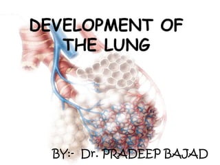
DEVELOPMENT OF
THE LUNG
BY:- Dr. PRADEEP BAJAD
ROLE OF EMBRYOLOGY IN DISEASE
MANAGEMENT
• To know the exact etiology and prognosis
of disease. eg. Tracheo-broncheal fistula
• To understand multisystem disorder
• To understand imgaing features.
• To get cellular level of information
regarding normal/abnormal tissue.
Stages of human lung development and their timing
Embryonic Period (Weeks 4--7)
A
LUNG ANLAGE
 The lung anlage appears at
day 26 as two ventral buds of
the foregut at the caudal end
of the laryngotracheal sulci.
 It will give rise to the left and
right lung. Both buds elongate,
grow into the surrounding
mesenchyme, and form the
left and right main bronchi
(day 32).
CONTINUED...
The terminal ends of the growing bronchial
tree start a repetitive process of growth and
mainly dichotomous branching.
 By day E37 the future conducting airways are
preformed to the lobar,
 By day E41 to the segmental,
And by day E48 to the subsegmental bronchi.
1.Agenesis - Bronchus and lung are absent.
2.Aplasia - Rudimentary bronchus is present and
limited to a blindend pouch without lung tissue.
3. Hypoplasia - Bronchial hypoplasia with variable
reduction of lung tissue .
Pulmonary underdevelopment
Agenesis occurs during the embryonic period
(approximately 4 weeks gestation).
Can occur bilaterally or unilaterally.
Presence of both bronchi and alveoli in an
underdeveloped lobe.
Due to early failure of the respiratory bud to develop
and/or branch (e.g. insufficient mesoderm, teratogens
such as RA or alcohol, or genetic mutation).
Agenesis of the Lungs
 Unilateral lung agenesis is compatible with life
(remaining side usually hyperexpands and
compensates).
 50% of children with pulmonary agenesis have
associated congenital anomalies.
 Cardiovascular (more frequent patent ductus
arteriosus and patent foramen ovale),
gastrointestinal, skeletal, and genitourinary
systems
CONTINUE…
Pulmonary hypoplasia
• Reduction in the number of lung
cells, airways, and alveoli that
results in a lower organ size and
weight.( U/l or b/l)
• Associated congenital anomalies -
cardiac, gastrointestinal,
genitourinary, and skeletal
malformations.(50-80%)
• Etiologies include prolonged
rupture of membranes, fetal renal
dysplasias and obstruction, and
fetal neuromuscular diseases.
Congenital Lung Cysts
Arise secondary to abnormal budding of the
primitive ventral foregut, early in fetal life.
Location - Mediastinum (Commonest – 70 %)
Pulmonary parenchyma. Rarely - Neck,
pericardium, or abdominal cavity
Symptoms
Incidental finding
Symptomatic infants – Respiratory distress
Older children - Infected cysts
Spontaneous pneumothorax – Rarely
CONTINUE…
Cysts (filled with fluid or air) are formed by the dilation
of terminal bronchi, due to branching irregularities in
later development
Complications
 Infection
 hemorrhage
 erosion into adjacent structures.
 Medical therapy
-antibiotics in children
with CCAM
complicated by
pneumpnia and
supportive care, ranging
from oxygen
supplementation to
mechanical ventilation,
in older children with
respiratory distress.
 Surgical Resection –
main line of treatment
TREATMENT
Tracheal bronchus
Tracheal Bronchus
A bronchial anomaly
originating fromthe
trachea.
Usually in the right
lateral wall of the
trachea.
2 cm above the carina.
Tracheal bronchus – Displaced (More frequent ) or
supernumerary
Prevalence –
•Right tracheal bronchus - 0.1%–2%.
•Left tracheal bronchus - 0 3. % – 1 %.
Symptoms – Asymptomatic usually. Consider
diagnosis in persistent/recurrent upper lobe
pneumonia or atelectasis or air trapping.
CONTINUE…
Bronchial atresia
Focal obliteration of a proximal segmental or
sub-segmental bronchus.
Lacks communication with the central airways
Development of distal structures is normal. Most often affects
segmental bronchi at or near their origin.
Bronchi distal to the stenosis become filled with mucus →
bronchocele.
May be acquired postnatally - Traumatic/ postinflammatory
insult .
Upper-lobe bronchi are more frequently affected.
Usually asymptomatic incidental finding in approximately 50% of
cases, mostly in young men.
 Dyspnea, pneumonia, and bronchial asthma have been
Splitting of foregut into esophagus and trachea
Langman’s fig 13-02
Initially, the lung bud is in
open communication with the
foregut then
tracheoesophageal ridges,
separate it from the foregut
These ridges fuse to form the
tracheoesophageal septum.
 The foregut is divided into a
dorsal portion esophagus,
and a ventral portion, the
trachea and lung buds .
Tracheo-esophageal fistulas
• Incomplete separation of
esophagous and trachea by
tracheosophageal septum
results in atresia of
esophagus with or without
tracheosophageal fistula.
• Defect likely in mesoderm
and usually associated with
other defects involving
mesoderm (cardiovascular
malformations, VATER /
VACTERL, etc.)
Langman’s fig 13-03
Continued..
Occur in approx 1/3000 births, most (90%) are that
shown in (A) above.
Complications
•PRENATAL: Polyhydramnios (due to inability to swallow amniotic
fluid in utero)
•POSTNATAL
– Gastrointestinal: Infants cough and choke when swallowing
because of accumulation of excessive saliva in mouth and
upper respiratory tract. Milk is regurgitated immediately after
feeding.
– Respiratory: Gastric contents may also reflux into the
trachea and lungs, causing choking and often leading to
pneumonitis.
5 weeks - pleuropericardial fold
forms
8 weeks - lungs grow and
expand into pleural cavity
6 weeks - pleuropericardial
membrane reaches midline
7 weeks -further maturation of
pericardium -expands pleural cavity
Moore & Persaud fig 10-4
Pleura and Formation of Lobes
The growing lung buds expand in caudolateral direction into
the pericardioperitoneal canals.
During week 5, the pleuropericardial folds meet and fuse
with the foregut mesenchyme.
During weeks 5–7, pleuroperitoneal membranes meet and
fuse with the posterior edge of the septum transversum and
close the pleural cavities.
The visceral pleura have formed by the splanchnic
mesoderm, which covers the outside of the lung.
Pleura and Formation of Lobes
Continue…
• The parietal pleura have formed by the somatic
mesoderm layer covering the inner surface of the body
wall. visceral pleura invaginations of the pleura start to
separate the lobar bronchi and give rise to the lobar
fissure and the lung lobes.
Branching continues to be regulated by epithelial-
mesenchymal interactions.
Respiratory tract is derived from foregut endoderm
and associated mesoderm
From endoderm:
epithelial lining of trachea, larynx, bronchi, alveoli
From splanchnic mesoderm:
cartilage, muscle, and connective tissue of tract and visceral
pleura.
Growth Factors- Transcription factors likeTTF-1, Gli2, and Gli3;
aswell as growth factors like FGF-10, TGF-β, BMP-4, SHH, EGF,
and VEGF.
Carlson fig 15-02
Organogenesis
Vasculogenesis of the Pulmonary Circulation
The mesenchyme surrounding the lung buds contains a
number of progenetor cells of endothelial cells.
The newly formed endothelial cells are connecting to each
other to form first capillary tubes. These capillaries coalesce
to form small blood vessels alongside the airways,so the
earliest pulmonary vessels form by vasculogenesis
Distal angiogenesis to form branches of vessels.
Development of the airways and arteries.
Row 1
Row 3
0
2
4
6
8
10
12
Col
um
n 1
Col
um
n 3
FETAL LUNG DEVELOPMENT
Pseudoglandular stage,(5-17 week)
 Formation of bronchial tree and large parts of prospective
respiratory parenchyma; birth of the acinus
 Histology- the epithelial tubules branch constantly and
penetrate into the surrounding mesenchyme. A loose
three-dimensional capillary network is located in the
mesenchyme.
Canalicular Period (16-26 weeks)
(1) the differentiation of the pulmonary epithelium and
formation of the typical air-blood barrier;
(2) the beginning of surfactant synthesis and
secretion;
(3) the “canalization” of the lung parenchyma by
capillaries.
At the end of the canalicular stage, the lung has
reached a state of development in which gas
exchange is possible in principle.
Before these developmental steps, a prematurely
born infanthas no chance to survive.
POSTNATAL LUNG DEVELOPMENT
POST NATAL LUNG DEVELOPMENT
Alveoli start to form in the weeks before birth and the
process lasts well into the postnatal period. At birth
the lung contains between zero and about 50 million
alveoli.
Changes at time of birth
 Lungs are fluid filled; fluid squeezed out and into
lymphatics and blood vessels, expelled via trachea
at delivery.
 Surfactant remains on surface, lowers air/blood
tension.
Alveolar stage 36 weeks to 1–2 years
• These airspaces are mostly of the classical
‘saccular’ type, i.e. their walls are made of
thick septa with a central layer of connective
tissue sandwiched between two capillary
networks.
• These septa present at birth have been
termed originally ‘primary septa’. The double
capillary network septa represent the basic
structures needed for alveolarization.
Alveolarization
C The alveoli are formed by
lifting off of new tissue ridges
from the existing primary
septa. this process produces a
large number of small buds
appearing along the primary
septa.
These buds correspond to low
ridges representing newly
forming septa, Soon these low
ridges increase in height and
subdivide the airspaces into
smaller units, the alveoli.
Alveolarization of the lung
• Primary septa stage - lung
parenchyma made of saccules
• Secondary septa stage - saccules
transformed into alveolar ducts and
alveolar sacs.
The essence of this stage is the restructuring
of the double capillary networks in the
parenchymal septa to the mature aspect
with a single capillary system by 3 steps –
a. Capillary Fusion and Differential Growth
b. Programmed Cell Death
c. Interalveolar Pores (Pores of Kohn)
MicrovascularMaturation Stage(Birth to 2--3 Years)
Circulatory changes
 During fetal life, blood flow through the lung is
limited to between 10 and 15 percent of the cardiac
output.
 At birth ductus arteriosus closes and the shunting
of the entire cardiac output through the lung.
 The ductus arteriosus, first obstructed by
muscular contraction, and is anatomically closed
within a few weeks by the fibroticorganization of an
intravascular clot and is known as ligamentum
arteriosus.
Late Development
18 months until body growth stops
The lung volume increases to the power of 1
to body weight, and the pulmonary
compartments augment linearly with lung
volume.
The surface area for gas exchange also
increase the power of 1 to body mass.
Surfactant proteins
Four major surfactant proteins: A, B, C, and D
a. Surfactant A: activates macrophages to elicit uterine
contractions, also important in host defense.
b. Surfactant B: organizes into tubular structures that are
much more efficient at reducing surface tension (specific
deficiency in Surfactant B can lead to respiratory
distress). It’s a phospholipoprotein formed by type 2
alveolar cells.
c. Surfactant C: enhances function of surfactant
phospholipids
d. Surfactant D: important in host defense.
This disease affects 2% of live newborn infants, with
prematurely born being most susceptible. 30% of all
neonatal disease results from HMD or its
complications
Surfactant deficiency causes RDS or HMD. The
lungs are underinflated and the alveoli contain a
fluid of high protein content, probably derived from
circulation substances and injured pulmonary
epithelium.
Hyaline membrane disease
CONTINUE…
In addition to prematurity, prolonged
intrauterine asphyxia may produce
irreversible changes in Type II alveolar cells,
rendering them incapable of producing
surfactant.
Prolonged, labored breathing damages
alveolar epithelium, leading to protein
deposition, or “hyaline” changes
Complications
 Alveolar rupture
 Infection
 Intracranial hemorrhage and periventricular leukomalacia
 Patent ductus arteriosus (PDA) with increasing left-to-
right shunt
 Pulmonary hemorrhage
 Necrotizing enterocolitis (NEC) and/or gastrointestinal (GI)
perforation
 Apnea of prematurity
development of lung

More Related Content

What's hot

Development of diaphragm
Development of diaphragmDevelopment of diaphragm
Development of diaphragmNeeta Chhabra
 
Development of repiratory system By Adhish Gautam
Development of repiratory system By Adhish GautamDevelopment of repiratory system By Adhish Gautam
Development of repiratory system By Adhish GautamCyril Skaria
 
development of lung, congenital malformations of lung
development of lung, congenital malformations of lungdevelopment of lung, congenital malformations of lung
development of lung, congenital malformations of lungDr Praman Kushwah
 
Mediastinum-RADIOLOGY
Mediastinum-RADIOLOGYMediastinum-RADIOLOGY
Mediastinum-RADIOLOGYNavdeep Shah
 
Congenital lung abnormalities
Congenital lung abnormalitiesCongenital lung abnormalities
Congenital lung abnormalitiesairwave12
 
Developemental anomaly of lungs
Developemental anomaly of lungsDevelopemental anomaly of lungs
Developemental anomaly of lungsDrAmbikaGupta
 
05.26.09(b): Development of the Respiratory System and Diaphragm
05.26.09(b): Development of the Respiratory System and Diaphragm05.26.09(b): Development of the Respiratory System and Diaphragm
05.26.09(b): Development of the Respiratory System and DiaphragmOpen.Michigan
 
Pulmonary sequestration ppt
Pulmonary sequestration pptPulmonary sequestration ppt
Pulmonary sequestration pptprapulla chandra
 
Development of aorta and pulmonary trunk
Development of aorta and pulmonary trunkDevelopment of aorta and pulmonary trunk
Development of aorta and pulmonary trunkDr Laxman Khanal
 
Developmental disorders of lungs
Developmental disorders of lungsDevelopmental disorders of lungs
Developmental disorders of lungsFiroz Hakkim
 
11 development of the heart
11   development of the heart11   development of the heart
11 development of the heartpuneet mahajan
 
CONGENITAL DISORDERS OF LUNG
CONGENITAL DISORDERS OF LUNGCONGENITAL DISORDERS OF LUNG
CONGENITAL DISORDERS OF LUNGAmeen Rageh
 
Radiology in newborn collected by Dr. Saiful islam MD
Radiology in newborn collected by Dr. Saiful islam MDRadiology in newborn collected by Dr. Saiful islam MD
Radiology in newborn collected by Dr. Saiful islam MDDr. Habibur Rahim
 
Pneumomediastinum
PneumomediastinumPneumomediastinum
PneumomediastinumGamal Agmy
 
Anatomy of lungs and development of lungs
Anatomy of lungs and development of lungsAnatomy of lungs and development of lungs
Anatomy of lungs and development of lungsDrhappyachu
 
Development of Respiratory System (Special Embryology)
Development of Respiratory System (Special Embryology)Development of Respiratory System (Special Embryology)
Development of Respiratory System (Special Embryology)Dr. Sherif Fahmy
 

What's hot (20)

Development of diaphragm
Development of diaphragmDevelopment of diaphragm
Development of diaphragm
 
Development of repiratory system By Adhish Gautam
Development of repiratory system By Adhish GautamDevelopment of repiratory system By Adhish Gautam
Development of repiratory system By Adhish Gautam
 
development of lung, congenital malformations of lung
development of lung, congenital malformations of lungdevelopment of lung, congenital malformations of lung
development of lung, congenital malformations of lung
 
Development & anomalies respiratory system embryology
Development & anomalies respiratory system embryologyDevelopment & anomalies respiratory system embryology
Development & anomalies respiratory system embryology
 
Mediastinum-RADIOLOGY
Mediastinum-RADIOLOGYMediastinum-RADIOLOGY
Mediastinum-RADIOLOGY
 
Congenital lung abnormalities
Congenital lung abnormalitiesCongenital lung abnormalities
Congenital lung abnormalities
 
Lecture 3 lungs & pleura
Lecture 3 lungs & pleuraLecture 3 lungs & pleura
Lecture 3 lungs & pleura
 
Developemental anomaly of lungs
Developemental anomaly of lungsDevelopemental anomaly of lungs
Developemental anomaly of lungs
 
05.26.09(b): Development of the Respiratory System and Diaphragm
05.26.09(b): Development of the Respiratory System and Diaphragm05.26.09(b): Development of the Respiratory System and Diaphragm
05.26.09(b): Development of the Respiratory System and Diaphragm
 
Pulmonary sequestration ppt
Pulmonary sequestration pptPulmonary sequestration ppt
Pulmonary sequestration ppt
 
blood supply of lungs
blood supply of lungsblood supply of lungs
blood supply of lungs
 
Development of aorta and pulmonary trunk
Development of aorta and pulmonary trunkDevelopment of aorta and pulmonary trunk
Development of aorta and pulmonary trunk
 
Developmental disorders of lungs
Developmental disorders of lungsDevelopmental disorders of lungs
Developmental disorders of lungs
 
11 development of the heart
11   development of the heart11   development of the heart
11 development of the heart
 
CONGENITAL DISORDERS OF LUNG
CONGENITAL DISORDERS OF LUNGCONGENITAL DISORDERS OF LUNG
CONGENITAL DISORDERS OF LUNG
 
Radiology in newborn collected by Dr. Saiful islam MD
Radiology in newborn collected by Dr. Saiful islam MDRadiology in newborn collected by Dr. Saiful islam MD
Radiology in newborn collected by Dr. Saiful islam MD
 
Pneumomediastinum
PneumomediastinumPneumomediastinum
Pneumomediastinum
 
Anatomy of lungs and development of lungs
Anatomy of lungs and development of lungsAnatomy of lungs and development of lungs
Anatomy of lungs and development of lungs
 
Development of Respiratory System (Special Embryology)
Development of Respiratory System (Special Embryology)Development of Respiratory System (Special Embryology)
Development of Respiratory System (Special Embryology)
 
Tree in-bud sign golden s sign
Tree in-bud sign golden s signTree in-bud sign golden s sign
Tree in-bud sign golden s sign
 

Similar to development of lung

Embryology Respiratory System.ppt
Embryology Respiratory System.pptEmbryology Respiratory System.ppt
Embryology Respiratory System.pptMASHUDMORAA
 
Lecture 1- Development of Respiratory System.ppt
Lecture 1- Development of Respiratory System.pptLecture 1- Development of Respiratory System.ppt
Lecture 1- Development of Respiratory System.pptnidhi sharma
 
DEVELOPMENT OF LUNG.pdf
DEVELOPMENT OF LUNG.pdfDEVELOPMENT OF LUNG.pdf
DEVELOPMENT OF LUNG.pdfdawsonfinger1
 
embryo respiratory4.pdf
embryo respiratory4.pdfembryo respiratory4.pdf
embryo respiratory4.pdfTofikMohammed3
 
3-Embryology Of Respiratory System.pdf
3-Embryology Of Respiratory System.pdf3-Embryology Of Respiratory System.pdf
3-Embryology Of Respiratory System.pdfMuhRidhoAkbar
 
development of respiratory sysytem.pptx
development of respiratory sysytem.pptxdevelopment of respiratory sysytem.pptx
development of respiratory sysytem.pptxDr Subodh Shah
 
L1-Dev of Res Sys.pptx
L1-Dev of Res Sys.pptxL1-Dev of Res Sys.pptx
L1-Dev of Res Sys.pptxIgnacioMontalv
 
Embryology and histology of the lung
Embryology and histology of the lungEmbryology and histology of the lung
Embryology and histology of the lungOrlando Joseph
 
Respiratory embryology.pptx
Respiratory embryology.pptxRespiratory embryology.pptx
Respiratory embryology.pptxPraise252443
 
Congenital cystic lesions of lung
Congenital cystic lesions of lungCongenital cystic lesions of lung
Congenital cystic lesions of lungK KHAING SAW LWIN
 
Lung malformation part 1
Lung malformation part 1Lung malformation part 1
Lung malformation part 1Faheem Andrabi
 
Fetal circulation
Fetal circulation Fetal circulation
Fetal circulation Komal Parmar
 
pediatrics hand out.ppt
pediatrics hand out.pptpediatrics hand out.ppt
pediatrics hand out.pptbayisahrsa
 

Similar to development of lung (20)

Embryology Respiratory System.ppt
Embryology Respiratory System.pptEmbryology Respiratory System.ppt
Embryology Respiratory System.ppt
 
Lecture 1- Development of Respiratory System.ppt
Lecture 1- Development of Respiratory System.pptLecture 1- Development of Respiratory System.ppt
Lecture 1- Development of Respiratory System.ppt
 
DEVELOPMENT OF LUNG.pdf
DEVELOPMENT OF LUNG.pdfDEVELOPMENT OF LUNG.pdf
DEVELOPMENT OF LUNG.pdf
 
embryo respiratory4.pdf
embryo respiratory4.pdfembryo respiratory4.pdf
embryo respiratory4.pdf
 
3-Embryology Of Respiratory System.pdf
3-Embryology Of Respiratory System.pdf3-Embryology Of Respiratory System.pdf
3-Embryology Of Respiratory System.pdf
 
development of respiratory sysytem.pptx
development of respiratory sysytem.pptxdevelopment of respiratory sysytem.pptx
development of respiratory sysytem.pptx
 
Embryology part 6
Embryology part 6Embryology part 6
Embryology part 6
 
L1-Dev of Res Sys.pptx
L1-Dev of Res Sys.pptxL1-Dev of Res Sys.pptx
L1-Dev of Res Sys.pptx
 
Embryology and histology of the lung
Embryology and histology of the lungEmbryology and histology of the lung
Embryology and histology of the lung
 
Respiratory embryology.pptx
Respiratory embryology.pptxRespiratory embryology.pptx
Respiratory embryology.pptx
 
Imaging of Urinary tract TB
Imaging of Urinary tract TBImaging of Urinary tract TB
Imaging of Urinary tract TB
 
Respiratorysystem.ppt
Respiratorysystem.pptRespiratorysystem.ppt
Respiratorysystem.ppt
 
Congenital cystic lesions of lung
Congenital cystic lesions of lungCongenital cystic lesions of lung
Congenital cystic lesions of lung
 
Lung malformation part 1
Lung malformation part 1Lung malformation part 1
Lung malformation part 1
 
Surgery 5th year, 1st lecture (Dr. Ahmed Al-Azzawi)
Surgery 5th year, 1st lecture (Dr. Ahmed Al-Azzawi)Surgery 5th year, 1st lecture (Dr. Ahmed Al-Azzawi)
Surgery 5th year, 1st lecture (Dr. Ahmed Al-Azzawi)
 
PAEDIATRIC LUNG DISEASE
PAEDIATRIC LUNG DISEASEPAEDIATRIC LUNG DISEASE
PAEDIATRIC LUNG DISEASE
 
The lung
The lungThe lung
The lung
 
The lung
The lungThe lung
The lung
 
Fetal circulation
Fetal circulation Fetal circulation
Fetal circulation
 
pediatrics hand out.ppt
pediatrics hand out.pptpediatrics hand out.ppt
pediatrics hand out.ppt
 

Recently uploaded

Pests of soyabean_Binomics_IdentificationDr.UPR.pdf
Pests of soyabean_Binomics_IdentificationDr.UPR.pdfPests of soyabean_Binomics_IdentificationDr.UPR.pdf
Pests of soyabean_Binomics_IdentificationDr.UPR.pdfPirithiRaju
 
Speech, hearing, noise, intelligibility.pptx
Speech, hearing, noise, intelligibility.pptxSpeech, hearing, noise, intelligibility.pptx
Speech, hearing, noise, intelligibility.pptxpriyankatabhane
 
Microphone- characteristics,carbon microphone, dynamic microphone.pptx
Microphone- characteristics,carbon microphone, dynamic microphone.pptxMicrophone- characteristics,carbon microphone, dynamic microphone.pptx
Microphone- characteristics,carbon microphone, dynamic microphone.pptxpriyankatabhane
 
《Queensland毕业文凭-昆士兰大学毕业证成绩单》
《Queensland毕业文凭-昆士兰大学毕业证成绩单》《Queensland毕业文凭-昆士兰大学毕业证成绩单》
《Queensland毕业文凭-昆士兰大学毕业证成绩单》rnrncn29
 
Environmental Biotechnology Topic:- Microbial Biosensor
Environmental Biotechnology Topic:- Microbial BiosensorEnvironmental Biotechnology Topic:- Microbial Biosensor
Environmental Biotechnology Topic:- Microbial Biosensorsonawaneprad
 
Dubai Calls Girl Lisa O525547819 Lexi Call Girls In Dubai
Dubai Calls Girl Lisa O525547819 Lexi Call Girls In DubaiDubai Calls Girl Lisa O525547819 Lexi Call Girls In Dubai
Dubai Calls Girl Lisa O525547819 Lexi Call Girls In Dubaikojalkojal131
 
THE ROLE OF PHARMACOGNOSY IN TRADITIONAL AND MODERN SYSTEM OF MEDICINE.pptx
THE ROLE OF PHARMACOGNOSY IN TRADITIONAL AND MODERN SYSTEM OF MEDICINE.pptxTHE ROLE OF PHARMACOGNOSY IN TRADITIONAL AND MODERN SYSTEM OF MEDICINE.pptx
THE ROLE OF PHARMACOGNOSY IN TRADITIONAL AND MODERN SYSTEM OF MEDICINE.pptxNandakishor Bhaurao Deshmukh
 
The dark energy paradox leads to a new structure of spacetime.pptx
The dark energy paradox leads to a new structure of spacetime.pptxThe dark energy paradox leads to a new structure of spacetime.pptx
The dark energy paradox leads to a new structure of spacetime.pptxEran Akiva Sinbar
 
basic entomology with insect anatomy and taxonomy
basic entomology with insect anatomy and taxonomybasic entomology with insect anatomy and taxonomy
basic entomology with insect anatomy and taxonomyDrAnita Sharma
 
BIOETHICS IN RECOMBINANT DNA TECHNOLOGY.
BIOETHICS IN RECOMBINANT DNA TECHNOLOGY.BIOETHICS IN RECOMBINANT DNA TECHNOLOGY.
BIOETHICS IN RECOMBINANT DNA TECHNOLOGY.PraveenaKalaiselvan1
 
Citronella presentation SlideShare mani upadhyay
Citronella presentation SlideShare mani upadhyayCitronella presentation SlideShare mani upadhyay
Citronella presentation SlideShare mani upadhyayupadhyaymani499
 
Radiation physics in Dental Radiology...
Radiation physics in Dental Radiology...Radiation physics in Dental Radiology...
Radiation physics in Dental Radiology...navyadasi1992
 
Pests of Bengal gram_Identification_Dr.UPR.pdf
Pests of Bengal gram_Identification_Dr.UPR.pdfPests of Bengal gram_Identification_Dr.UPR.pdf
Pests of Bengal gram_Identification_Dr.UPR.pdfPirithiRaju
 
Bioteknologi kelas 10 kumer smapsa .pptx
Bioteknologi kelas 10 kumer smapsa .pptxBioteknologi kelas 10 kumer smapsa .pptx
Bioteknologi kelas 10 kumer smapsa .pptx023NiWayanAnggiSriWa
 
Best Call Girls In Sector 29 Gurgaon❤️8860477959 EscorTs Service In 24/7 Delh...
Best Call Girls In Sector 29 Gurgaon❤️8860477959 EscorTs Service In 24/7 Delh...Best Call Girls In Sector 29 Gurgaon❤️8860477959 EscorTs Service In 24/7 Delh...
Best Call Girls In Sector 29 Gurgaon❤️8860477959 EscorTs Service In 24/7 Delh...lizamodels9
 
Pests of jatropha_Bionomics_identification_Dr.UPR.pdf
Pests of jatropha_Bionomics_identification_Dr.UPR.pdfPests of jatropha_Bionomics_identification_Dr.UPR.pdf
Pests of jatropha_Bionomics_identification_Dr.UPR.pdfPirithiRaju
 
Call Girls in Munirka Delhi 💯Call Us 🔝8264348440🔝
Call Girls in Munirka Delhi 💯Call Us 🔝8264348440🔝Call Girls in Munirka Delhi 💯Call Us 🔝8264348440🔝
Call Girls in Munirka Delhi 💯Call Us 🔝8264348440🔝soniya singh
 
OECD bibliometric indicators: Selected highlights, April 2024
OECD bibliometric indicators: Selected highlights, April 2024OECD bibliometric indicators: Selected highlights, April 2024
OECD bibliometric indicators: Selected highlights, April 2024innovationoecd
 
Four Spheres of the Earth Presentation.ppt
Four Spheres of the Earth Presentation.pptFour Spheres of the Earth Presentation.ppt
Four Spheres of the Earth Presentation.pptJoemSTuliba
 

Recently uploaded (20)

Pests of soyabean_Binomics_IdentificationDr.UPR.pdf
Pests of soyabean_Binomics_IdentificationDr.UPR.pdfPests of soyabean_Binomics_IdentificationDr.UPR.pdf
Pests of soyabean_Binomics_IdentificationDr.UPR.pdf
 
Speech, hearing, noise, intelligibility.pptx
Speech, hearing, noise, intelligibility.pptxSpeech, hearing, noise, intelligibility.pptx
Speech, hearing, noise, intelligibility.pptx
 
Microphone- characteristics,carbon microphone, dynamic microphone.pptx
Microphone- characteristics,carbon microphone, dynamic microphone.pptxMicrophone- characteristics,carbon microphone, dynamic microphone.pptx
Microphone- characteristics,carbon microphone, dynamic microphone.pptx
 
Volatile Oils Pharmacognosy And Phytochemistry -I
Volatile Oils Pharmacognosy And Phytochemistry -IVolatile Oils Pharmacognosy And Phytochemistry -I
Volatile Oils Pharmacognosy And Phytochemistry -I
 
《Queensland毕业文凭-昆士兰大学毕业证成绩单》
《Queensland毕业文凭-昆士兰大学毕业证成绩单》《Queensland毕业文凭-昆士兰大学毕业证成绩单》
《Queensland毕业文凭-昆士兰大学毕业证成绩单》
 
Environmental Biotechnology Topic:- Microbial Biosensor
Environmental Biotechnology Topic:- Microbial BiosensorEnvironmental Biotechnology Topic:- Microbial Biosensor
Environmental Biotechnology Topic:- Microbial Biosensor
 
Dubai Calls Girl Lisa O525547819 Lexi Call Girls In Dubai
Dubai Calls Girl Lisa O525547819 Lexi Call Girls In DubaiDubai Calls Girl Lisa O525547819 Lexi Call Girls In Dubai
Dubai Calls Girl Lisa O525547819 Lexi Call Girls In Dubai
 
THE ROLE OF PHARMACOGNOSY IN TRADITIONAL AND MODERN SYSTEM OF MEDICINE.pptx
THE ROLE OF PHARMACOGNOSY IN TRADITIONAL AND MODERN SYSTEM OF MEDICINE.pptxTHE ROLE OF PHARMACOGNOSY IN TRADITIONAL AND MODERN SYSTEM OF MEDICINE.pptx
THE ROLE OF PHARMACOGNOSY IN TRADITIONAL AND MODERN SYSTEM OF MEDICINE.pptx
 
The dark energy paradox leads to a new structure of spacetime.pptx
The dark energy paradox leads to a new structure of spacetime.pptxThe dark energy paradox leads to a new structure of spacetime.pptx
The dark energy paradox leads to a new structure of spacetime.pptx
 
basic entomology with insect anatomy and taxonomy
basic entomology with insect anatomy and taxonomybasic entomology with insect anatomy and taxonomy
basic entomology with insect anatomy and taxonomy
 
BIOETHICS IN RECOMBINANT DNA TECHNOLOGY.
BIOETHICS IN RECOMBINANT DNA TECHNOLOGY.BIOETHICS IN RECOMBINANT DNA TECHNOLOGY.
BIOETHICS IN RECOMBINANT DNA TECHNOLOGY.
 
Citronella presentation SlideShare mani upadhyay
Citronella presentation SlideShare mani upadhyayCitronella presentation SlideShare mani upadhyay
Citronella presentation SlideShare mani upadhyay
 
Radiation physics in Dental Radiology...
Radiation physics in Dental Radiology...Radiation physics in Dental Radiology...
Radiation physics in Dental Radiology...
 
Pests of Bengal gram_Identification_Dr.UPR.pdf
Pests of Bengal gram_Identification_Dr.UPR.pdfPests of Bengal gram_Identification_Dr.UPR.pdf
Pests of Bengal gram_Identification_Dr.UPR.pdf
 
Bioteknologi kelas 10 kumer smapsa .pptx
Bioteknologi kelas 10 kumer smapsa .pptxBioteknologi kelas 10 kumer smapsa .pptx
Bioteknologi kelas 10 kumer smapsa .pptx
 
Best Call Girls In Sector 29 Gurgaon❤️8860477959 EscorTs Service In 24/7 Delh...
Best Call Girls In Sector 29 Gurgaon❤️8860477959 EscorTs Service In 24/7 Delh...Best Call Girls In Sector 29 Gurgaon❤️8860477959 EscorTs Service In 24/7 Delh...
Best Call Girls In Sector 29 Gurgaon❤️8860477959 EscorTs Service In 24/7 Delh...
 
Pests of jatropha_Bionomics_identification_Dr.UPR.pdf
Pests of jatropha_Bionomics_identification_Dr.UPR.pdfPests of jatropha_Bionomics_identification_Dr.UPR.pdf
Pests of jatropha_Bionomics_identification_Dr.UPR.pdf
 
Call Girls in Munirka Delhi 💯Call Us 🔝8264348440🔝
Call Girls in Munirka Delhi 💯Call Us 🔝8264348440🔝Call Girls in Munirka Delhi 💯Call Us 🔝8264348440🔝
Call Girls in Munirka Delhi 💯Call Us 🔝8264348440🔝
 
OECD bibliometric indicators: Selected highlights, April 2024
OECD bibliometric indicators: Selected highlights, April 2024OECD bibliometric indicators: Selected highlights, April 2024
OECD bibliometric indicators: Selected highlights, April 2024
 
Four Spheres of the Earth Presentation.ppt
Four Spheres of the Earth Presentation.pptFour Spheres of the Earth Presentation.ppt
Four Spheres of the Earth Presentation.ppt
 

development of lung

  • 1. DEVELOPMENT OF THE LUNG BY:- Dr. PRADEEP BAJAD
  • 2. ROLE OF EMBRYOLOGY IN DISEASE MANAGEMENT • To know the exact etiology and prognosis of disease. eg. Tracheo-broncheal fistula • To understand multisystem disorder • To understand imgaing features. • To get cellular level of information regarding normal/abnormal tissue.
  • 3. Stages of human lung development and their timing
  • 4. Embryonic Period (Weeks 4--7) A LUNG ANLAGE  The lung anlage appears at day 26 as two ventral buds of the foregut at the caudal end of the laryngotracheal sulci.  It will give rise to the left and right lung. Both buds elongate, grow into the surrounding mesenchyme, and form the left and right main bronchi (day 32).
  • 5. CONTINUED... The terminal ends of the growing bronchial tree start a repetitive process of growth and mainly dichotomous branching.  By day E37 the future conducting airways are preformed to the lobar,  By day E41 to the segmental, And by day E48 to the subsegmental bronchi.
  • 6. 1.Agenesis - Bronchus and lung are absent. 2.Aplasia - Rudimentary bronchus is present and limited to a blindend pouch without lung tissue. 3. Hypoplasia - Bronchial hypoplasia with variable reduction of lung tissue . Pulmonary underdevelopment
  • 7. Agenesis occurs during the embryonic period (approximately 4 weeks gestation). Can occur bilaterally or unilaterally. Presence of both bronchi and alveoli in an underdeveloped lobe. Due to early failure of the respiratory bud to develop and/or branch (e.g. insufficient mesoderm, teratogens such as RA or alcohol, or genetic mutation). Agenesis of the Lungs
  • 8.  Unilateral lung agenesis is compatible with life (remaining side usually hyperexpands and compensates).  50% of children with pulmonary agenesis have associated congenital anomalies.  Cardiovascular (more frequent patent ductus arteriosus and patent foramen ovale), gastrointestinal, skeletal, and genitourinary systems CONTINUE…
  • 9. Pulmonary hypoplasia • Reduction in the number of lung cells, airways, and alveoli that results in a lower organ size and weight.( U/l or b/l) • Associated congenital anomalies - cardiac, gastrointestinal, genitourinary, and skeletal malformations.(50-80%) • Etiologies include prolonged rupture of membranes, fetal renal dysplasias and obstruction, and fetal neuromuscular diseases.
  • 10. Congenital Lung Cysts Arise secondary to abnormal budding of the primitive ventral foregut, early in fetal life. Location - Mediastinum (Commonest – 70 %) Pulmonary parenchyma. Rarely - Neck, pericardium, or abdominal cavity Symptoms Incidental finding Symptomatic infants – Respiratory distress Older children - Infected cysts Spontaneous pneumothorax – Rarely
  • 11. CONTINUE… Cysts (filled with fluid or air) are formed by the dilation of terminal bronchi, due to branching irregularities in later development Complications  Infection  hemorrhage  erosion into adjacent structures.
  • 12.  Medical therapy -antibiotics in children with CCAM complicated by pneumpnia and supportive care, ranging from oxygen supplementation to mechanical ventilation, in older children with respiratory distress.  Surgical Resection – main line of treatment TREATMENT
  • 13. Tracheal bronchus Tracheal Bronchus A bronchial anomaly originating fromthe trachea. Usually in the right lateral wall of the trachea. 2 cm above the carina.
  • 14. Tracheal bronchus – Displaced (More frequent ) or supernumerary Prevalence – •Right tracheal bronchus - 0.1%–2%. •Left tracheal bronchus - 0 3. % – 1 %. Symptoms – Asymptomatic usually. Consider diagnosis in persistent/recurrent upper lobe pneumonia or atelectasis or air trapping. CONTINUE…
  • 15. Bronchial atresia Focal obliteration of a proximal segmental or sub-segmental bronchus. Lacks communication with the central airways Development of distal structures is normal. Most often affects segmental bronchi at or near their origin. Bronchi distal to the stenosis become filled with mucus → bronchocele. May be acquired postnatally - Traumatic/ postinflammatory insult . Upper-lobe bronchi are more frequently affected. Usually asymptomatic incidental finding in approximately 50% of cases, mostly in young men.  Dyspnea, pneumonia, and bronchial asthma have been
  • 16. Splitting of foregut into esophagus and trachea Langman’s fig 13-02 Initially, the lung bud is in open communication with the foregut then tracheoesophageal ridges, separate it from the foregut These ridges fuse to form the tracheoesophageal septum.  The foregut is divided into a dorsal portion esophagus, and a ventral portion, the trachea and lung buds .
  • 17. Tracheo-esophageal fistulas • Incomplete separation of esophagous and trachea by tracheosophageal septum results in atresia of esophagus with or without tracheosophageal fistula. • Defect likely in mesoderm and usually associated with other defects involving mesoderm (cardiovascular malformations, VATER / VACTERL, etc.) Langman’s fig 13-03
  • 18. Continued.. Occur in approx 1/3000 births, most (90%) are that shown in (A) above. Complications •PRENATAL: Polyhydramnios (due to inability to swallow amniotic fluid in utero) •POSTNATAL – Gastrointestinal: Infants cough and choke when swallowing because of accumulation of excessive saliva in mouth and upper respiratory tract. Milk is regurgitated immediately after feeding. – Respiratory: Gastric contents may also reflux into the trachea and lungs, causing choking and often leading to pneumonitis.
  • 19. 5 weeks - pleuropericardial fold forms 8 weeks - lungs grow and expand into pleural cavity 6 weeks - pleuropericardial membrane reaches midline 7 weeks -further maturation of pericardium -expands pleural cavity Moore & Persaud fig 10-4 Pleura and Formation of Lobes
  • 20. The growing lung buds expand in caudolateral direction into the pericardioperitoneal canals. During week 5, the pleuropericardial folds meet and fuse with the foregut mesenchyme. During weeks 5–7, pleuroperitoneal membranes meet and fuse with the posterior edge of the septum transversum and close the pleural cavities. The visceral pleura have formed by the splanchnic mesoderm, which covers the outside of the lung. Pleura and Formation of Lobes
  • 21. Continue… • The parietal pleura have formed by the somatic mesoderm layer covering the inner surface of the body wall. visceral pleura invaginations of the pleura start to separate the lobar bronchi and give rise to the lobar fissure and the lung lobes. Branching continues to be regulated by epithelial- mesenchymal interactions.
  • 22. Respiratory tract is derived from foregut endoderm and associated mesoderm From endoderm: epithelial lining of trachea, larynx, bronchi, alveoli From splanchnic mesoderm: cartilage, muscle, and connective tissue of tract and visceral pleura. Growth Factors- Transcription factors likeTTF-1, Gli2, and Gli3; aswell as growth factors like FGF-10, TGF-β, BMP-4, SHH, EGF, and VEGF. Carlson fig 15-02 Organogenesis
  • 23. Vasculogenesis of the Pulmonary Circulation The mesenchyme surrounding the lung buds contains a number of progenetor cells of endothelial cells. The newly formed endothelial cells are connecting to each other to form first capillary tubes. These capillaries coalesce to form small blood vessels alongside the airways,so the earliest pulmonary vessels form by vasculogenesis Distal angiogenesis to form branches of vessels.
  • 24. Development of the airways and arteries. Row 1 Row 3 0 2 4 6 8 10 12 Col um n 1 Col um n 3
  • 25. FETAL LUNG DEVELOPMENT Pseudoglandular stage,(5-17 week)  Formation of bronchial tree and large parts of prospective respiratory parenchyma; birth of the acinus  Histology- the epithelial tubules branch constantly and penetrate into the surrounding mesenchyme. A loose three-dimensional capillary network is located in the mesenchyme.
  • 26. Canalicular Period (16-26 weeks) (1) the differentiation of the pulmonary epithelium and formation of the typical air-blood barrier; (2) the beginning of surfactant synthesis and secretion; (3) the “canalization” of the lung parenchyma by capillaries. At the end of the canalicular stage, the lung has reached a state of development in which gas exchange is possible in principle. Before these developmental steps, a prematurely born infanthas no chance to survive.
  • 27. POSTNATAL LUNG DEVELOPMENT POSTNATAL LUNG DEVELOPMENT POST NATAL LUNG DEVELOPMENT
  • 28. Alveoli start to form in the weeks before birth and the process lasts well into the postnatal period. At birth the lung contains between zero and about 50 million alveoli. Changes at time of birth  Lungs are fluid filled; fluid squeezed out and into lymphatics and blood vessels, expelled via trachea at delivery.  Surfactant remains on surface, lowers air/blood tension.
  • 29. Alveolar stage 36 weeks to 1–2 years • These airspaces are mostly of the classical ‘saccular’ type, i.e. their walls are made of thick septa with a central layer of connective tissue sandwiched between two capillary networks. • These septa present at birth have been termed originally ‘primary septa’. The double capillary network septa represent the basic structures needed for alveolarization.
  • 30. Alveolarization C The alveoli are formed by lifting off of new tissue ridges from the existing primary septa. this process produces a large number of small buds appearing along the primary septa. These buds correspond to low ridges representing newly forming septa, Soon these low ridges increase in height and subdivide the airspaces into smaller units, the alveoli.
  • 31. Alveolarization of the lung • Primary septa stage - lung parenchyma made of saccules • Secondary septa stage - saccules transformed into alveolar ducts and alveolar sacs.
  • 32. The essence of this stage is the restructuring of the double capillary networks in the parenchymal septa to the mature aspect with a single capillary system by 3 steps – a. Capillary Fusion and Differential Growth b. Programmed Cell Death c. Interalveolar Pores (Pores of Kohn) MicrovascularMaturation Stage(Birth to 2--3 Years)
  • 33. Circulatory changes  During fetal life, blood flow through the lung is limited to between 10 and 15 percent of the cardiac output.  At birth ductus arteriosus closes and the shunting of the entire cardiac output through the lung.  The ductus arteriosus, first obstructed by muscular contraction, and is anatomically closed within a few weeks by the fibroticorganization of an intravascular clot and is known as ligamentum arteriosus.
  • 34. Late Development 18 months until body growth stops The lung volume increases to the power of 1 to body weight, and the pulmonary compartments augment linearly with lung volume. The surface area for gas exchange also increase the power of 1 to body mass.
  • 35. Surfactant proteins Four major surfactant proteins: A, B, C, and D a. Surfactant A: activates macrophages to elicit uterine contractions, also important in host defense. b. Surfactant B: organizes into tubular structures that are much more efficient at reducing surface tension (specific deficiency in Surfactant B can lead to respiratory distress). It’s a phospholipoprotein formed by type 2 alveolar cells. c. Surfactant C: enhances function of surfactant phospholipids d. Surfactant D: important in host defense.
  • 36. This disease affects 2% of live newborn infants, with prematurely born being most susceptible. 30% of all neonatal disease results from HMD or its complications Surfactant deficiency causes RDS or HMD. The lungs are underinflated and the alveoli contain a fluid of high protein content, probably derived from circulation substances and injured pulmonary epithelium. Hyaline membrane disease
  • 37. CONTINUE… In addition to prematurity, prolonged intrauterine asphyxia may produce irreversible changes in Type II alveolar cells, rendering them incapable of producing surfactant. Prolonged, labored breathing damages alveolar epithelium, leading to protein deposition, or “hyaline” changes
  • 38. Complications  Alveolar rupture  Infection  Intracranial hemorrhage and periventricular leukomalacia  Patent ductus arteriosus (PDA) with increasing left-to- right shunt  Pulmonary hemorrhage  Necrotizing enterocolitis (NEC) and/or gastrointestinal (GI) perforation  Apnea of prematurity