
Recommended
More Related Content
What's hot
What's hot (20)
Similar to Nasopharynx gross anatomy and applied anatomy in dental and medical aspects
Similar to Nasopharynx gross anatomy and applied anatomy in dental and medical aspects (20)
Recently uploaded
Recently uploaded (20)
Nasopharynx gross anatomy and applied anatomy in dental and medical aspects
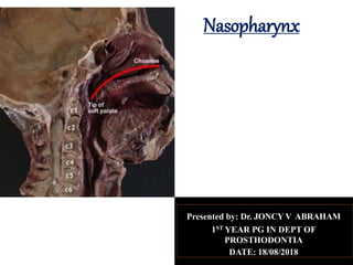
- 1. Nasopharynx Presented by: Dr. JONCY V ABRAHAM 1ST YEAR PG IN DEPT OF PROSTHODONTIA DATE: 18/08/2018
- 2. CONTENTS INTRODUCTION DEVELOPMENT OF PHARYNX STRUCTURE OF PHARYNX ANATOMICAL EXTENSION OF NASOPHARYNX BLOOD SUPPLY NERVE SUPPLY LYMPHATIC DRAINAGE HISTOLOGY APPLIED ANATOMY REFRENCES
- 3. INTRODUCTION • PHARYNX • Pharynx is a wide musculomembranous tube situated behind the nasal cavities, the mouth and the larynx. • DIMENSIONS OF PHARYNX • Length • 12-14 cm in length extending from the cranial base to the level of sixth cervical vertebra and the lower border of the cricoid cartilage. • Width • Greatest superiorly measuring 3.5 cm. • At its junction with the oesophagus it is reduced to about 1.5 cm this being the narrowest part of the alimentary canal.
- 4. INTERIOR OF PHARYNX- POSTERIOR VIEW
- 5. DEVELOPMENT The primitive gut extends from the buccopharyngeal membrane cranially to the cloacal membrane caudally. Divided into 4 parts- 1-The pharynx 2-The foregut 3-The midgut 4-the hindgut The pharynx extends from the buccopharyngeal membrane to the tracheobronchial diverticulum.
- 6. • Divided into 1- upper part nasopharynx 2-middle part the oropharynx 3-the lower part the laryngopharynx
- 7. STRUCTURE OF PHARYNX • The wall of pharynx is composed of five layers from within outwards: 1- MUCOSA 2- SUBMUCOSA 3-PHARYNGOBASILAR FASCIA 4-MUSCULAR COAT 5-BUCCOPHARYNGEAL FASCIA
- 8. 3-PHARYNGO-BASILAR FASCIA- • The intermediate fibrous layer which is thick above where the muscular fibres are absent. • Firmly connected to A- Basilar occipital and petrous temporal bones medial to carotid canal. B –Curving under the auditory tube and forward to the posterior border of the medial pterygoid plate and pterygomandibular raphae. C- As it descends its thickness diminishes. D- Posteriorly attached to pharyngeal tubercle and descends as Medial Pharyngeal raphae of constrictors .
- 9. 4- THE MUSCULAR COAT- 3 PAIRS OF CONSTRICTOR- • SUPERIOR CONSTRICTOR • MIDDLE CONSTRICTOR • INFERIOR CONSTRICTOR INNER LONGITUDNAL LAYERS STYLOPHARYNGEUS SALPINGOPHARYNGEUS PALATOPHARYNGEUS.
- 10. 5-BUCCOPHARYNGEAL FASCIA: Covers the outer surface of constrictors of pharynx. Extends forward across the pterygomandibular raphae to cover th Buccinator.
- 11. •Parts of Pharynx • Cavity of pharynx is divided into 3 parts a. The nasal part, NASOPHARYNX b. The oral part, OROPHARYNX c. The laryngeal part ,LARYNGOPHARYNX
- 12. NASOPHARYNX Behind the nasal cavity. Extends from SKULL BASE superiorly to the SOFT PALATE inferiorly. Communicates with nasal cavity with two posterior nasal apertures which are 25mm vertically and 12.5mm transversely and are separated by posterior edge of nasal septum. Nasal and oral part communicate through the PHARYNGEAL ISTHMUS,which is closed during swallowing by elevation of palate and contraction of PALATOPHARYNGEAL SPHINCHTER. LATERAL WALL presents pharyngeal opening of the AUDITORY TUBE, • 10-12.5mm behind and and a little below the posterior end of the inferior nasal concha.
- 13. NASOPHARYNX
- 14. ANATOMICAL EXTENT OF NASOPHARYNX Roof Floor Anterior wall Posterior wall Lateral wall
- 15. ROOF OF NASOPHARYNX Supported mainly by basilar occipital bone posteriorly. Posterior part of the body of the sphenoid anteriorly. Pharyngeal tonsil, a lymphoid mass which lies in the submucosa of the upper part of this surface and it is best developed in childhood.
- 16. PHARYNGEAL TONSIL Visible during the later fetal months. Increases in size up to 6 or 7 years and then usually begins to atrophy. In a child of 18 months it is a forward directed pyramidal prominence, with its apex near the nasal septum and its base at the junction of nasopharyngeal roof and posterior wall. It consist of folds radiating antero-laterally from a median recess,the PHARYNGEAL BURSA, which ascends backward into its substance. It represents attachment of notochord to the pharyngeal endoderm during embryonic life. The mucosal folds are mainly diffuse lymphoid tissue,but also contain deep mucous glands.
- 17. APPLIED ANATOMY In young children lymphoid hypertrophy in nose and nasopharynx(ADENOIDS),with or without enlargement of the palatine tonsil, may obstruct nasal respiration. The mouth has to be kept open to breathe.(MOUTH BREATHING). The hard palate and alveolar arch are then habitually out of contact with the lingual dorsum. Develop an abnormally high arch and forward projection. The hard palate becomes transversely narrow and projecting alveolar process afford little room for the permanent teeth which leads to crowding and overhang the lower teeth.
- 18. Maxillary surfaces appears pinched together,with narrowing of nasal cavities and maxillary air sinuses. Upper lip is drawn up exposing the projecting upper incisors. The face is lengthened by dropping of the lower jaw leading to . CAHRACTERISTIC FACIAL EXPRESSION (ADENOID FACIES)
- 20. CLINICAL SIGNIFICANCE OF NASOPHARYNGEAL BURSA An abscess can form in the bursa –TORNWALDT’S DISEASE. A TORNWALDT’S CYST develops if the embryonic remanant gets obstructed.
- 22. FLOOR OF NASOPHARYNX Formed by soft palate anteriorly. Deficient posteriorly called as nasopharyngeal isthmus via which it communicates with the oropharynx. PHARYNGEAL ISTHMUS remains closed during swallowing by the elevation of palate and contraction of the PALATOPHARYNGEAL SPHINCTER.
- 23. PASSAVANT’S RIDGE • The Superior pharyngeal constrictor muscle contracts to narrow the nasopharyngeal space. • TENSOR VELI PALATINI muscle tenses the soft palate to prevent distortion. • The LEVATOR VELI PALATINI, PALATOPHARYNGEUS AND SALPINGOPHARYNGEUS elevate it postero-superiorly.
- 24. • The lateral and posterior walls around the nasopharyngeal isthmus are then made taut by contraction of the palatopharyngeal sphincter muscle fibres( consist of skeletal muscle fibres of the most superior aspect of the palatopharyngeus muscle. These fibres forms an incomplete circle along the lateral and posterior walls of the nasopharyngeal isthmus at the level of C1 vertebra.) • THE LATTER ACTION FORMS PASSAVANT’S RIDGE. • These series of action prevent communication b/w the nasopharynx and the oropharynx during swallowing.
- 25. CLINICAL SIGNIFICANCE Failure to close the naso-oropharyngeal communication results in a condition known as velo-pharyngeal insufficiency. Can be caused by variety of disorders (Structural, Genetic, Functional or Accquired.) Very often associated with a Cleft palate. Parents usually bring in infants with this complication due to food and liquid coming through the nose during feeding and vomiting. Older individuals may present with recurrent sinus and ear infections due to ingested contents flowing back into the nasal sinus and the ostium pharyngeum respectively
- 26. TREATMENT PHYSICAL THERAPY SPEECH THERAPY OPERATIVE TECHNIQUE-. 1. Sphincter palatoplasty. 2. Posterior wall augmentation. 3. Pharyngeal flap. NON-OPERATIVE TECHNIQUES- 1. SPEECH BULB PROSTHESIS 2. PALATAL LIFT PROSTHESIS
- 29. ANTERIOR EXTENT OF NASOPHARYNX Anteriorly it communicates with the nasal cavity,through the two posterior nasal apertures,which are each 25mm vertically,12.5mm transversely. Seperated by posterior edge of nasal septum.
- 32. POSTERIOR WALL OF NASOPHARYNX Bounded by Atlas vertebra Dens of C2 Vertebra Superior constrictor Buccopharyngeal fascia Retropharyngeal space Prevertebral fascia
- 33. • Superior constrictor muscle: A quadrilateral sheet ,thinner and paler than others. • ORIGIN: • 1 –attached anteriorly to the PTERYGOID HAMULUS. • • 2- PTERYGOMANDIBULAR RAPHAE. • 3- POSTERIOR END OF THE MYLOHYOID LINE of the mandible. • 4-SIDE OF POSTERIOR PART OF THE TONGUE. • INSERTION: • 1-MEDIAN PHARYNGEAL RAPHAE • 2-SOME FIBRES ARE PROLONGED BY AN APONEUROSIS TO THE PHARYNGEAL TUBERCLE ON THE BASILAR PART OF OCCIPITAL BONE
- 35. RELATIONS OF SUPERIOR CONSTRICTOR • EXTERNALLY: • PREVERTEBRAL FASCIA AND MUSCLES • THE ASCENDING PHARYNGEAL ARTERY • THE PHARYNGEAL VENOUS PLEXUS • GLOSSOPHARYNGEAL AND LINGUAL NERVES • STYLOGLOSSUS AND MIDDLE CONSTRICTOR • MEDIAL PTERYGOID • STYLOHYOID LIGAMENT • STYLOPHARYNGEUS • INTERNAL CAROTID ARTERY • SYMPATHETIC TRUNK • HYPOGLOSSAL NERVE • INTERNAL JUGULAR VEIN • STYLOID PROCESS
- 37. INTERNAL RELATION OF SPC PALOTOPHARYNGEUS THE TONSILLAR CAPSULE PHARYNGOBASILLAR FASCIA
- 38. • SUPERIORLY: IT IS SEPARATED FROM THE CRANIAL BASE BY A CRESENTRIC INTERVAL CONTAINING LEVATOR VELI PALITINI. TENSOR VELI PALITINI. THE AUDITORY TUBE.
- 39. • INFERIORLY: ITS BORDER IS SEPERATED FROM MIDDLE CONSTRICTOR BY • 1- STYLOPHARYNGEUS • 2- GLOSSOPHARYNGEAL NERVE
- 40. • ANTERIORLY: SEPARATED FROM BUCCINATOR BY THE PTERYGOMANDIBULAR RAPHAE
- 41. NERVE SUPPLY AND ACTION • NERVE SUPPLY: THE PHARYNGEAL BRANCH OF VAGUS NERVE VIA THE PHARYNGEAL PLEXUS . • ACTION OF SPC: CONSTRICT WALL OF PHARYNX DURING SWALLOWING.
- 42. BUCCOPHARYNGEAL FASCIA The buccopharyngeal fascia is a fascia in the head. Parallel to the carotid sheath and along its medial aspect the pretracheal fascia gives off a thin lamina, the buccopharyngeal fascia, which closely invests the constrictor muscles of the pharynx and is continued forward from the constrictor pharyngis superior onto the buccinator. It is attached to the prevertebral layer by loose connective tissue only, and thus an easily distended space, the retropharyngeal space, is found between them.
- 44. RETROPHARYNGEAL SPACE The retropharyngeal space is a potential space of the head and neck, bounded by the buccopharyngeal fascia anteriorly and the alar fascia posteriorly. Together with the lateral pharyngeal space, these spaces are termed the parapharyngeal spaces. It contains the retropharyngeal lymph nodes.
- 45. PRE-VERTEBRAL FASCIA The prevertebral fascia is fixed above to the base of the skull, and below it extends behind the esophagus into the posterior mediastinal cavity of the thorax. It descends in front of the longus colli muscles. The prevertebral fascia is prolonged downward and laterally behind the carotid vessels and in front of the scalene muscles.
- 47. LATERAL WALL OF PHARYNX Each lateral wall presents a pharyngeal opening of the auditory tube 10- 12.5mm behind and little below the posterior end of the inferior nasal concha. It is somewhat triangular in shape,this opening is bounded above and behind by the TUBAL ELEVATION.Formed over the underlying pharyngeal end of the cartilage of the tube.
- 48. A vertical SALPINGOPHARYNGEAL FOLD OF MUCOSA descends from the tubal elevation , covering the SALPINGOPHARYNGEUS MUSCLE in the wall of the pharynx. A smaller SALPINGOPALATINE FOLD ,extends from the anterosuperior angle of the tubal elevation to the soft palate. The LEVATOR VELI PALITINI entering the soft palate produces an elevation of the mucosa immediately below the tubal opening.
- 50. LATERAL PHARYNGEAL RECESS/ FOSSA OF ROSENMULLER The anatomy of the fossa was first described in 1808 by Johann Christian Rosenmüller. The fossa of Rosenmüller is a bilateral projection of the nasopharynx just below the skull base. It is also called the lateral pharyngeal recess or simply the pharyngeal recess. The fossa is covered by nasopharyngeal mucosa . The lateral pharyngeal recess, or the fossa of Rosenmüller, is located behind the torus tubarius
- 51. BOUNDARIES OF THE FOSSA OF ROSSENMULLER • ANTERIORLY: • 1-Eustachian tube. • 2-Levator palatini muscle. • POSTERIOR: • 1-Posterior wall of nasopharynx. • 2-Retropharyngeal space.
- 52. • LATERAL: • 1-Parapharyngeal space. • 2-Tensor veli palatini muscle. • 3-Mandibular nerve • 4-Pre styloid compartment of Parapharyngeal space. • INFERIORLY: • 1-Upper edge of the superior constrictor muscle. • MEDIALLY: • 1- Nasopharynx
- 53. •POSTERO-LATERAL OR APEX- • 1-CAROTID CANAL OPENING AND PETROUS APEX POSTERIORLY. • 2-FORAMEN OVALE AND SPINOSUM LATERALLY. •SUPERIORLY: • 1-FORAMEN LACERUM • 2-FLOOR OF CAROTID CANAL
- 54. RADIO-ANATOMY
- 55. CLINICAL SIGNIFICANCE OF FOSSA OF ROSENMULLER 1-NASOPHARYNGEAL CARCINOMA: . It was determined to be the most common site of origin of nasopharyngeal carcinoma. 50% of cases. Loh et al attempted to study the anatomy of the fossa on 23 CT scans. They found that the fossa projects at about a 45-degree angle from the sagittal plane and ranges in length from 1.7 mm to 18.8 mm with a relatively narrow orifice. This led them to conclude that the fossa was often too deep and narrow for clinical inspection with a nasopharyngoscope and could constitute a blind spot in the postnasal space, especially the floor of the fossa. This had clinical implications in the early detection of nasopharyngeal carcinoma. Deep infiltration of NPC was most commonly to the intracranial region, usually through the foramen lacerum and the foramen ovale.(Trotter's syndrome is a cluster of symptoms associated with certain types of advanced nasopharyngeal carcinoma)
- 57. Frequency of lymph node manifestration • Upper jugular-94% • Middle jugular-85% • Retropharyngeal node- 80% • Posterior cervical - 46% • Lower jugular-19% • Supraclavicular -17% • Submental-17%
- 58. • LYMPHATIC SPREAD-Most common to upper, middle deep cervical & retropharyngeal lymph nodes.
- 59. TREATMENT OF NPC The main treatment for NPC is radiation therapy. It is often given in combination with chemotherapy. This approach may be called concomitant chemoradiotherapy Surgery for NPC is occasionally used, mainly to remove lymph nodes after chemoradiotherapy or to treat NPC that has come back after initial treatment.
- 60. SINUS OF MORGAGINI • A large gap between the upper concave border of the superior constrictor and the base of the skull is semi-lunar and is known as the SINUS OF MORGAGINI. • It is closed by the upper strong part of the pharyngo basilar fascia. • STRUCTURES PASSING THROUGH THIS GAP: 1- THE AUDITORY TUBE 2-THE LEVATOR VELI PALITINI MUSCLE 3-THE ASCENDING PALATINE ARTERY
- 61. SINUS OF MORGAGNI Space between base of skull & sup.connstictor. Through it enters- Eustachian tube Tensor &Levator veli palatini muscle Asc. Palatine artery. a-mucosa b-pharyngobasilar fascia c-muscular coat d-buccopharyngeal fascia
- 62. CLINICAL SIGNIFICANCE OF SINUS OF MORGAGINI • In nasopharyngeal carcinoma, the tumor may extend laterally and involve this sinus. • It can easily breach into the PARAPHARYNGEAL SPACE.
- 63. BLOOD SUPPLY • ARTERIES THAT SUPPLY UPPER PARTS OF THE PHARYNX: 1-THE ASCENDING PHARYNGEAL ARTERY 2-THE ASCENDING PALATINE AND TONSILLAR BRANCHES OF FACIAL ARTERY 3-NUMEROUS BRANCHES OF MAXILLARY AND LINGUAL ARTERIES.(all these vessels are from the external carotid artery)
- 64. •ARTERIES SUPPLYING LOWER PART OF THE PHARYNX 1-PHARYNGEAL BRANCHES FROM THE INFERIOR THYROID ARTERY(originating from the thyrocervical trunk of the subclavian artery.)
- 65. VEINS VEINS OF THE PHARYNX FORMS A PLEXUS,WHICH DRAINS SUPERIORLY INTO THE PTERYGOID PLEXUS IN THE INFRATEMPORAL FOSSA. INFERIORLY INTO THE FACIAL AND INTERNAL JUGULAR VEIN.
- 66. LYMPHATICS • Lymphatic vessels from the pharynx drain into the deep cervical nodes and include RETROPHARYNGEAL (between nasopharynx and vertebral column), PARATRACHEAL AND INFRAHYOID NODES. • The palatine tonsils drain through the pharyngeal wall into THE JUGULODIGASTRIC NODE in the region where the facial vein drains into the internal jugular vein (and inferior to the posterior belly of the digastric muscle).
- 68. NERVE SUPPLY • Motor and most sensory innervation (except for the nasal region) of the pharynx is mainly through branches of the vagus [ X ] and glossopharyngeal [ IX ] nerves, which form a plexus in the outer fascia of the pharyngeal wall. • PHARYNGEAL PLEXUS: The pharyngeal plexus is formed by: ■ the pharyngeal branch of the vagus nerve [ X ] ■ branches from the external laryngeal nerve from the superior laryngeal branch of the vagus nerve [ X ] and ■ pharyngeal branches of the glossopharyngeal nerve [ IX ] .
- 69. • The pharyngeal branch of the vagus nerve [ X ] originates from the upper part of its inferior ganglion above the origin of the superior laryngeal nerve and is the major motor nerve of the pharynx. • All muscles of the pharynx are innervated by the vagus nerve [ X ] mainly through the pharyngeal plexus, except for the stylopharyngeus, which is innervated directly by a branch of the glossopharyngeal nerve [ IX ] .
- 70. SENSORY NERVE SUPPLY • Each subdivision of the pharynx has a different sensory innervation: 1-The nasopharynx is innervated by a pharyngeal branch of the maxillary nerve [ V 2 ] that originates in the pterygopalatine fossa and passes through the palatovaginal canal in the sphenoid bone to reach the roof of the pharynx. 2-The oropharynx is innervated by the glossopharyngeal nerve [ IX ] via the pharyngeal plexus. 3- The laryngopharynx is innervated by the vagus nerve [ X ].
- 72. HISTOLOGY 1-Nasopharyngeal epithelium is- • CILIATED PSEUDO STRATIFIED COLUMNAR EPITHELIUM containing • GOBLET CELLS AND RECEIVING SUBMUCOUS GLANDS. 2- The oro and laryngo pharynx it is- • NON-KERATINIZED STRATIFIED SQUAMOUS EPITHELIUM. 3- A narrow transitional zone is present of CUBOIDAL EPITHELIUM, the cilia being imperfect or absent.
- 73. FUNCTIONAL CO-RELATION 1-The mucus helps maintain epithelial moisture and traps particulate material and pathogens moving through the airway. 2-The oropharynx, are also subject to the abrasive swallowing of food. To prevent the destruction of the respiratory epithelium in these areas, it changes to stratified squamous epithelium, which is better suited to the constant sloughing and abrasion. 3-The squamous layer of the oropharynx is continuous with the esophagus.
- 74. REFERENCES Gray’s Anatomy 37th edition. DiFIORE’s Atlas of Histology. Langman’s Medical Embryology11th edition. Grant’s Atlas of Anatomy. Fank.H.Netter Atlas of Human Anatomy.-4th Edition Gray’s Anatomy for Students 2nd Edition. BD Chaurasia’s Human Anatomy Vol 3- 5th edition.