SlideShare a Scribd company logo
1 of 66
ROLE OF USG IN GB
PATHOLOGIES
Dr.Pratibha Malik
Moderator-
Dr. Revathi
Always look for cholecytectomy
scar first!!
Indications
Normal Anatomy
Pathogical Findings:GB
gallstones
sludge
cholecystitis
polyps
gallbladder cancer
Biliary tract
Bile duct stones
klatskin tumour
08/03/14
CONTENTS
08/03/14
INDICATIONS
 Abdominal pain
 vomiting
 fever
 jaundice
 abnormal labs
(bilirubin,
transaminases)
08/03/14
Technique and Preparation
Curvilinear probes :2-5
MHz
Views:
 Subcostal sagittal
 intercostal sagittal
 coronal
 left lateral decubitus
 oblique views
 reverse trendelenberg
 Indications
 Normal Anatomy
 Pathogical Findings:GB
 gallstones
 sludge
 cholecystitis
 polyps
 gallbladder cancer
 Biliary tract
 Bile duct stones
 klatskin tumour
Contents
NORMAL ANATOMY OF THE
BILLIARY SYSTEM
NORMAL APPERANCE OF GB
Normal Gallbladder is seen as a sonolucent pear shaped
structure with slim wall (2 mm or so). Best seen with overnight
fasting or at least 4-5 hours fasting. It is seen usually at the
inferior aspect of the liver right lobe.
  The main hepatic fissure appears as an echogenic
line that extends from the neck of the gallbladder to
the portal vein and serves as a landmark.
 The complex of the gallbladder, main hepatic fissure,
and portal vein (in the short-axis) has the
appearance of an exclamation point
08/03/14
08/03/14
"Mickey Mouse" view of
Portal Triad
08/03/14
08/03/14
NORMAL VARIANTS
Phrygian cap
Figure (A) A folded gallbladder is
difficult to examine with the patient
supine.
(B) Turning the patient, right side
raised, unfolds the gallbladder,
enabling the lumen to be satisfactorily
examined.
The gallbladder may be ‘folded’ (the so-
called Phrygian cap).
CONTENTS
 Indications
 Normal Anatomy
 Pathogical Findings:GB
 Gallstones
 sludge
 cholecystitis
 polyps
 gallbladder cancer
 Biliary tract
 Bile duct stones
 klatskin tumour
Gold standard for diagnosis:
3 sonographic criteria-
 Echogenic focus
 Cast acoustic shadow
 Seek gravitational dependence
CHOLELITHIASIS
Cholelithiasis with posterior acoustic shadowing and 
normal GB wall thickness
CONTENTS
 Indications
 Normal Anatomy
 Pathogical Findings:GB
 gallstones
 sludge
 cholecystitis
 polyps
 gallbladder cancer
 Biliary tract
 Bile duct stones
 klatskin tumour
Sludge is identified on ultrasound as slightly hyperechoic material forming a
meniscus within the gallbladder lumen.
Sludge may be a precursor to gallstones and has been related to pathology
such as acute cholecystitis and acute pancreatitis
08/03/14
08/03/14
DD
ECHOGENIC LESIONS
IN GALL BLADDER
DD
MOBILE OR POSTERIOR
SHADOWING
•STONES
•SLUDGE
FIXED
•POLYP
•ADHESIVE STONE
•SLOW MOVING TUMEFACTIVE
SLUDGE
CONTENTS
 Indications
 Normal Anatomy
 Pathogical Findings:GB
 gallstones
 sludge
 cholecystitis
 polyps
 gallbladder cancer
 Biliary tract
 Bile duct stones
 klatskin tumour
GB inflammation
CALCULUS CHOLECYSTITIS VS ACALCULUS
 Acute cholecystitis refers to the acute inflammation of the
gallbladder . It is the primary complication of cholelithiasis
 90 to 95% of cases are due to calculous obstruction of the gallbladder
neck or cystic duct.
 Most sensitive US finding :
1. Presence Of Cholelithiasis
2. Sonographic Murphy Sign.
3. Gallbladder Wall Thickening (>3mm)
4. Pericholecystic fluid.
 Other less specific imaging findings include gallbladder distension(>4
cm) and sludge.
99m
Tc-HIDA scintigraphy
 HIDA cholescintigraphy in acute cholecystitis will demonstrate
nonvisualization of the gallbladder.
Figure: Acute cholecystitis: (A) TS of an oedematous, thickened gallbladder wall
with a stone. (B) LS with a thickened wall (arrows). Stones and debris are present.
(C) and (D) TS and LS demonstrating pericholecystic fluid.
Sonographic Murphy’s sign
08/03/14
Sonographic Murphy’s sign is positive when the point of maximal tenderness is
identified in the right upper quadrant while the gallbladder is identified on the
ultrasound monitor.
Differential diagnosis of acute cholecystitis
choledocholithiasis
pancreatitis
peptic ulcer disease
acute hepatitis
 Complications
 gangrenous cholecystitis
 emphysematous cholecystitis
 gallbladder perforation
 pericholecystic abscess
 cholecystoenteric fistula
 liver abscess
 Gangrenous and emphysematous cholecystitis - serious complications of acute
cholecystitis that may be identified with ultrasound by the presence of air within
the gallbladder wall or lumen . Air on ultrasound is represented by “comet-
tail” artifacts.
 Gallbladder perforation may also be diagnosed by ultrasound. Findings of
perforation include significant amounts of pericholecystic fluid that may contain
echogenic material which may be walled off from the rest of the abdomen.
Gallbladder gangrene/mucosal sloughing. Longitudinal
ultrasound of a patient who had acute cholecystitis
secondary to stone (arrow) impacted in the
gallbladder neck. Note the intraluminal membranes
(arrowheads) that are associated with gangrene of
the gallbladder.
Chronic calculus cholecystitis
USG FINDINGS
 Thickened echogenic wall
 Contracted GB
 Cholelithisis
Chronic calculus cholecystitis
 When the gallbladder is entirely filled with stones, a wall echo shadow
(WES) sign is seen
 The WES triad is another sign of chronic cholecystitis; here the WES
stands for Wall (of the GB), Echo (of the calculus) and Shadow
(caused by the stones).
ACALCULUS CHOLECYSTITIS
 Acute acalculous cholecystitis (AAC) refers
development of cholecystitis either in a gallbladder without
gallstones or in a gallbladder with gallstones where the
stones are not the contribuatry factor to the development
of cholecystitis .
AETIOLOGY
 It usually occurs in critically ill or
 injured patients.
Figure :(A) Acalculous cholecystitis. The gallbladder wall is markedly thickened
and tender on scanning.
(B) Gravity-dependent sludge with a thick, oedematous wall. No stones were
present.
PORCELAIN GB
 A porcelain gallbladder refers to extensive calcium
encrustation of the gallbladder wall.
 The term porcelain gallbladder has been used to
emphasize the blue discoloration and brittle
consistency of the gallbladder wall at surgery
 Association between porcelain gallbladder and
gallbladder adenocarcinoma- 22-30 % .
 Cholecystectomy routinely performed when a
porcelain gallbladder is identified.
Porcelain gallbladder. (A) Sagittal imageof the gallbladder shows a densely
echogenic anterior wall (arrow) with a sharp shadow that obliterates the
gallbladder lumen and posterior wall. (B) Transverse ultrasound of the
gallbladder in the same patient. The anterior wall is bright, but, without enough
reflection or attenuation to eliminate visualization of the lumen and posterior wall
(arrow), which is also echogenic and casts a posterior acoustic shadow.
CONTENTS
 Indications
 Normal Anatomy
 Pathogical Findings:GB
 gallstones
 sludge
 cholecystitis
 polyps
 gallbladder cancer
 Biliary tract
 Bile duct stones
 klatskin tumour
GB POLYPS
 Gallbladder polyps are outgrowths of the gallbladder mucosal wall.
 Do not cast an acoustic shadow.
 Remain fixed on turning the patient ;so distinguishable from stones
 Majority are not neoplastic but are hyperplastic or represent lipid
deposits(cholesterolosis).
 Gallbladder (GB) polyps are incidentally detected in approximately
4%–7% of patients who undergo ultrasonography
GB POLYPS
GB polyps are classified into 2 groups:
Neoplastic
 Adenomas
 Adenocarcinomas
Nonneoplastic
 Cholesterol polyps
 Inflammatory polyps
 Adenomyomatosis
GB polyps are soft tissue masses attached to the wall of the gallbladder and
differentiated from gallstones by their lack of mobility and shadowing
On CD demonstration of supplying vessel
pathognomic of polyp.
INCREASE RISK OF MALIGNANCY IN
POLYP
 Diameters > 10 mm
 Sessile Polyps
 Single Polyps
 Polyps With Adjacent Wall Thickening Or Invasion
 Increased Patient Age
 Size Of At Least 10 mm Is The Most Well-established
predictor of malignancy
CONTENTS
 Indications
 Normal Anatomy
 Pathogical Findings:GB
 gallstones
 sludge
 cholecystitis
 polyps
 gallbladder cancer
 Biliary tract
 Bile duct stones
 klatskin tumour
CARCINOMA GB
 Gall bladder carcinoma is the most common biliary
tract cancer.
 Delayed presentation and early spread of tumor
make it one of the lethal tumors with poor prognosis.
RISK FACTOR FOR CA GB
 Gallstones
 History of chronic cholecystitis
 Porcelain gallbladder.
 Choledochal cysts,
 Anomalous pancreaticobiliary duct junctions
 Gallbladder polyps > 1 cm in size.
 Peak incidence in the 6th
-7th
decades of life
 3-5 times more predominant in females
USG FINDINGS IN CA GB
 A Mass Completely Occupying Or Replacing The Gallbladder
Lumen
 Focal Or Diffuse Asymmetric Gallbladder Wall Thickening
 An Intraluminal Polypoid Lesion.
 Invasion of adjacent liver parenchyma
 Hepatic metastasis
 Periportal/ peripancreatic lymphadenopathy
 Sonographically : Heterogeneous, Predominantly Hypoechoic
Tumor Fills Much Or All of the gallbladder lumen.
CA GB WITH LIVER INVASION
08/03/14
 The normal gallbladder wall measures less than 4 mm.  the gallbladder wall is measured at the
most narrow point of the anterior wall in the short-axis.  Care must be taken to not measure the
wall at an oblique angle
THICKENING OF GALL BLADDER
WALL
DD
FOCAL
ADENOMYOMATOSIS
GALL BLADDER CANCER
DIFFUSE
ACUTE CHOLECYSTITIS
CHRONIC CHOLECYSTITIS
XANTHOGRANULOMATOUS
CHOLECYSTTIS
ADENOMYOMATOSIS
GB WALL EDEMA
GB CANCER
OTHERS-
ASCITIS,CHF,HYPOALBUMINEMIA
CONTENTS
 Indications
 Normal Anatomy
 Pathogical Findings:GB
 gallstones
 sludge
 cholecystitis
 polyps
 gallbladder cancer
 Biliary tract
 Bile duct stones
 klatskin tumour
BILIARY TRACT
08/03/14
BILE DUCTS
 Sonographically, the CBD appears as an anechoic tubular
structure in the main portal triad, anterior to and following the
course of the main portal vein
 Conventionally, the upper limit of normal for the common bile
duct as measured by ultrasound is considered to be 6 mm.
 Biliary duct obstruction: caused by stones, pancreatic
pathology (e.g. mass), or stricture is detected measuring a
CBD larger than 6-7 mm.
2000 Am. Coll. of Gastroenterology
08/03/14
Intrahepatic duct stones
The Mirizzi Syndrome
 Form of obstructive jaundice-described by
Mirizzi.
 Caused by a stone or stones impacted in the
neck of the gallbladder or the cystic duct, such
that the common hepatic duct is narrowed.
 Rare complication of gallstones-occurs in about
0.1% to 0.7% of patients with cholelitiasis.
Csendes classification of
Mirizzi syndrome
Figure: Mirizzi syndrome: a large stone in the
neck of the gallbladder (arrow) is compressing the bile duct,
causing intrahepatic duct dilatation. The lower end of the CBD
remains normal in calibre.
COMPONENTS OF MS
There must be 4 components for the syndrome to occur:
1. Anatomy- placing the cystic duct parallel to the common
hepatic duct
2. Impaction of a stone in the cystic dust or gallbladder neck
3. Obstruction of the common hepatic duct from the stone
itself, or from the resultant inflammatory response
4. Intermittent or constant jaundice occasionally causing
cholangitis, and with longstanding obstruction, biliary
cirrhosis
INTRAHEPATIC GALL STONES
INTRAHEPATIC VS EXTRAHEPATIC CALCULUS
•Caroli's disease is characterized by congenital
segmental dilation of the intrahepatic bile ducts producing
primary intrahepatic gallstones.
•Secondary intrahepatic gall stone formation occurs due to
chronic obstruction of CBD and CHD
•It's believed that most patients suffering a chronic illness
have excessive amounts of gallstones in the liver.
• Gallstones found in gallbladder tend to be hardened and
relatively large while stones found in liver tends to be soft
and noncalcified
•Intrahepatic Gallstones cause liver congestion and elevted
liver enzymes
Primary intrahepatic stones exclusively involving the
intrahepatic biliary tree-related to chronic parasitic infestation of
the biliary tree- (ascariasis )
Mixed intrahepatic stone- Associated with extrahepatic lithiasis
Secondary intrahepatic stones related to ananatomical
condition precipitating stasis or infection
TYPES OF INTRAHEPATIC CALCULUS
EXTRAHEPATIC CALCULUS
CALCULI IN THE DISTAL CBD
CONTENTS
 Indications
 Normal Anatomy
 Pathogical Findings:GB
 gallstones
 sludge
 cholecystitis
 polyps
 gallbladder cancer
 Biliary tract
 Bile duct stones
 klatskin tumour
KLATSKIN TUMOUR
 A Klatskin tumour is a term that was traditionally given
to a hilar cholangiocarcinoma (occuring at the
bifurcation of the common hepatic duct).
Typically, these tumours are
 Small In Size
 Poorly Differentiated
 Exhibit Aggressive Biologic Behaviour
 And tend to obstruct the intrahepatic bile ducts
 25% of all cholangiocarcinoma
Risk factors :
 Primary sclerosing cholangitis
 Choledochal cysts
 Parasitic infections
 Hepatolithiasis (also called Oriental cholangiohepatitis)
 Toxin exposures
 Genetic Factors
ULTRASOUND FINDINGS
 Presence of a hilar mass with obstruction
 Increased echogenicity relative to surrounding
liver ~79%
 Reduced echogenicity ~ 19 %
 Mixed echogenicity ~ 2 % 
 Segmental dilatation or nonunion of R and L ducts
 Polypoid intraluminal masses,
 Nodular smooth masses with mural thickening.
 Should do doppler, as this is helpful to assess
Vascular invasion (unresectable)
Figure : Cholangiocarcinoma. (A) Irregular mass at the porta,
causing biliary obstruction—a Klatskin tumour.
(B) MRI of the same patient, confirming the mass at the porta.
KLATSKIN TUMOUR
KLATSKIN TUMOUR
COLOUR DOPPLER TO LOOK FOR
VASCULAR INVASION
08/03/14
THICKENING OF BILE DUCT WALL
FOCAL STRICTURE
•BACTERIAL CHOLANGITIS
•CHOLANGIOCARCINOMA
•CHOLEDOCAL CYSTS
•METASTASIS
•BENIGN STRICTURE
NO STRICTURE
•POLYPOID CANCER
•STONES
•PERIAMPULLARY CANCER
•SENILE CHANGE
•POSTCHOLECYSTECTOMY
DILATATION
•CHOLEDOCHAL CYST
•ASCARIASIS
08/03/14
Pearls and Pitfalls
 The gallbladder is a mobile organ; remember to change patient positioning
and/or probe placement to find the organ of interest.
 Distinguish gallstones from polyps and septations or folds by always scanning
through the whole organ and in both longitudinal and transverse planes.
 During the biliary exam, use color Doppler to help distinguish nonvascular from
vascular structures.
 Ultrasound findings must be interpreted in the context of the clinical
presentation; findings suggestive of acute cholecystitis (e.g., gallstone or
thickened wall) may be present in patients in a nondiseased state.
 The common bile duct can be dilated in the absence of pathology in older
patients and postcholecystectomy patients.
 Measure the anterior wall of the gallbladder. The posterior wall may appear
artificially thickened because of acoustic enhancement or artifact from bowel
gas.
USG in gall bladder pathologies

More Related Content

What's hot

Bile duct Ultrasound
Bile duct UltrasoundBile duct Ultrasound
Bile duct UltrasoundSafi. Khan
 
radiology.Plain abd.(dr.kawa)
radiology.Plain abd.(dr.kawa)radiology.Plain abd.(dr.kawa)
radiology.Plain abd.(dr.kawa)student
 
Liver segments on ultrasound
Liver segments on ultrasoundLiver segments on ultrasound
Liver segments on ultrasoundDurre Sabih
 
Pancreas ultrasound
Pancreas ultrasoundPancreas ultrasound
Pancreas ultrasoundSafi. Khan
 
Radiological approach to gastric ulcer disease
Radiological approach to gastric ulcer diseaseRadiological approach to gastric ulcer disease
Radiological approach to gastric ulcer diseaseNavneet Ranjan
 
Spleen Ultrasound anatomy structure scanning techniques and pathologies
Spleen Ultrasound anatomy structure scanning techniques and pathologies Spleen Ultrasound anatomy structure scanning techniques and pathologies
Spleen Ultrasound anatomy structure scanning techniques and pathologies Safi. Khan
 
Ultrasound of pancrease in Radiology
Ultrasound of pancrease in RadiologyUltrasound of pancrease in Radiology
Ultrasound of pancrease in RadiologyMahesh Kumar
 
Gall bladder Ultrasound
Gall bladder UltrasoundGall bladder Ultrasound
Gall bladder UltrasoundSafi. Khan
 
The Radiology of Malrotation
The Radiology of MalrotationThe Radiology of Malrotation
The Radiology of Malrotationtboulden
 
Imaging of the large bowel
Imaging of the large bowelImaging of the large bowel
Imaging of the large bowelArchana Koshy
 
Presentation1.pptx, ultrasound examination of the adrenal glands and kidneys.
Presentation1.pptx, ultrasound examination of the adrenal glands and kidneys.Presentation1.pptx, ultrasound examination of the adrenal glands and kidneys.
Presentation1.pptx, ultrasound examination of the adrenal glands and kidneys.Abdellah Nazeer
 
Doppler ultrasound of the kidneys
Doppler ultrasound of the kidneysDoppler ultrasound of the kidneys
Doppler ultrasound of the kidneysSamir Haffar
 
Presentation1.pptx, radiological imaging of scrotal diseases.
Presentation1.pptx, radiological imaging of scrotal diseases.Presentation1.pptx, radiological imaging of scrotal diseases.
Presentation1.pptx, radiological imaging of scrotal diseases.Abdellah Nazeer
 
Ultrasound imaging of Bowel pathology
Ultrasound imaging of Bowel pathologyUltrasound imaging of Bowel pathology
Ultrasound imaging of Bowel pathologyairwave12
 
Liver ultrasound tips and tricks
Liver ultrasound tips and tricksLiver ultrasound tips and tricks
Liver ultrasound tips and tricksDr-mahmoud Desoky
 

What's hot (20)

Bile duct Ultrasound
Bile duct UltrasoundBile duct Ultrasound
Bile duct Ultrasound
 
Barium swallow diseases
Barium swallow diseasesBarium swallow diseases
Barium swallow diseases
 
radiology.Plain abd.(dr.kawa)
radiology.Plain abd.(dr.kawa)radiology.Plain abd.(dr.kawa)
radiology.Plain abd.(dr.kawa)
 
Liver segments on ultrasound
Liver segments on ultrasoundLiver segments on ultrasound
Liver segments on ultrasound
 
Pancreas ultrasound
Pancreas ultrasoundPancreas ultrasound
Pancreas ultrasound
 
Radiological approach to gastric ulcer disease
Radiological approach to gastric ulcer diseaseRadiological approach to gastric ulcer disease
Radiological approach to gastric ulcer disease
 
Spleen Ultrasound anatomy structure scanning techniques and pathologies
Spleen Ultrasound anatomy structure scanning techniques and pathologies Spleen Ultrasound anatomy structure scanning techniques and pathologies
Spleen Ultrasound anatomy structure scanning techniques and pathologies
 
Spleen ultrasound
Spleen ultrasoundSpleen ultrasound
Spleen ultrasound
 
Doppler of the portal system
Doppler of the portal systemDoppler of the portal system
Doppler of the portal system
 
Ultrasound of pancrease in Radiology
Ultrasound of pancrease in RadiologyUltrasound of pancrease in Radiology
Ultrasound of pancrease in Radiology
 
Gall bladder Ultrasound
Gall bladder UltrasoundGall bladder Ultrasound
Gall bladder Ultrasound
 
The Radiology of Malrotation
The Radiology of MalrotationThe Radiology of Malrotation
The Radiology of Malrotation
 
Imaging of the large bowel
Imaging of the large bowelImaging of the large bowel
Imaging of the large bowel
 
Liver ultrasound
Liver ultrasoundLiver ultrasound
Liver ultrasound
 
Prostate ultrasound (basic)
Prostate ultrasound (basic)Prostate ultrasound (basic)
Prostate ultrasound (basic)
 
Presentation1.pptx, ultrasound examination of the adrenal glands and kidneys.
Presentation1.pptx, ultrasound examination of the adrenal glands and kidneys.Presentation1.pptx, ultrasound examination of the adrenal glands and kidneys.
Presentation1.pptx, ultrasound examination of the adrenal glands and kidneys.
 
Doppler ultrasound of the kidneys
Doppler ultrasound of the kidneysDoppler ultrasound of the kidneys
Doppler ultrasound of the kidneys
 
Presentation1.pptx, radiological imaging of scrotal diseases.
Presentation1.pptx, radiological imaging of scrotal diseases.Presentation1.pptx, radiological imaging of scrotal diseases.
Presentation1.pptx, radiological imaging of scrotal diseases.
 
Ultrasound imaging of Bowel pathology
Ultrasound imaging of Bowel pathologyUltrasound imaging of Bowel pathology
Ultrasound imaging of Bowel pathology
 
Liver ultrasound tips and tricks
Liver ultrasound tips and tricksLiver ultrasound tips and tricks
Liver ultrasound tips and tricks
 

Similar to USG in gall bladder pathologies

ACUTE ABDOMEN SONOGRAPHIC FINDINGS ppt
ACUTE ABDOMEN SONOGRAPHIC FINDINGS pptACUTE ABDOMEN SONOGRAPHIC FINDINGS ppt
ACUTE ABDOMEN SONOGRAPHIC FINDINGS pptPrafulla Pinjarkar
 
膽道系統炎症之影像診斷
膽道系統炎症之影像診斷膽道系統炎症之影像診斷
膽道系統炎症之影像診斷calaf0618
 
Biliary pathologies
Biliary pathologiesBiliary pathologies
Biliary pathologiesNavdeep Shah
 
presentation_THurs.docx
presentation_THurs.docxpresentation_THurs.docx
presentation_THurs.docxDakaneMaalim
 
Gall bladder stone disease surgical perspective
Gall bladder stone disease surgical perspectiveGall bladder stone disease surgical perspective
Gall bladder stone disease surgical perspectivevishalvaishnavi2
 
Rapid review of radiology abdomen
Rapid review of radiology abdomenRapid review of radiology abdomen
Rapid review of radiology abdomenDouble M
 
Cholecystitis and Choldocholithiasis
Cholecystitis and CholdocholithiasisCholecystitis and Choldocholithiasis
Cholecystitis and CholdocholithiasisNAVANEETA KUSUM
 
Approach to intestinal obstructions.pptx
Approach to intestinal obstructions.pptxApproach to intestinal obstructions.pptx
Approach to intestinal obstructions.pptxyashwanthnaik8
 
Acute abdomen
Acute abdomenAcute abdomen
Acute abdomenairwave12
 
Bohomolets Surgery 4th year Lecture #6
Bohomolets Surgery 4th year Lecture #6Bohomolets Surgery 4th year Lecture #6
Bohomolets Surgery 4th year Lecture #6Dr. Rubz
 
Radiology 5th year, 2nd lecture/cont. (Dr. Nasrin Alatrushi)
Radiology 5th year, 2nd lecture/cont. (Dr. Nasrin Alatrushi)Radiology 5th year, 2nd lecture/cont. (Dr. Nasrin Alatrushi)
Radiology 5th year, 2nd lecture/cont. (Dr. Nasrin Alatrushi)College of Medicine, Sulaymaniyah
 
Imaging of dangerous cholecystitis
Imaging of dangerous cholecystitisImaging of dangerous cholecystitis
Imaging of dangerous cholecystitisAhmed Bahnassy
 
Pediatric High and Low intestinal Obstruction.pptx
Pediatric High and Low intestinal Obstruction.pptxPediatric High and Low intestinal Obstruction.pptx
Pediatric High and Low intestinal Obstruction.pptxDr Muhammad Tahir Javed
 

Similar to USG in gall bladder pathologies (20)

Sahad gb
Sahad gbSahad gb
Sahad gb
 
Imaging of Acute Abdomen
Imaging of Acute Abdomen Imaging of Acute Abdomen
Imaging of Acute Abdomen
 
Usg gb & biliary t
Usg gb & biliary tUsg gb & biliary t
Usg gb & biliary t
 
ACUTE ABDOMEN SONOGRAPHIC FINDINGS ppt
ACUTE ABDOMEN SONOGRAPHIC FINDINGS pptACUTE ABDOMEN SONOGRAPHIC FINDINGS ppt
ACUTE ABDOMEN SONOGRAPHIC FINDINGS ppt
 
膽道系統炎症之影像診斷
膽道系統炎症之影像診斷膽道系統炎症之影像診斷
膽道系統炎症之影像診斷
 
Biliary pathologies
Biliary pathologiesBiliary pathologies
Biliary pathologies
 
presentation_THurs.docx
presentation_THurs.docxpresentation_THurs.docx
presentation_THurs.docx
 
Gall bladder stone disease surgical perspective
Gall bladder stone disease surgical perspectiveGall bladder stone disease surgical perspective
Gall bladder stone disease surgical perspective
 
Rapid review of radiology abdomen
Rapid review of radiology abdomenRapid review of radiology abdomen
Rapid review of radiology abdomen
 
Hepato Biliary.pptx
Hepato Biliary.pptxHepato Biliary.pptx
Hepato Biliary.pptx
 
Acute abdomen
Acute abdomenAcute abdomen
Acute abdomen
 
Cholecystitis and Choldocholithiasis
Cholecystitis and CholdocholithiasisCholecystitis and Choldocholithiasis
Cholecystitis and Choldocholithiasis
 
Approach to intestinal obstructions.pptx
Approach to intestinal obstructions.pptxApproach to intestinal obstructions.pptx
Approach to intestinal obstructions.pptx
 
inflammatory bowel disease (IBD)
inflammatory bowel disease (IBD)inflammatory bowel disease (IBD)
inflammatory bowel disease (IBD)
 
Intestinal obstruction
Intestinal obstructionIntestinal obstruction
Intestinal obstruction
 
Acute abdomen
Acute abdomenAcute abdomen
Acute abdomen
 
Bohomolets Surgery 4th year Lecture #6
Bohomolets Surgery 4th year Lecture #6Bohomolets Surgery 4th year Lecture #6
Bohomolets Surgery 4th year Lecture #6
 
Radiology 5th year, 2nd lecture/cont. (Dr. Nasrin Alatrushi)
Radiology 5th year, 2nd lecture/cont. (Dr. Nasrin Alatrushi)Radiology 5th year, 2nd lecture/cont. (Dr. Nasrin Alatrushi)
Radiology 5th year, 2nd lecture/cont. (Dr. Nasrin Alatrushi)
 
Imaging of dangerous cholecystitis
Imaging of dangerous cholecystitisImaging of dangerous cholecystitis
Imaging of dangerous cholecystitis
 
Pediatric High and Low intestinal Obstruction.pptx
Pediatric High and Low intestinal Obstruction.pptxPediatric High and Low intestinal Obstruction.pptx
Pediatric High and Low intestinal Obstruction.pptx
 

Recently uploaded

Call Girls in Delhi Triveni Complex Escort Service(🔝))/WhatsApp 97111⇛47426
Call Girls in Delhi Triveni Complex Escort Service(🔝))/WhatsApp 97111⇛47426Call Girls in Delhi Triveni Complex Escort Service(🔝))/WhatsApp 97111⇛47426
Call Girls in Delhi Triveni Complex Escort Service(🔝))/WhatsApp 97111⇛47426jennyeacort
 
Premium Call Girls In Jaipur {8445551418} ❤️VVIP SEEMA Call Girl in Jaipur Ra...
Premium Call Girls In Jaipur {8445551418} ❤️VVIP SEEMA Call Girl in Jaipur Ra...Premium Call Girls In Jaipur {8445551418} ❤️VVIP SEEMA Call Girl in Jaipur Ra...
Premium Call Girls In Jaipur {8445551418} ❤️VVIP SEEMA Call Girl in Jaipur Ra...parulsinha
 
Most Beautiful Call Girl in Bangalore Contact on Whatsapp
Most Beautiful Call Girl in Bangalore Contact on WhatsappMost Beautiful Call Girl in Bangalore Contact on Whatsapp
Most Beautiful Call Girl in Bangalore Contact on WhatsappInaaya Sharma
 
Call Girls Service Jaipur {9521753030 } ❤️VVIP BHAWNA Call Girl in Jaipur Raj...
Call Girls Service Jaipur {9521753030 } ❤️VVIP BHAWNA Call Girl in Jaipur Raj...Call Girls Service Jaipur {9521753030 } ❤️VVIP BHAWNA Call Girl in Jaipur Raj...
Call Girls Service Jaipur {9521753030 } ❤️VVIP BHAWNA Call Girl in Jaipur Raj...khalifaescort01
 
Night 7k to 12k Chennai City Center Call Girls 👉👉 7427069034⭐⭐ 100% Genuine E...
Night 7k to 12k Chennai City Center Call Girls 👉👉 7427069034⭐⭐ 100% Genuine E...Night 7k to 12k Chennai City Center Call Girls 👉👉 7427069034⭐⭐ 100% Genuine E...
Night 7k to 12k Chennai City Center Call Girls 👉👉 7427069034⭐⭐ 100% Genuine E...hotbabesbook
 
Call Girls Service Jaipur {8445551418} ❤️VVIP BHAWNA Call Girl in Jaipur Raja...
Call Girls Service Jaipur {8445551418} ❤️VVIP BHAWNA Call Girl in Jaipur Raja...Call Girls Service Jaipur {8445551418} ❤️VVIP BHAWNA Call Girl in Jaipur Raja...
Call Girls Service Jaipur {8445551418} ❤️VVIP BHAWNA Call Girl in Jaipur Raja...parulsinha
 
Independent Call Girls Service Mohali Sector 116 | 6367187148 | Call Girl Ser...
Independent Call Girls Service Mohali Sector 116 | 6367187148 | Call Girl Ser...Independent Call Girls Service Mohali Sector 116 | 6367187148 | Call Girl Ser...
Independent Call Girls Service Mohali Sector 116 | 6367187148 | Call Girl Ser...karishmasinghjnh
 
8980367676 Call Girls In Ahmedabad Escort Service Available 24×7 In Ahmedabad
8980367676 Call Girls In Ahmedabad Escort Service Available 24×7 In Ahmedabad8980367676 Call Girls In Ahmedabad Escort Service Available 24×7 In Ahmedabad
8980367676 Call Girls In Ahmedabad Escort Service Available 24×7 In AhmedabadGENUINE ESCORT AGENCY
 
Call Girl in Indore 8827247818 {LowPrice} ❤️ (ahana) Indore Call Girls * UPA...
Call Girl in Indore 8827247818 {LowPrice} ❤️ (ahana) Indore Call Girls  * UPA...Call Girl in Indore 8827247818 {LowPrice} ❤️ (ahana) Indore Call Girls  * UPA...
Call Girl in Indore 8827247818 {LowPrice} ❤️ (ahana) Indore Call Girls * UPA...mahaiklolahd
 
Coimbatore Call Girls in Thudiyalur : 7427069034 High Profile Model Escorts |...
Coimbatore Call Girls in Thudiyalur : 7427069034 High Profile Model Escorts |...Coimbatore Call Girls in Thudiyalur : 7427069034 High Profile Model Escorts |...
Coimbatore Call Girls in Thudiyalur : 7427069034 High Profile Model Escorts |...chennailover
 
Manyata Tech Park ( Call Girls ) Bangalore ✔ 6297143586 ✔ Hot Model With Sexy...
Manyata Tech Park ( Call Girls ) Bangalore ✔ 6297143586 ✔ Hot Model With Sexy...Manyata Tech Park ( Call Girls ) Bangalore ✔ 6297143586 ✔ Hot Model With Sexy...
Manyata Tech Park ( Call Girls ) Bangalore ✔ 6297143586 ✔ Hot Model With Sexy...vidya singh
 
(Low Rate RASHMI ) Rate Of Call Girls Jaipur ❣ 8445551418 ❣ Elite Models & Ce...
(Low Rate RASHMI ) Rate Of Call Girls Jaipur ❣ 8445551418 ❣ Elite Models & Ce...(Low Rate RASHMI ) Rate Of Call Girls Jaipur ❣ 8445551418 ❣ Elite Models & Ce...
(Low Rate RASHMI ) Rate Of Call Girls Jaipur ❣ 8445551418 ❣ Elite Models & Ce...parulsinha
 
Night 7k to 12k Navi Mumbai Call Girl Photo 👉 BOOK NOW 9833363713 👈 ♀️ night ...
Night 7k to 12k Navi Mumbai Call Girl Photo 👉 BOOK NOW 9833363713 👈 ♀️ night ...Night 7k to 12k Navi Mumbai Call Girl Photo 👉 BOOK NOW 9833363713 👈 ♀️ night ...
Night 7k to 12k Navi Mumbai Call Girl Photo 👉 BOOK NOW 9833363713 👈 ♀️ night ...aartirawatdelhi
 
Call Girls Raipur Just Call 9630942363 Top Class Call Girl Service Available
Call Girls Raipur Just Call 9630942363 Top Class Call Girl Service AvailableCall Girls Raipur Just Call 9630942363 Top Class Call Girl Service Available
Call Girls Raipur Just Call 9630942363 Top Class Call Girl Service AvailableGENUINE ESCORT AGENCY
 
Call Girl In Pune 👉 Just CALL ME: 9352988975 💋 Call Out Call Both With High p...
Call Girl In Pune 👉 Just CALL ME: 9352988975 💋 Call Out Call Both With High p...Call Girl In Pune 👉 Just CALL ME: 9352988975 💋 Call Out Call Both With High p...
Call Girl In Pune 👉 Just CALL ME: 9352988975 💋 Call Out Call Both With High p...chetankumar9855
 
Mumbai ] (Call Girls) in Mumbai 10k @ I'm VIP Independent Escorts Girls 98333...
Mumbai ] (Call Girls) in Mumbai 10k @ I'm VIP Independent Escorts Girls 98333...Mumbai ] (Call Girls) in Mumbai 10k @ I'm VIP Independent Escorts Girls 98333...
Mumbai ] (Call Girls) in Mumbai 10k @ I'm VIP Independent Escorts Girls 98333...Ishani Gupta
 
Call Girls Ahmedabad Just Call 9630942363 Top Class Call Girl Service Available
Call Girls Ahmedabad Just Call 9630942363 Top Class Call Girl Service AvailableCall Girls Ahmedabad Just Call 9630942363 Top Class Call Girl Service Available
Call Girls Ahmedabad Just Call 9630942363 Top Class Call Girl Service AvailableGENUINE ESCORT AGENCY
 
Russian Call Girls Service Jaipur {8445551418} ❤️PALLAVI VIP Jaipur Call Gir...
Russian Call Girls Service  Jaipur {8445551418} ❤️PALLAVI VIP Jaipur Call Gir...Russian Call Girls Service  Jaipur {8445551418} ❤️PALLAVI VIP Jaipur Call Gir...
Russian Call Girls Service Jaipur {8445551418} ❤️PALLAVI VIP Jaipur Call Gir...parulsinha
 

Recently uploaded (20)

Call Girls in Delhi Triveni Complex Escort Service(🔝))/WhatsApp 97111⇛47426
Call Girls in Delhi Triveni Complex Escort Service(🔝))/WhatsApp 97111⇛47426Call Girls in Delhi Triveni Complex Escort Service(🔝))/WhatsApp 97111⇛47426
Call Girls in Delhi Triveni Complex Escort Service(🔝))/WhatsApp 97111⇛47426
 
Premium Call Girls In Jaipur {8445551418} ❤️VVIP SEEMA Call Girl in Jaipur Ra...
Premium Call Girls In Jaipur {8445551418} ❤️VVIP SEEMA Call Girl in Jaipur Ra...Premium Call Girls In Jaipur {8445551418} ❤️VVIP SEEMA Call Girl in Jaipur Ra...
Premium Call Girls In Jaipur {8445551418} ❤️VVIP SEEMA Call Girl in Jaipur Ra...
 
Most Beautiful Call Girl in Bangalore Contact on Whatsapp
Most Beautiful Call Girl in Bangalore Contact on WhatsappMost Beautiful Call Girl in Bangalore Contact on Whatsapp
Most Beautiful Call Girl in Bangalore Contact on Whatsapp
 
Call Girls Service Jaipur {9521753030 } ❤️VVIP BHAWNA Call Girl in Jaipur Raj...
Call Girls Service Jaipur {9521753030 } ❤️VVIP BHAWNA Call Girl in Jaipur Raj...Call Girls Service Jaipur {9521753030 } ❤️VVIP BHAWNA Call Girl in Jaipur Raj...
Call Girls Service Jaipur {9521753030 } ❤️VVIP BHAWNA Call Girl in Jaipur Raj...
 
Night 7k to 12k Chennai City Center Call Girls 👉👉 7427069034⭐⭐ 100% Genuine E...
Night 7k to 12k Chennai City Center Call Girls 👉👉 7427069034⭐⭐ 100% Genuine E...Night 7k to 12k Chennai City Center Call Girls 👉👉 7427069034⭐⭐ 100% Genuine E...
Night 7k to 12k Chennai City Center Call Girls 👉👉 7427069034⭐⭐ 100% Genuine E...
 
Call Girls Service Jaipur {8445551418} ❤️VVIP BHAWNA Call Girl in Jaipur Raja...
Call Girls Service Jaipur {8445551418} ❤️VVIP BHAWNA Call Girl in Jaipur Raja...Call Girls Service Jaipur {8445551418} ❤️VVIP BHAWNA Call Girl in Jaipur Raja...
Call Girls Service Jaipur {8445551418} ❤️VVIP BHAWNA Call Girl in Jaipur Raja...
 
🌹Attapur⬅️ Vip Call Girls Hyderabad 📱9352852248 Book Well Trand Call Girls In...
🌹Attapur⬅️ Vip Call Girls Hyderabad 📱9352852248 Book Well Trand Call Girls In...🌹Attapur⬅️ Vip Call Girls Hyderabad 📱9352852248 Book Well Trand Call Girls In...
🌹Attapur⬅️ Vip Call Girls Hyderabad 📱9352852248 Book Well Trand Call Girls In...
 
Call Girls in Gagan Vihar (delhi) call me [🔝 9953056974 🔝] escort service 24X7
Call Girls in Gagan Vihar (delhi) call me [🔝  9953056974 🔝] escort service 24X7Call Girls in Gagan Vihar (delhi) call me [🔝  9953056974 🔝] escort service 24X7
Call Girls in Gagan Vihar (delhi) call me [🔝 9953056974 🔝] escort service 24X7
 
Independent Call Girls Service Mohali Sector 116 | 6367187148 | Call Girl Ser...
Independent Call Girls Service Mohali Sector 116 | 6367187148 | Call Girl Ser...Independent Call Girls Service Mohali Sector 116 | 6367187148 | Call Girl Ser...
Independent Call Girls Service Mohali Sector 116 | 6367187148 | Call Girl Ser...
 
8980367676 Call Girls In Ahmedabad Escort Service Available 24×7 In Ahmedabad
8980367676 Call Girls In Ahmedabad Escort Service Available 24×7 In Ahmedabad8980367676 Call Girls In Ahmedabad Escort Service Available 24×7 In Ahmedabad
8980367676 Call Girls In Ahmedabad Escort Service Available 24×7 In Ahmedabad
 
Call Girl in Indore 8827247818 {LowPrice} ❤️ (ahana) Indore Call Girls * UPA...
Call Girl in Indore 8827247818 {LowPrice} ❤️ (ahana) Indore Call Girls  * UPA...Call Girl in Indore 8827247818 {LowPrice} ❤️ (ahana) Indore Call Girls  * UPA...
Call Girl in Indore 8827247818 {LowPrice} ❤️ (ahana) Indore Call Girls * UPA...
 
Coimbatore Call Girls in Thudiyalur : 7427069034 High Profile Model Escorts |...
Coimbatore Call Girls in Thudiyalur : 7427069034 High Profile Model Escorts |...Coimbatore Call Girls in Thudiyalur : 7427069034 High Profile Model Escorts |...
Coimbatore Call Girls in Thudiyalur : 7427069034 High Profile Model Escorts |...
 
Manyata Tech Park ( Call Girls ) Bangalore ✔ 6297143586 ✔ Hot Model With Sexy...
Manyata Tech Park ( Call Girls ) Bangalore ✔ 6297143586 ✔ Hot Model With Sexy...Manyata Tech Park ( Call Girls ) Bangalore ✔ 6297143586 ✔ Hot Model With Sexy...
Manyata Tech Park ( Call Girls ) Bangalore ✔ 6297143586 ✔ Hot Model With Sexy...
 
(Low Rate RASHMI ) Rate Of Call Girls Jaipur ❣ 8445551418 ❣ Elite Models & Ce...
(Low Rate RASHMI ) Rate Of Call Girls Jaipur ❣ 8445551418 ❣ Elite Models & Ce...(Low Rate RASHMI ) Rate Of Call Girls Jaipur ❣ 8445551418 ❣ Elite Models & Ce...
(Low Rate RASHMI ) Rate Of Call Girls Jaipur ❣ 8445551418 ❣ Elite Models & Ce...
 
Night 7k to 12k Navi Mumbai Call Girl Photo 👉 BOOK NOW 9833363713 👈 ♀️ night ...
Night 7k to 12k Navi Mumbai Call Girl Photo 👉 BOOK NOW 9833363713 👈 ♀️ night ...Night 7k to 12k Navi Mumbai Call Girl Photo 👉 BOOK NOW 9833363713 👈 ♀️ night ...
Night 7k to 12k Navi Mumbai Call Girl Photo 👉 BOOK NOW 9833363713 👈 ♀️ night ...
 
Call Girls Raipur Just Call 9630942363 Top Class Call Girl Service Available
Call Girls Raipur Just Call 9630942363 Top Class Call Girl Service AvailableCall Girls Raipur Just Call 9630942363 Top Class Call Girl Service Available
Call Girls Raipur Just Call 9630942363 Top Class Call Girl Service Available
 
Call Girl In Pune 👉 Just CALL ME: 9352988975 💋 Call Out Call Both With High p...
Call Girl In Pune 👉 Just CALL ME: 9352988975 💋 Call Out Call Both With High p...Call Girl In Pune 👉 Just CALL ME: 9352988975 💋 Call Out Call Both With High p...
Call Girl In Pune 👉 Just CALL ME: 9352988975 💋 Call Out Call Both With High p...
 
Mumbai ] (Call Girls) in Mumbai 10k @ I'm VIP Independent Escorts Girls 98333...
Mumbai ] (Call Girls) in Mumbai 10k @ I'm VIP Independent Escorts Girls 98333...Mumbai ] (Call Girls) in Mumbai 10k @ I'm VIP Independent Escorts Girls 98333...
Mumbai ] (Call Girls) in Mumbai 10k @ I'm VIP Independent Escorts Girls 98333...
 
Call Girls Ahmedabad Just Call 9630942363 Top Class Call Girl Service Available
Call Girls Ahmedabad Just Call 9630942363 Top Class Call Girl Service AvailableCall Girls Ahmedabad Just Call 9630942363 Top Class Call Girl Service Available
Call Girls Ahmedabad Just Call 9630942363 Top Class Call Girl Service Available
 
Russian Call Girls Service Jaipur {8445551418} ❤️PALLAVI VIP Jaipur Call Gir...
Russian Call Girls Service  Jaipur {8445551418} ❤️PALLAVI VIP Jaipur Call Gir...Russian Call Girls Service  Jaipur {8445551418} ❤️PALLAVI VIP Jaipur Call Gir...
Russian Call Girls Service Jaipur {8445551418} ❤️PALLAVI VIP Jaipur Call Gir...
 

USG in gall bladder pathologies

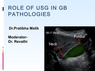
  • 1. ROLE OF USG IN GB PATHOLOGIES Dr.Pratibha Malik Moderator- Dr. Revathi
  • 2. Always look for cholecytectomy scar first!! Indications Normal Anatomy Pathogical Findings:GB gallstones sludge cholecystitis polyps gallbladder cancer Biliary tract Bile duct stones klatskin tumour 08/03/14 CONTENTS
  • 3. 08/03/14 INDICATIONS  Abdominal pain  vomiting  fever  jaundice  abnormal labs (bilirubin, transaminases)
  • 4. 08/03/14 Technique and Preparation Curvilinear probes :2-5 MHz Views:  Subcostal sagittal  intercostal sagittal  coronal  left lateral decubitus  oblique views  reverse trendelenberg
  • 5.  Indications  Normal Anatomy  Pathogical Findings:GB  gallstones  sludge  cholecystitis  polyps  gallbladder cancer  Biliary tract  Bile duct stones  klatskin tumour Contents
  • 6. NORMAL ANATOMY OF THE BILLIARY SYSTEM
  • 7. NORMAL APPERANCE OF GB Normal Gallbladder is seen as a sonolucent pear shaped structure with slim wall (2 mm or so). Best seen with overnight fasting or at least 4-5 hours fasting. It is seen usually at the inferior aspect of the liver right lobe.
  • 8.   The main hepatic fissure appears as an echogenic line that extends from the neck of the gallbladder to the portal vein and serves as a landmark.  The complex of the gallbladder, main hepatic fissure, and portal vein (in the short-axis) has the appearance of an exclamation point 08/03/14
  • 10. "Mickey Mouse" view of Portal Triad 08/03/14
  • 12. NORMAL VARIANTS Phrygian cap Figure (A) A folded gallbladder is difficult to examine with the patient supine. (B) Turning the patient, right side raised, unfolds the gallbladder, enabling the lumen to be satisfactorily examined. The gallbladder may be ‘folded’ (the so- called Phrygian cap).
  • 13. CONTENTS  Indications  Normal Anatomy  Pathogical Findings:GB  Gallstones  sludge  cholecystitis  polyps  gallbladder cancer  Biliary tract  Bile duct stones  klatskin tumour
  • 14. Gold standard for diagnosis: 3 sonographic criteria-  Echogenic focus  Cast acoustic shadow  Seek gravitational dependence
  • 16. CONTENTS  Indications  Normal Anatomy  Pathogical Findings:GB  gallstones  sludge  cholecystitis  polyps  gallbladder cancer  Biliary tract  Bile duct stones  klatskin tumour
  • 17. Sludge is identified on ultrasound as slightly hyperechoic material forming a meniscus within the gallbladder lumen. Sludge may be a precursor to gallstones and has been related to pathology such as acute cholecystitis and acute pancreatitis 08/03/14
  • 18. 08/03/14 DD ECHOGENIC LESIONS IN GALL BLADDER DD MOBILE OR POSTERIOR SHADOWING •STONES •SLUDGE FIXED •POLYP •ADHESIVE STONE •SLOW MOVING TUMEFACTIVE SLUDGE
  • 19. CONTENTS  Indications  Normal Anatomy  Pathogical Findings:GB  gallstones  sludge  cholecystitis  polyps  gallbladder cancer  Biliary tract  Bile duct stones  klatskin tumour
  • 21. CALCULUS CHOLECYSTITIS VS ACALCULUS  Acute cholecystitis refers to the acute inflammation of the gallbladder . It is the primary complication of cholelithiasis  90 to 95% of cases are due to calculous obstruction of the gallbladder neck or cystic duct.  Most sensitive US finding : 1. Presence Of Cholelithiasis 2. Sonographic Murphy Sign. 3. Gallbladder Wall Thickening (>3mm) 4. Pericholecystic fluid.  Other less specific imaging findings include gallbladder distension(>4 cm) and sludge. 99m Tc-HIDA scintigraphy  HIDA cholescintigraphy in acute cholecystitis will demonstrate nonvisualization of the gallbladder.
  • 22. Figure: Acute cholecystitis: (A) TS of an oedematous, thickened gallbladder wall with a stone. (B) LS with a thickened wall (arrows). Stones and debris are present. (C) and (D) TS and LS demonstrating pericholecystic fluid.
  • 23. Sonographic Murphy’s sign 08/03/14 Sonographic Murphy’s sign is positive when the point of maximal tenderness is identified in the right upper quadrant while the gallbladder is identified on the ultrasound monitor. Differential diagnosis of acute cholecystitis choledocholithiasis pancreatitis peptic ulcer disease acute hepatitis
  • 24.  Complications  gangrenous cholecystitis  emphysematous cholecystitis  gallbladder perforation  pericholecystic abscess  cholecystoenteric fistula  liver abscess  Gangrenous and emphysematous cholecystitis - serious complications of acute cholecystitis that may be identified with ultrasound by the presence of air within the gallbladder wall or lumen . Air on ultrasound is represented by “comet- tail” artifacts.  Gallbladder perforation may also be diagnosed by ultrasound. Findings of perforation include significant amounts of pericholecystic fluid that may contain echogenic material which may be walled off from the rest of the abdomen.
  • 25. Gallbladder gangrene/mucosal sloughing. Longitudinal ultrasound of a patient who had acute cholecystitis secondary to stone (arrow) impacted in the gallbladder neck. Note the intraluminal membranes (arrowheads) that are associated with gangrene of the gallbladder.
  • 26. Chronic calculus cholecystitis USG FINDINGS  Thickened echogenic wall  Contracted GB  Cholelithisis
  • 28.  When the gallbladder is entirely filled with stones, a wall echo shadow (WES) sign is seen  The WES triad is another sign of chronic cholecystitis; here the WES stands for Wall (of the GB), Echo (of the calculus) and Shadow (caused by the stones).
  • 29. ACALCULUS CHOLECYSTITIS  Acute acalculous cholecystitis (AAC) refers development of cholecystitis either in a gallbladder without gallstones or in a gallbladder with gallstones where the stones are not the contribuatry factor to the development of cholecystitis . AETIOLOGY  It usually occurs in critically ill or  injured patients.
  • 30. Figure :(A) Acalculous cholecystitis. The gallbladder wall is markedly thickened and tender on scanning. (B) Gravity-dependent sludge with a thick, oedematous wall. No stones were present.
  • 31. PORCELAIN GB  A porcelain gallbladder refers to extensive calcium encrustation of the gallbladder wall.  The term porcelain gallbladder has been used to emphasize the blue discoloration and brittle consistency of the gallbladder wall at surgery  Association between porcelain gallbladder and gallbladder adenocarcinoma- 22-30 % .  Cholecystectomy routinely performed when a porcelain gallbladder is identified.
  • 32. Porcelain gallbladder. (A) Sagittal imageof the gallbladder shows a densely echogenic anterior wall (arrow) with a sharp shadow that obliterates the gallbladder lumen and posterior wall. (B) Transverse ultrasound of the gallbladder in the same patient. The anterior wall is bright, but, without enough reflection or attenuation to eliminate visualization of the lumen and posterior wall (arrow), which is also echogenic and casts a posterior acoustic shadow.
  • 33. CONTENTS  Indications  Normal Anatomy  Pathogical Findings:GB  gallstones  sludge  cholecystitis  polyps  gallbladder cancer  Biliary tract  Bile duct stones  klatskin tumour
  • 34. GB POLYPS  Gallbladder polyps are outgrowths of the gallbladder mucosal wall.  Do not cast an acoustic shadow.  Remain fixed on turning the patient ;so distinguishable from stones  Majority are not neoplastic but are hyperplastic or represent lipid deposits(cholesterolosis).  Gallbladder (GB) polyps are incidentally detected in approximately 4%–7% of patients who undergo ultrasonography
  • 35. GB POLYPS GB polyps are classified into 2 groups: Neoplastic  Adenomas  Adenocarcinomas Nonneoplastic  Cholesterol polyps  Inflammatory polyps  Adenomyomatosis
  • 36. GB polyps are soft tissue masses attached to the wall of the gallbladder and differentiated from gallstones by their lack of mobility and shadowing
  • 37. On CD demonstration of supplying vessel pathognomic of polyp.
  • 38. INCREASE RISK OF MALIGNANCY IN POLYP  Diameters > 10 mm  Sessile Polyps  Single Polyps  Polyps With Adjacent Wall Thickening Or Invasion  Increased Patient Age  Size Of At Least 10 mm Is The Most Well-established predictor of malignancy
  • 39. CONTENTS  Indications  Normal Anatomy  Pathogical Findings:GB  gallstones  sludge  cholecystitis  polyps  gallbladder cancer  Biliary tract  Bile duct stones  klatskin tumour
  • 40. CARCINOMA GB  Gall bladder carcinoma is the most common biliary tract cancer.  Delayed presentation and early spread of tumor make it one of the lethal tumors with poor prognosis.
  • 41. RISK FACTOR FOR CA GB  Gallstones  History of chronic cholecystitis  Porcelain gallbladder.  Choledochal cysts,  Anomalous pancreaticobiliary duct junctions  Gallbladder polyps > 1 cm in size.  Peak incidence in the 6th -7th decades of life  3-5 times more predominant in females
  • 42. USG FINDINGS IN CA GB  A Mass Completely Occupying Or Replacing The Gallbladder Lumen  Focal Or Diffuse Asymmetric Gallbladder Wall Thickening  An Intraluminal Polypoid Lesion.  Invasion of adjacent liver parenchyma  Hepatic metastasis  Periportal/ peripancreatic lymphadenopathy  Sonographically : Heterogeneous, Predominantly Hypoechoic Tumor Fills Much Or All of the gallbladder lumen.
  • 43. CA GB WITH LIVER INVASION
  • 44. 08/03/14  The normal gallbladder wall measures less than 4 mm.  the gallbladder wall is measured at the most narrow point of the anterior wall in the short-axis.  Care must be taken to not measure the wall at an oblique angle THICKENING OF GALL BLADDER WALL DD FOCAL ADENOMYOMATOSIS GALL BLADDER CANCER DIFFUSE ACUTE CHOLECYSTITIS CHRONIC CHOLECYSTITIS XANTHOGRANULOMATOUS CHOLECYSTTIS ADENOMYOMATOSIS GB WALL EDEMA GB CANCER OTHERS- ASCITIS,CHF,HYPOALBUMINEMIA
  • 45.
  • 46. CONTENTS  Indications  Normal Anatomy  Pathogical Findings:GB  gallstones  sludge  cholecystitis  polyps  gallbladder cancer  Biliary tract  Bile duct stones  klatskin tumour
  • 48. 08/03/14 BILE DUCTS  Sonographically, the CBD appears as an anechoic tubular structure in the main portal triad, anterior to and following the course of the main portal vein  Conventionally, the upper limit of normal for the common bile duct as measured by ultrasound is considered to be 6 mm.  Biliary duct obstruction: caused by stones, pancreatic pathology (e.g. mass), or stricture is detected measuring a CBD larger than 6-7 mm. 2000 Am. Coll. of Gastroenterology
  • 50. The Mirizzi Syndrome  Form of obstructive jaundice-described by Mirizzi.  Caused by a stone or stones impacted in the neck of the gallbladder or the cystic duct, such that the common hepatic duct is narrowed.  Rare complication of gallstones-occurs in about 0.1% to 0.7% of patients with cholelitiasis.
  • 52. Figure: Mirizzi syndrome: a large stone in the neck of the gallbladder (arrow) is compressing the bile duct, causing intrahepatic duct dilatation. The lower end of the CBD remains normal in calibre.
  • 53. COMPONENTS OF MS There must be 4 components for the syndrome to occur: 1. Anatomy- placing the cystic duct parallel to the common hepatic duct 2. Impaction of a stone in the cystic dust or gallbladder neck 3. Obstruction of the common hepatic duct from the stone itself, or from the resultant inflammatory response 4. Intermittent or constant jaundice occasionally causing cholangitis, and with longstanding obstruction, biliary cirrhosis
  • 55. INTRAHEPATIC VS EXTRAHEPATIC CALCULUS •Caroli's disease is characterized by congenital segmental dilation of the intrahepatic bile ducts producing primary intrahepatic gallstones. •Secondary intrahepatic gall stone formation occurs due to chronic obstruction of CBD and CHD •It's believed that most patients suffering a chronic illness have excessive amounts of gallstones in the liver. • Gallstones found in gallbladder tend to be hardened and relatively large while stones found in liver tends to be soft and noncalcified •Intrahepatic Gallstones cause liver congestion and elevted liver enzymes
  • 56. Primary intrahepatic stones exclusively involving the intrahepatic biliary tree-related to chronic parasitic infestation of the biliary tree- (ascariasis ) Mixed intrahepatic stone- Associated with extrahepatic lithiasis Secondary intrahepatic stones related to ananatomical condition precipitating stasis or infection TYPES OF INTRAHEPATIC CALCULUS
  • 58. CONTENTS  Indications  Normal Anatomy  Pathogical Findings:GB  gallstones  sludge  cholecystitis  polyps  gallbladder cancer  Biliary tract  Bile duct stones  klatskin tumour
  • 59. KLATSKIN TUMOUR  A Klatskin tumour is a term that was traditionally given to a hilar cholangiocarcinoma (occuring at the bifurcation of the common hepatic duct). Typically, these tumours are  Small In Size  Poorly Differentiated  Exhibit Aggressive Biologic Behaviour  And tend to obstruct the intrahepatic bile ducts  25% of all cholangiocarcinoma
  • 60. Risk factors :  Primary sclerosing cholangitis  Choledochal cysts  Parasitic infections  Hepatolithiasis (also called Oriental cholangiohepatitis)  Toxin exposures  Genetic Factors
  • 61. ULTRASOUND FINDINGS  Presence of a hilar mass with obstruction  Increased echogenicity relative to surrounding liver ~79%  Reduced echogenicity ~ 19 %  Mixed echogenicity ~ 2 %   Segmental dilatation or nonunion of R and L ducts  Polypoid intraluminal masses,  Nodular smooth masses with mural thickening.  Should do doppler, as this is helpful to assess Vascular invasion (unresectable)
  • 62. Figure : Cholangiocarcinoma. (A) Irregular mass at the porta, causing biliary obstruction—a Klatskin tumour. (B) MRI of the same patient, confirming the mass at the porta. KLATSKIN TUMOUR
  • 63. KLATSKIN TUMOUR COLOUR DOPPLER TO LOOK FOR VASCULAR INVASION
  • 64. 08/03/14 THICKENING OF BILE DUCT WALL FOCAL STRICTURE •BACTERIAL CHOLANGITIS •CHOLANGIOCARCINOMA •CHOLEDOCAL CYSTS •METASTASIS •BENIGN STRICTURE NO STRICTURE •POLYPOID CANCER •STONES •PERIAMPULLARY CANCER •SENILE CHANGE •POSTCHOLECYSTECTOMY DILATATION •CHOLEDOCHAL CYST •ASCARIASIS
  • 65. 08/03/14 Pearls and Pitfalls  The gallbladder is a mobile organ; remember to change patient positioning and/or probe placement to find the organ of interest.  Distinguish gallstones from polyps and septations or folds by always scanning through the whole organ and in both longitudinal and transverse planes.  During the biliary exam, use color Doppler to help distinguish nonvascular from vascular structures.  Ultrasound findings must be interpreted in the context of the clinical presentation; findings suggestive of acute cholecystitis (e.g., gallstone or thickened wall) may be present in patients in a nondiseased state.  The common bile duct can be dilated in the absence of pathology in older patients and postcholecystectomy patients.  Measure the anterior wall of the gallbladder. The posterior wall may appear artificially thickened because of acoustic enhancement or artifact from bowel gas.