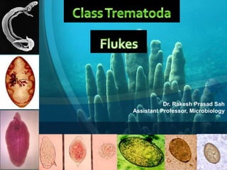
Trematodes by Dr. Rakesh Prasad Sah
•
50 likes•3,072 views

Trematodes by Dr. Rakesh Prasad Sah

Recommended
More Related Content
What's hot
What's hot (20)
Similar to Trematodes by Dr. Rakesh Prasad Sah
Similar to Trematodes by Dr. Rakesh Prasad Sah (20)
More from Dr. Rakesh Prasad Sah
More from Dr. Rakesh Prasad Sah (20)
Recently uploaded
Recently uploaded (20)
Trematodes by Dr. Rakesh Prasad Sah
- 1. Dr. Rakesh Prasad Sah Assistant Professor, Microbiology
- 2. • Multicellular eukaryotic helminths • Unsegmented leaf-like organisms • Also called as “FLUKES” • Size: 1mm to several centimeters • MONECIOUS except for schistosomes (DIECIOUS)
- 3. trematodes or flukes - when they say ‘flat’ worms they mean it • Small doro-ventrally flattened worms, • No body cavity, • Incomplete digestive tract • . • No circulatory or respiratory organs • simple ladder nervous system.
- 4. • Two suckers oral & ventral – Oral sucker (nourishment) – Ventral sucker or acetabulum (to attach to the host) • Most of trematodes are hermaphrodite (self fertilization occurs) except the schistosomes
- 5. General Rules • All are hermaphroditic • All require two intermediate hosts • Eggs of all trematodes are operculated • The infective stage is metacercariae Schistosomes
- 6. Taxonomical Classification of trematodes Superfamily Family Genus Species Schistosomatoedea Schistosomatoedea Schistosoma S. haematobium S. mansoni S. japonicum S. intercalatum Paramphistomatoidea Paramphistomatoidea Watsonius W. watsoni Gastrodiscoides Gastrodiscoides G. hominis Echinostomatoidea Fasciolidae Fasciola F. hepatica Fasciolopsis F. buski Opisthorchioidea Opisthorchiidae Clonorchis C. sinensis Opisthorchis O. felineus Heterophydiae Hetrophyes H. heterophyes Metagonimus M. yokogawai Plagiorchioidea Troglotrematidae Paragonimus P. westermani
- 7. Classification of trematodes according to their habitat Habitat Trematode 1 Blood trematodes (Blood flukes) In the vesical and pelvic venous plexuses S. hematobium In the inferior mesentric vein S. mansoni In the superior mesentric vein S. japonicum 2 Hepatic trematodes (Liver flukes) F. hepatica, C. sinensis, Opisthorchis sp. 3 Intestinal trematodes (Intestinal flukes) Small intestine F. buski, H. heterophyes, M. yokogawai, W. watsoni Larger intestine G. hominis 4 Lung trematodes (Lung flukes) P. westermani
- 8. Blood Flukes (Shistosomes) • Causative agent of disease schistosomiaisis. • Diecious trematodes • Males are broder than females • Formerly known as Bilharzia after Theodor Bilharz who discovered the parasite in the blood of mesenteric veins of a young man in Cairo in 1851.
- 9. MALE& FEMALE SHISTOSOMULAE IN BLOOD VESSEL
- 10. Differentiating charcters of S. haematobium, S. mansoni, and S. japonicum Character S. haematobium S. mansoni S. japonicum Geographical distribution East Mediterranean and African countries, India & Turkey Africa, Americas and the East Mediterranean China, Philippines, Indonesia and Thailand Host Involved Definitive host Man Man Man and domestic animals Intermediate host Bulinus sp. Biomphalaria sp. Oncomelania sp.
- 11. Differentiating charcters of S. haematobium, S. mansoni, and S. japonicum Character S. haematobium S. mansoni S. japonicum Morphology of Worms Male Size 10-15mm X 0.8mm 6-12mm X 1mm 12-20 mm X 0.5mm Integume nt Covered with minute tuerculations Covered with monre conspicuous tuberculations Covered with minute spines Female Size 20mm X 0.25mm 7-17 mm X 0.25mm 26 mm X 0.3mm Ovary Present in the posterior third of the body Present in the anteriour half of the body Present in the middle of the body Uterus Contains 20-200eggs Contains 1-3 eggs Contains 50 or more eggs
- 12. Differentiating characters of S. haematobium, S. mansoni, and S. japonicum Character S. haematobium S. mansoni S. japonicum Egg Size 110-170µm X 40-70µm 115-180µm X 40-70µm 70-100µm X 55- 65µm Shape Elongated Elongated Oval Shell Thin, smooth shell Thin, smooth shell Smooth, relatively thick Anterior end Rounded Pointed and curved Rounded Posterior end Terminal spine present Lateral spine present Lateral knob present Eggs discharged in Urine Faeces Faeces
- 16. Pathogenesis • Following skin penetration by cercariae A pruritic rash and dermatitis may occur. Intestinal bilharziasis • S. mansoni • Intestinal Bilharziais or schistosomal dysentery • Intestinal symptoms – Abdominal pain & bloody diarrhoea.
- 18. Acute schistosomiasis or katyama fever • occurs with S. japonicum and S. mansoni and rarely with S. heematobium. • Symptoms – High fever – Hepatosplenomegaly – Lymphadenopathy – Dysentery
- 19. Haematuria • Painless haematuria • Initially microscopic, but becomes gross if infection is heavy. • Occurs in case of S. haematobium infection. • Calculi may form in bladder due to deposition of oxalate and uric acid crystals around eggs. • Chronic schistosomiasis urinary bladder cancer.
- 20. Lab diagnosis • Microscopy – Detection of characteristic eggs. • Cystoscopy and biopsy of lesions in bladder • Serology – IFAT – ELISA – CFT – RIA
- 21. Fasciola hepatica (LIVER FLUKE) • The sheep liver fluke and the common liver fluke • Parasite of herbivorous animals. • Lives in biliary passages of liver. • Occasionally found in man. • Exist in 6 morphological forms – adult, miracidium, sporocyst, 1st generation redia, 2nd generation redia, cercaria & metacercaria. • Leaf like, 30mm x 15mm, grey or brown. • Anteriorly conical projection, posteriorly round.
- 22. • 5yr life span in sheep & 10yr in Man. • Eggs are operculated, bile stained and measure 140 um x 80 um. • Eggs immature when passed in faeces, can develop only in water. • contains immature larva (miracidium) • Shell of egg is thin with smooth surface. • Can not be differentiated from egg of F. buski.
- 25. Clinical features • Fascioliasis • Fever • abdominal pain • hepatomegaly • Ascites and Jaundice • Eosinophilia – acute disease. • In untreated patients – intermitent biliary obstruction, cholecystitis, cholelithiasis & development of strictures.
- 26. Lab diagnosis • Stool examination – operculated eggs • Eosinophilia • ELISA
- 27. Clonorchis sinensis (Chinese Liver Fluke) • Parasite of man, dogs and cats in the southeast of Asia • Common: China, Korea ,Japan, Taiwan and Vietnam • Adult worm located in the biliary tract.
- 31. Clinical features • Clonorchiasis • Epigastric pain • Indigestion • Liver necrosis and tenderness • Liver dysfunctions related to fluke burden • Diarrhea, hepatomegaly, jaundice and ascites, in heavier infections
- 32. • Diagnosis eggs in the feces or bile • Eosinophilia • ELISA • CFT
- 33. Fasciola buski (INTESTINAL FLUKES) • Also k/a large or Giant Intestinal Fluke • Largest trematode • Assam and west bengal in India • Exist in 6 forms – adult, miracidum, sporocyst, redia, cercaria, metacercaria. • Adult live in small intestine of humans and pig. • Size 20 – 75 mm x 8-20 mm x 0.5 – 3mm. • 25,000 eggs per day
- 34. 5 cm
- 35. • Eggs measure 130 – 150 um x 60 – 90 um • Bile stained.
- 38. Clinical features • Fasciolopsis • Damage traumatic, mechanical & toxic effects • Ulceration at the site of attachment • Worm partial obstruction of bowel • Diarrhoea, anorexia, nausea, vomiting, abodominal pain, fever, & mild anaemia.
- 39. Lab diagnosis • Stool examination -Demonstration of operculated eggs. • TREATMENT, PREVENTION AND CONTROL – Praziquantel – 25mg /kg body wt TID for 1 day. – Prevention of water pollution. – Destruction of snails
- 40. Paragonimus westermani (Lung Fluke) • Oriental lung fluke • Discovered in lungs of Bengal tigers in 1878 by Kerbert • Most commonly encountered in parts of Asia, Africa and South America. • Endemic in India Assam, Bengal, Tamil Nadu & Kerela. • Reddish brown oval worm • 10mm length, 5mm breadth and 4 mm thickness
- 41. • Definitive Host: – Man, – Cat – Dog – Fox – Tiger – Leopard and – Monkey • Intermediate Host – 1st Int host • Freshwater snail – 2nd Int host • Freshwater crayfish or a Crab
- 44. Pathogenesis • Migrate by penetrating through the intestinal wall peritoneal cavity abdominal wall diaphragm lungs. • Immature worms settle close to the bronchi grow sexually mature hermaphrodite worms eggs
- 45. Pathogenesis • Inflammatory reaction to the worms & eggs granuloma formation, cystic dilation of bronchi, abscesses and pneumonitis. • Cough with chest pain and haemoptysis • Blood-stained rusty brown sputum • Sputum contain yellowish brown eggs of parasite. • Chronic case may be misdiagnosed as TB.
- 46. Clinical features • Sometimes migrating larva lose their way and are lodged in ectopic sites such as – liver, – peritoneum, – mesenteric lymph nodes, – brain, – heart and – subcutaneous tissues of groin. •Headache, •Fever, •Paralysis and •Convulsive seizures when parasites lodge in brain or spinal cord.
- 47. DIAGNOSIS • Symptoms • History of eating improperly cooked crab-meat in endemic areas • Eggs in the sputum • ELISA • Intradermal test