
Recommended
More Related Content
What's hot
What's hot (20)
Similar to Blood groups,blood transfusion,hazards,blood bank
Similar to Blood groups,blood transfusion,hazards,blood bank (20)
More from Ranadhi Das
Recently uploaded
Recently uploaded (20)
Blood groups,blood transfusion,hazards,blood bank
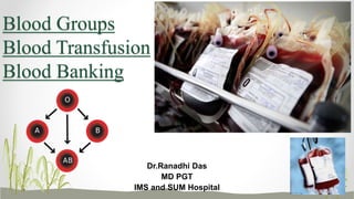
- 1. Blood Groups Blood Transfusion Blood Banking Dr.Ranadhi Das MD PGT IMS and SUM Hospital
- 2. Austrian Karl Landsteiner(1901) discovered – •Human blood possess different antigenic and immune properties •Blood clumping was an immunological reaction. •Nobel Prize in Physiology and Medicine in 1930. •Adriano Sturli and Alfred von Decastello who were working under Landsteiner discovered type AB a year later in 1902. •Janský is credited with the first classification of blood into the four types (A, B, AB, O)in 1907, which remains in use today. Introduction
- 3. They are group specific substrate on the surface of RBC and enable the different individual to be identified from each other just like fingerprints. CLASSIFICATION- Major blood group system- ABO system, Rh system, MNS system Minor blood group system- Kell, Duffy, Lutheran, Lewis, Deigo, kidd Etc. Thirty-five major blood group systems were recognized by the International Society of Blood Transfusion (ISBT) in October 2012. Blood Group System
- 4. I. Discovery II. Antigen and Antibodies III. Inheritance ABO Blood Group System
- 5. • Karl Landsteiner(1901) classified human blood into A,B,O groups. • Von Decastello and Sturli (1902) discovered AB blood group. • Von Dungern and Hirszfeld(1911) divided group A into 2 subgroups A1 and A2. Discovery
- 6. • Appear in the sixth week of fetal life. • Present on red cell membrane and in other tissues like salivary glands, pancreas, kidney, liver, lung, semen, testes, amniotic fluid.( EXCEPT CNS) • Antibodies formed in children after few months of birth. Antigens
- 7. Glucose Galactose N-acetylglucosamine Galactose RBC Fucose N-acetylgalactosamine N-acetyl-galactosaminyl Transferase (product of A gene- GENES at 9q34.1) Formation of the A antigen
- 8. Formation of the B antigen Glucose Galactose N-acetylglucosamine Galactose RBC Fucose Galactose D-galactosyl Transferase(product of B gene- GENES at 19 q13.2)
- 9. • Mostly IgM type. • Produced by bone marrow and lymph gland cells . • Known as cold antibodies. Anti-A and Anti-B agglutinins
- 10. ABO Group Antigen (agglutinogen) Present Antigen Missing Antibody (agglutinin) Present A A B Anti-B B B A Anti-A O None A and B Anti-A&B AB A and B None None Antigen and Antibody Present in ABO Blood Group
- 11. LANDSTEINER’s LAW- 1.If an agglutinogen (antigen) is present on red blood cell membrane ,the corresponding agglutinin (antibody) must be absent in the plasma. 2.If an agglutinogen (antigen) is absent on red blood cell membrane, then corresponding agglutinin (antibody) must be present in the plasma. 2nd law doesnot hold good for Rh factor (Rh factor absent anti D absent)
- 12. Inheritance of ABO blood group Follows Mendelian Law. In 1924, Felix Bernstein proposed presently accepted theory. Heterozygous: AO or BO Homozygous: AA or BB Mom Dad Offspring Blood Group AA BB 100% AB BO OO 50% each of B or O OO OO 100% O OO AO 50% each of A or O
- 13. If baby is Parents must have Mother blood group Father would not be O O+O No matter which AB AB A+B No matter which O A A+O A+A B or O B or O B B+O B+B A or O A or O PaternityDispute
- 14. The Rhesus Blood Group System I. Discovery of Rh system II. Antigen and antibodies III. Inheritance IV. Haemolytic disease of newborn
- 15. •History of the Rh System • Levine and Stetson(1939) described a hemolytic transfusion reaction in an obstetric patient following delivery of stillborn infant. • An antibody was isolated from the mother’s serum. It was postulated that the fetus and the father possessed a common factor that the mother lacked. •While the mother carry the fetus, the mother was exposed to this factor and developed antibody against the transfused red cells from the father and resulted in transfusion reaction. •At that time ,the responsible antibody was not named.
- 16. • Landsteiner and Wiener(1940) injected RBC’s of rhesus monkeys in rabbits developed antibodies. • The antibody was named as anti-Rh. • When resulting antiserum was mixed with human RBC’s, then agglutination occurred. •The RBC antigen responsible for this reaction was called as Rh factor. •The antibody discovered by Levin and Stetson in the mother was subsequently re- examined and found identical in activity as the anti-Rh antibody found by Landsteiner and Weiner. •So this work led to discovery of Rh system.
- 17. •Anti-rhesus formed by the animals was renamed anti-LW (Landsteiner and Wiener). •Fisher and Race(1943 and 1944) discovered C, E, c, e Ag and their corresponding antibodies anti-C , anti -E ,anti- c ,anti -e.
- 18. Rh Antigens • Rh system involves 6 Rh antigens D,d,C,c,E,e Rh positive Presence of rh D 85% occurrence Rh negative Absence of rhD 15% Occurrence •Integral membrane proteins with an active phoshpholipid component. •Present on red blood cells. •D antigen is commonest and most immunogenic.
- 19. • IgG type. • Known as warm antibodies. • Produced by exposure to foreign Rh antigen . Rh Antibodies
- 20. • D Ag is inherited as a dominant gene D. • Genotype of individual may be: DD Dd dd Inheritance
- 21. Examples- Heterozygous Rh + Homozygous Rh- 50% hetrozygous rh+ 50% homozygous rh-
- 22. • ABO Incompatibility- immediate reaction as antibodies are naturally present. • Rh Incompatibility- First exposure- Primary response Immunological memory Second exposure- Immediate and severe response. Incompatibility
- 23. • Transfusion reactions are the adverse reactions in the body, which occur due to transfusion error that involves transfusion of incompatible (mismatched) blood. • The reactions may be mild causing only fever and hives (skin disorder characterized by itching) or may be severe leading to renal failure, shock and death. Transfusion Reactions Due To ABO Incompatibility
- 24. • Erythroblastosis Fetalis (“Hemolytic Disease of the Newborn”) Erythroblastosis fetalis is a disease of the fetus and newborn child characterized by agglutination and phagocytosis of the fetus’s red blood cells. In most instances of erythroblastosis fetalis, the mother is Rh negative and the father Rh positive. • The baby has inherited the Rh-positive antigen from the father, and the mother develops anti-Rh agglutinins from exposure to the fetus’s Rh antigen. • In turn, the mother’s agglutinins diffuse through the placenta into the fetus and cause red blood cell agglutination.
- 25. I.Erythroblastosis fetalis- • Erythroblastosis Mechanism- Rapid production of red blood cells by haematopoeitic tissues of baby in order to replace haemolysed RBC’s. • Anaemia • Hydrops fetalis- Grossly edemataous, Intra uterine fetal death usually occurs. II.Icterus gravis neonatorum- 1. Infant born at term 2. Jaundice within 24 hrs 3. No anemia ( compensated by normoblastic response in bone marrow) 4. Nucleated RBC in circulation 5. Severely damaged liver 6. splenomegaly 7. Kernicterus- unconjugated bilirubin is deposited in basal ganglia Manifestations
- 27. I. Anti-D Prophylaxis – • Based on principle antibody mediated immunosuppression. • Inject anti-D antibody in mother soon after child birth within 72 hrs. • RhoGAM –Ɣ glodulin rich in anti D • Fetal Rh typing by amniocentesis or chorionic villi sampling, then give Rh immunoglobulin to expectant mother between 28 to 30 weeks. II. Exchange transfusion – About 400 ml of Rh – ve blood is transfused over a period of 1.5 hr or more while the neonates taking out the Rh +ve blood. III. Phototherapy – Any new-born with a total serum bilirubin greater than 18mg/dL should receive phototherapy. Prevention And Treatment
- 28. • THE Ii SYSTEM- Weiner et al (1956) 2 antigens –I and i Difference- • At birth , RBC’s are rich in i Ag ,not I Ag. • In first second years of life, gradual changeover from i to I. • THE DUFFY SYSTEM Cutbush , Mollison and Parkin(1950). System has 3 blood groups-Fya ,Fyb and Fyab. Fyab blood groups are resistant to plasmodium vivax whereas Fya and Fyb are susceptible to vivax malaria. Other Blood Group Systems
- 29. • KELL SYSTEM- Coombs ,Mourant , Race(1946). Antigen - K Rare In India(.3 To .7%) • MN SYSTEM - Landsteiner and Levine(1927). Required for- • Paternity tests • Anthropological and genetic studies. • Bombay Blood Group- Dr.Y.M. Bhenda(1952) Also known as HH blood group. They do not express H antigen, as a result they cannot form A Ag or B Ag on their RBC. They can donate blood to anybody but can receive blood only from Bombay blood group people.
- 30. I. In blood transfusion. II. In preventing Hemolytic Diseases of Newborn due to Rh Incompatibility. III. In paternity disputes. IV. In medicolegal cases. V. Anthropological study VI. In knowing susceptibility to diseases. A group- Gastric cancer, Pancreatic cancer, Diabetes mallitus B group- Gastrointestinal disorder O group- Peptic ulcer, Rheumatic fever Clinical Implications
- 31. I. Indications II. Donor and Recepeints III. Precautions IV. Hazards Blood Transfusion
- 32. Richard Lower pioneered the first blood transfusion from animal to human in 1665 at the Royal Society( Banned in 1668). Proper transfusion done by Landsteiner and Weiner In 1840 Dr. Blundell, performed the first successful whole blood transfusion to treat haemophilia. In 1914 Lewisham brought the concept of transfusion during 1st world war Introduction
- 33. I. Blood loss ( Hemorrhage, Surgery) II. For quick restoration of Hb. III. Exchange transfusion ( erythroblastosis fetalis) IV. Bone marrow failure (aplastic anaemia, leukaemia ) V. To correct clotting factor deficiency-hemophilia, purpurae,clotting defects. VI. Restore plasma volume- Burn VII. Acute poisoning (carbon monoxide) VIII. Acute infections or fever when gamma globulin is needed. IX. Shock X. To improve oxygen transport to tissue- severe anemia Indications
- 34. • Donor selection- I. Healthy and age between 18 to 60 years. II. Hemoglobin > 12 gm% III. Weight more than 45 kg. IV. Females should not be Pregnant, lactating and menstruating. V. No diseases like AIDS, Viral Hepatitis ,Malaria, Syphilis. VI. No past H/O jaundice , HTN,TB, and cardiac diseases. Jehovah Witnesses (1870,Pittsburgh,Pennsylvania,USA)- Christian community which refuses blood transfusion. Donor And Recepient
- 35. Blood transfusion-who can receive blood from whom? Blood Group Antigens Antibodies Can give blood to Can receive blood from AB (universal recipient) A and B None AB AB, A, B, O A A B A and AB A and O B B A B and AB B and O O (universal donor) None A and B AB, A, B, O O
- 36. • For safe and compatible blood transfusion- • ABO and Rh typing • Cross matching • Antibody screening Investigations
- 37. • Processing and Testing of Blood Donated blood is usually subjected to processing after it is collected, to make it suitable for use in specific patient populations. Collected blood is then separated into blood components by centrifugation: red blood cells, plasma, platelets, albumin protein, clotting factor concentrates, cryoprecipitate, fibrinogen concentrate and immunoglobulins (antibodies) All donated blood is tested for infections like HIVs, Hepatitis B, Hepatitis C, Syphilis All donated blood is also tested for ABO and Rh groups, along with the presence of any red blood cell antibodies. Pathogen Reduction treatment done.
- 38. Determination Of Blood Group Blood Typing
- 39. Under Microscope Human RBC before (left) and after (right) adding serum containing antibodies. No agglutination Agglutination
- 40. II. Cross Matching- Major cross matching -Donor’s RBCs are mixed with recepient’s plasma. Minor cross matching- Recepient’s RBC’s are mixed with donor’s plasma. When no agglutination then only donors blood can be transfused. Cross Matching
- 41. III. Antibody Screening – Antibody Screening
- 42. IV. Rh positive blood should never be transfused to Rh negative blood. V. Check blood bag for correct label. VI. Transfuse blood at slow rate(not >20 drops/min) under proper aseptic measures. VII. Careful watch on recipient's condition. VIII. Stop transfusion if reaction occurs.
- 43. Transfusion reactions • Hemolysis- Massive destruction of RBC of donor release of Hb in blood hemolytic jaundice. • Renal vasoconstriction- Due to release of toxic substances from hemolysed RBC. • Circulatory Shock- Loss of RBC and release of toxins from hemolytic RBC lead to fall of BP. Decrease renal blood flow oliguria • Hemoglobinuria- Free Hb binds with haptoglobin present in plasma pass through glomerular membrane and passed through urine. • Renal tubular damage- Free Hb precipitate in tubules as acid hematin. • Anuria- Absence of urine due to vasoconstriction, shock, hypotension, renal tubular damage. • Uremia- increased nitrogenous substances and K⁺ renal failure coma death. • Transmission of blood borne diseases- AIDS, Hepatitis, Malaria, Syphilis • Fever, Chill, Rigor • Hyperkalemia • Hypocalcemia • Tightness of chest
- 44. • Replacement of more than 50% of circulating blood volume in less than 3 hrs Or, transfusion @ >150ml/min Or, > 10 units of blood within 24 hrs. • Blood loss- • < 20% of total blood volume - well tolerated by the body • 20%-40% of total blood volume - changes in vital signs • > 40% of total blood volume - Hemorrhagic shock Massive Blood Transfusion
- 45. • Plasma, normal saline • Hydroxy ethyle starch • Low molecular weight dextran (Lomodex) • Synthetic Blood- • Fluosol-DA (colorless, Oxygen carrying capacity 7 days, dev in Japan) • Coconut water Recent advances- Synthetic bio-molecule blood substitutes- • Perflurocarbon based- Oxycyte, NVX-108 • Hemoglobin based oxygen carrier- Hemopure, Hemospan. • Hyperbranched Polymer protected porphyrine- Polyheme Blood Substitutes
- 46. • Safely given- 1:1:1 protocol- ‘O’ negative packed cell : FFP : Random donor platelet. • Advance trauma centre equipped with – Thromboelastography (gives idea about the amount of blood loss and efficiency of blood coagulation). Blood loss-Unknown amount
- 47. • It is a centre where blood gathered as a result of blood donation, stored and preserved for later use in blood transfusion. • Equipped with proper testing for blood and blood products. • 1unit = 450 ml of CPD mixed blood. • Stored at 4°C with 63 ml of anticoagulants. • CPD – Citrate(anticoagulant by chelating calcium), Phosphate(synthesize ATP), Dextrose(nutrition). • Blood can be kept 21 days at 4°C in blood bank. • Blood donation- Weight- 45-55 kg 350ml/ 49 ml anticoagulant Weight- > 55 kg 450ml/ 63 ml anticoagulant Blood Bank
- 48. • RBC- with CPD 35 days, with SAGM (saline, adenine, glucose, manitol) 42 days • Platelet- 5 days • Plasma- 1 year • Clotting factors Cryoprecipitate- 1 year Drawback in old blood- (Lesion of storage) • Na+K+ ATPase pump stops working leading to Hyperkalemia • Fall in serum Calcium level swelling of the RBC • Build up of lactic acids • Decrease in glucose consumption • Decrease in ATP level • Decrease in pH. • Diminish activity of 2,3 DPG reduce the ability to deliver oxygen • Decrease platelet. Storage