
Recommended
More Related Content
What's hot
What's hot (20)
Similar to Peroperative PNS CT - A 10-steps evaluation
Similar to Peroperative PNS CT - A 10-steps evaluation (20)
Recently uploaded
Recently uploaded (20)
Peroperative PNS CT - A 10-steps evaluation
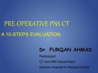
- 1. PRE-OPERATIVE PNS CT A 10-STEPS EVALUATION Dr. FURQAN AHMAD Radiologist CT and MRI Department Doctors Hospital & Medical Centre
- 3. POST-FESS
- 4. OBJECTIVE 1. To illustrate important anatomic variants and landmarks on pre-operative sinus CT 2. With a focus on critical variants and landmarks that predispose patients to surgical complications
- 5. Normal Variant Septal deviation Nasal obstruction limiting endoscopic visualization and access Implication – may need septoplasty STEP 1 - NASAL SEPTUM
- 6. DNS and ENLARGED IT
- 8. CORONAL CT OF OMC
- 10. Landmark for access to the posterior ethmoidal air cells Normal Variants Concha bullosa Paradoxical middle turbinate Implication Critical attachment to base of skull at cribriform plate. Preserved as a landmark for revision surgery STEP 2 - BASAL LAMELLA of the MIDDLE TURBINATE
- 11. CONCHA BULLOSA
- 12. PARADOXICAL CURVE of MT
- 13. Landmark for the osteomeatal complex (OMC), lying just posterior to it. The role of the uncinate process is that of facilitating drainage of the frontal recess. STEP 3 – UNCINATE PROCESS
- 14. Landsberg aand Friedman classification of superior uncinate process insertion. a) Type 1 - insertion into lamina papyracea, b) Type 2 - insertion into the posterior wall of the agger nasi cell, c) insertion into the lamina papyracea and junction of the middle turbinate with the cribriform plate, e) Type 5 - insertion into the skull base, f) Type 6 - insertion into the middle turbinate.
- 15. Normal Variants Lateral deviation Implication There is a high risk of entry into the orbit STEP 3 – UNCINATE PROCESS
- 16. INFUNDIBULAR NARROWING by UNCINATE PROCESS
- 17. The ethmoid bulla - largest and most constant anterior ethmoid air cell. Implication laterally - risk of penetration especially of a dehiscent lamina papyracea with risk of damaging orbital contents, superiorly - penetration into floor of anterior cranial fossa. STEP 4 – ETHMOID AIR CELLS
- 18. Sagittal CT demonstrating an ethmoid bulla (arrow) complicated by sinusitis and an effusion. The ethmoid bulla is bound superiorly by the floor of the anterior cranial fossa and laterally by the lamina papyracea.
- 19. AGGER NASI CELL the most anterior ethmoid air cell lying just anterior to attachment of middle turbinate and frontal recess Implication – if large may cause medial displacement of the middle turbinate causing narrowing of the frontal recess. STEP 4 – ETHMOID AIR CELLS
- 20. AGGER NASI
- 21. HALLER CELL infraorbital cell extending to floor of orbit Implication may cause narrowing of maxillary sinus ostium/ethmoid infundibulum. STEP 4 – ETHMOID AIR CELLS
- 22. HALLER CELLS and INFUNDIBULAR NARROWING
- 23. The frontal sinus drainage pathway (FSDP) has 2 compartments: The frontal air cells constitute the superior compartment. The inferior compartment is a narrow passageway formed by either the ethmoid infundibulum or the middle meatus STEP 5 – FRONTAL/KUHN'S AIR CELLS
- 25. FRONTAL and SE RECESSES
- 26. SAGITTAL CT FRONTAL RECESS
- 27. AGGAR NASI CELLS
- 28. TYPE 1 FRONTAL CELL
- 29. TYPE 2 FRONTAL CELLS
- 30. TYPE 2 FRONTAL CELLS
- 31. TYPE 3 FRONTAL CELLS
- 32. TYPE 3 FRONTAL CELLS and FRONTAL BULLA
- 33. TYPE 4 FRONTAL CELL
- 34. TYPE 4 FRONTAL CELL
- 35. Coronal plane Bone integrity Keros classification Keros I - 1 - 3mm Keros II - 3 - 7mm Keros III - 7 - 16mm Asymmetric Keros STEP 6 – CRIBRIFORM PLATE
- 36. Implication Higher risk of intracranial penetration with increasing depth Damaging the anterior and/or posterior ethmoidal artery. STEP 6 – CRIBRIFORM PLATE
- 37. KEROS TYPE 1
- 38. KEROS TYPE 2
- 39. KEROS TYPE 3
- 43. Coronal and axial planes Remote orbital fracture Orbital prolapse into ethmoid sinus Haller cells Uncinate process contacting orbital wall STEP 7 – LAMINA PAPYRACEA
- 44. LP DEHISCENCE
- 45. LP UP APPOSITION
- 46. ONODI CELL - posterior ethmoid air cell (5%) Implication Potential damage to (a) optic nerve (in 5%), and less commonly, (b) the internal carotid artery STEP 8 – ONDOI CELLS
- 47. ONODI CELLS
- 48. ONODI CELLS and OPTIC NERVES
- 49. SPHENOID BONE and SKULL BASE
- 50. AXIAL CT SPHENOETHMOIDAL RECESS
- 51. SAGITTAL CT SPHENOETHMOIDAL RECESS
- 52. Sagittal and axial planes Conchal, presellar and sellar pneumatization. Pneumatization into skull base and anterior clinoid STEP 9 – SPHENOID SINUS
- 53. CHONCHAL SS
- 54. PRESELLAR SS
- 55. SELLAR SS
- 56. Dehiscence of carotid canal Sinus septation inserting onto carotid canal Optic nerve dehiscence within sphenoid sinus STEP 9 – SPHENOID SINUS
- 60. DEHISCENT OPTIC NERVE CANAL
- 61. DEHISCENT SS and CEPHALOCELE
- 62. CT CISTERNOGRAM
- 63. Coronal plane Identify ethmoid notch Supraorbital pneumatization STEP 10 – ANTERIOR ETHMOIDAL ARTERY
- 65. VULNERABLE ANTERIOR ETHMOIDAL ARTERY
- 67. DEHISCENT LEFT POSTERIOR ETHMOIDAL ARTERY
- 68. Components of the “CLOSE” Mnemonic Critical Anatomic Structure Ideal Imaging Plane Items to Evaluate and Document/Report Cribriform plate Coronal Keros classification (type I–III) Asymmetric Keros Bony dehiscence of skull base Lamina papyracea Coronal, axial Remote orbital fracture Orbital prolapse into ethmoid sinus Presence of Haller cell Uncinate process contacting orbital wall Onodi cell Coronal Presence or absence of Onodi cell Dehiscence of optic nerve within Onodi cell Sphenoid sinus pneumatization Sagittal, axial Pneumatization pattern (conchal, presellar, sellar) Pneumatization into skull base & anterior clinoid Dehiscence of carotid canal Sinus septation inserting onto carotid canal Optic nerve dehiscence within sphenoid sinus Anterior) ethmoidal artery Coronal Identify ethmoidal notch Presence of supraorbital pneumatization
- 69. The radiologist's goal is to report on five key points: 1. the extent of sinus opacification/disease 2. opacification of sinus drainage pathways 3. anatomical variants 4. critical variants 5. condition of surrounding soft tissues of the neck, brain and orbits Radiologist’s goal
- 70. THANK YOU