2. vol25_no6_jum_745-814_online.q
5/17/06
1:25 PM
Page 778
Fetal Hydrocephalus With Idiopathic Thrombocytopenic Purpura
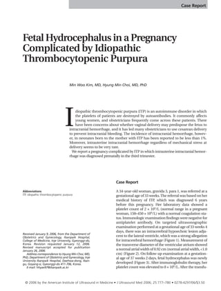
Figure 1. Transverse axial sonogram at a gestational age of 33 weeks 4 days. An
intracerebral hypoechoic lesion (arrow) is shown adjacent to the lateral ventricle.
sion of 12 U of platelet concentrate, the pregnancy was terminated by cesarean delivery under
general anesthesia. A female neonate weighing
2540 g was delivered with Apgar scores of 7 at 1
minute and 8 at 5 minutes.
The neonate had moderate physical activity
and crying. Head circumference was 34.5 cm
(75th–90th percentile). The fontanel was soft and
flat, and there was no evidence of caput or molding on the head. Multiple bruises were noted on
the face and scalp, and petechiae were present
on the whole body skin. The palate was perforated (cleft palate), and petechiae were also noted
Figure 2. Transverse axial sonogram at 33 weeks 4 days (transventricular view).
The transverse diameter of the ventricular atrium has a normal atrial width.
778
on it. Laboratory data showed a platelet count
of 1 × 10⁹/L (normal range in a neonate,
150–350 × 109/L) with a prolonged prothrombin time. Immunologic examination findings
were negative for antiplatelet antibody and
platelet-associated immunoglobulin G. On
brain ultrasonography, moderate to severe
hydrocephalus was noted, and there was no
evidence of intraventricular hemorrhage
(Figure 4). Computed tomography revealed diffuse ischemic and encephalomalatic changes
in both hemispheres and severe hydrocephalus
(Figure 5).
The neonate had been treated with intravenous
immunoglobulin therapy for 2 days (1 g ⋅ kg–1 ⋅
d–1). Because the platelet count remained low
during hospitalization, and the compression of
brain parenchyma due to hydrocephalus was not
remarkable, extraventricular drainage was not
performed. The infant was discharged on the
25th day. Since then, we have been following her
in the outpatient department. Her platelet count
was first normalized to 163 × 109/L on the 90th
day, and the last data showed a platelet count of
151 × 109/L on the 114th day. Neurologic signs
such as delay in eye contact and frequent vomiting subsequently developed, but the infant was
still alive.
Discussion
Idiopathic thrombocytopenic purpura is an
autoimmune disorder in which platelets are
destroyed in the reticuloendothelial system by
the antibody of the immunoglobulin G group
against platelet surface antigen. It affects about
5% of the population and often develops in
young women. The incidence of ITP in pregnant
women has been reported as approximately 1 to
2 per 1000 pregnancies.1,2
The autoantibody can cross the placenta and
induce neonatal thrombocytopenia. The risk of
severe thrombocytopenia in neonates born to
mothers with ITP seems to be very rare. The incidence of severe thrombocytopenia, defined as a
platelet count at birth of less than 20 × 109/L,
ranges from 1% to 5%.3
The diagnosis of ITP is based on the exclusion
of other causes of thrombocytopenia. The presence of antiplatelet antibodies is regarded as a
hallmark of ITP, but antigen-specific assays
detect platelet-associated and plasma autoantibodies in about 50% to 75% of patients.4 The varJ Ultrasound Med 2006; 25:777–780
3. vol25_no6_jum_745-814_online.q
5/17/06
1:25 PM
Page 779
Kim and Choi
ious types of assays proved to be poor in sensitivity, specificity, and reproducibility.5 In addition, the antibody can also be seen in various
other diseases, such as systemic lupus erythematosus and human immunodeficiency virus
infection. As a result, the presence of the autoantibody cannot be used as a diagnostic criterion
yet. In our case, maternal immune assay results
were negative for the antiplatelet antibody.
The purpose of management in pregnant
women with ITP is to reduce the hemorrhagic
complications by restoring hematologic profiles rather than to normalize the platelet count.
The treatment options are corticosteroid therapy, intravenous immunoglobulin or anti-RhD,
and plasmapheresis. To our knowledge, however, there have been no prospective trials guiding the treatment of pregnant patients with
this disorder. In our case, the maternal platelet
count was not sufficiently elevated after corticosteroid, immunoglobulin, and anti-RhD
(WinRho) therapy.
The fetus with severe thrombocytopenia is at
increased risk of hemorrhage, including
intracranial hemorrhage, which usually occurs
in association with labor and delivery. Also, there
are controversies with regard to the mode of
delivery in pregnancies with ITP Some reports
.
have shown that neonatal intracranial hemorrhage are unrelated to the mode of delivery,
which should be based on obstetric considerations only.6–8 In general, obstetricians prefer
cesarean delivery to lower the risk of intracranial
hemorrhage.
Some interventions have been used to determine the fetal blood count, including cordocentesis, scalp sampling, and umbilical cord
sampling. Some authors, however, emphasized
the risk of these procedures, including doubts
of reliability, and did not recommend performing these prenatal interventions.9,10 Recently,
with advances in techniques and procedures,
in utero therapies as well as diagnostic procedures have been reported to show promising
success rates.
In a review of the literature, Cook et al11
reported that the incidence rates of intracranial
hemorrhage among neonates with severe
thrombocytopenia (<50 × 109/L) were 4% after
cesarean delivery and 5% after vaginal delivery.
In our case, it is suspected that the intracranial
hemorrhage caused the hydrocephalus. The
mechanism by which intracranial hemorrhage
J Ultrasound Med 2006; 25:777–780
Figure 3. Transverse axial sonogram at 37 weeks 2 days (transventricular view).
Note that bilateral hydrocephalus has newly developed (arrow).
led to hydrocephalus has been suggested (or suspected) to be that arachnoid granulations were
obstructed by the breakdown products from the
hemorrhage. Neurodevelopmental outcome is
thought to be poor. With regard to neonatal prognosis, Boynton et al12 reported outcomes of 50
preterm neonates with posthemorrhagic hydrocephalus. The mortality rate was 7%; seizures
developed in 38% of the patients; and limitations
in motor function developed in 49%.12
There have been some reports about hydrocephalus caused by alloimmune thrombocytopenia, but hydrocephalus occurring after
Figure 4. Brain sonogram on the day of birth. Moderate to severe hydrocephalus
is shown, which is symmetric and compresses the brain parenchyma. There is no
sign of intracerebral or intraventricular hemorrhage.
779
4. vol25_no6_jum_745-814_online.q
5/17/06
1:25 PM
Page 780
Fetal Hydrocephalus With Idiopathic Thrombocytopenic Purpura
References
1.
2.
Warner MN, Moore JC, Warkentin TE, Santos AV, Kelton
JG. A prospective study of protein-specific assays used to
investigate idiopathic thrombocytopenic purpura. Br J
Haematol 1999; 104:442–447.
5.
Cines DB, Blanchette VS. Immune thrombocytopenic purpura. N Engl J Med 2002; 346:995–1008.
6.
George JN, El-Harake MA, Raskob GE. Chronic idiopathic
thrombocytopenic purpura. N Engl J Med 1994; 331:
1207–1211.
7.
Letsky EA, Greaves M. Guidelines on the investigation and
management of thrombocytopenia in pregnancy and
neonatal alloimmune thrombocytopenia. Br J Haematol
1996; 95:21–26.
8.
780
Kelton JG. Idiopathic thrombocytopenic purpura complicating pregnancy. Blood Rev 2000; 16:43–46.
4.
intracranial hemorrhage in fetuses of mothers
with ITP has rarely been reported. In 1985,
Morales and Stroup13 reported 1 case of intracranial hemorrhage in utero that developed in a
neonate with isoimmune thrombocytopenia and
commented that a similar condition can arise
from maternal ITP Only 1 case, reported by
.
Tampakoudis et al,14 demonstrated a similar
condition. They described a fetal intracranial
hemorrhage that was first diagnosed at the
beginning of the third trimester, possibly secondary to maternal ITP Similar to our case, no
.
sign of hemorrhage other than hydrocephalus
was found in the neonatal evaluation after
cesarean delivery. The neonate died 2 months
after birth.14
In the absence of other neonatal diagnostic
assay results, whether neonatal thrombocytopenia is attributable only to the maternal ITP is
unclear. However, it can be an important cause of
intracranial hemorrhage and subsequent hydrocephalus that spontaneously occur in a pregnancy complicated by ITP In addition, because the
.
infant in this case lived, we believe that our case
may present important information about the
prognosis of this condition.
Gil KK, Kelton JG. Management of idiopathic thrombocytopenic purpura in pregnancy. Semin Hematol 2000;
37:275–289.
3.
Figure 5. Computed tomogram taken on the second postnatal day shows severe
hydrocephalus and diffuse ischemic and encephalomalatic changes in both hemispheres.
al-Mofada SM, Osman ME, Kides E, al-Momen AK, al
Herbish AS, al-Mobaireek K. Risk of thrombocytopenia in
the infants of mothers with idiopathic thrombocytopenia.
Am J Perinatal 1994; 11:423–426.
Schwartz KA. Gestational thrombocytopenia and immune
thrombocytopenias in pregnancy. Hematol Oncol Clin
North Am 2000; 14:1101–1116.
9.
Burrows RF, Kelton JG. Pregnancy in patients with idiopathic thrombocytopenic purpura: assessing the risks for
the infant at delivery. Obstet Gynecol Surv 1993; 48:781–
788.
10.
Silver RM, Branch W, Scott JR. Maternal thrombocytopenia
in pregnancy: time for a reassessment. Am J Obstet Gynecol
1995; 173:479–482.
11.
Cook RL, Miller RC, Katz VL, Cefalo RC. Immune thrombocytopenic purpura in pregnancy: a reappraisal of management. Obstet Gynecol 1991; 78:578–583.
12.
Boynton BR, Boynton CA, Merritt TA, Vaucher YE, James
HE, Bejar RF. Ventriculoperitoneal shunts in low birth
weight infants with intracranial hemorrhage: neurodevelopmental outcome. Neurosurgery 1986; 18:141–145.
13.
Morales WJ, Stroup M. Intracranial hemorrhage in utero
due to isoimmune neonatal thrombocytopenia. Obstet
Gynecol 1985; 65(suppl):20S–21S.
14.
Tampakoudis P, Bili H, Lazaridis E, Anastasiadou E, Andreou
A, Mantalenakis S. Prenatal diagnosis of intracranial hemorrhage secondary to maternal idiopathic thrombocytopenic purpura: a case report. Am J Perinatol 1995; 12:
268–270.
J Ultrasound Med 2006; 25:777–780