Vip Call Girls Anna Salai Chennai 👉 8250192130 ❣️💯 Top Class Girls Available
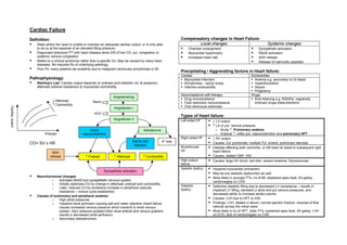
Cardiac failure ( long case approach ) summary
1. Cardiac Failure
Definition: Compensatory changes in Heart Failure:
State where the heart is unable to maintain an adequate cardiac output, or is only able Local changes Systemic changes
to do so at the expense of an elevated filling pressure. Chamber enlargement Sympathetic activation
Diagnosed whenever PT with heart disease devts S/S of low CO, pul. congestion or Myocardial hypertrophy RAAS activation
systemic venous congestion. Increased heart rate ADH release
Refers to a clinical syndrome rather than a specific Dx. May be caused by many heart Release of natriuretic peptides
diseases. Mx requires Rx of underlying aetiology.
Poor Px: many patients die suddenly due to malignant ventricular arrhythmias or MI.
Precipitating / Aggravating factors in Heart failure:
Cardiac Extracardiac
Pathophysiology: Myocardial infarction Anemia e.g. secondary to GI bleed
Starling’s Law: Cardiac output depends on preload (end-diastolic vol. & pressure), Arrhythmias – tachy/ brady Hyperthyroidism
afterload (arterial resistance) & myocardial contractility. Infective endocarditis Sepsis
Pregnancy
Noncompliance with therapy Drugs
Angiotensinog
Drug noncompliance fluid-retaining e.g. NSAIDs, negatively
↓ Afterload Renin Fluid restriction noncompliance inotropic drugs (beta-blockers)
↑ Contractility
Over-strenuous exercises
Cardiac output
Angiotensin I
ACE
Types of Heart failure:
Angiotensin II Left-sided HF ↓ LV output
↑ LA or pul. venous pressure
Direct Aldosterone o Acute ↑: Pulmonary oedema
Preload vasoconstriction o Gradual ↑: reflex pul. vasoconstriction and pulmonary HPT
+
Right sided HF ↓ RV output
Salt & H20 K loss
CO= SV x HR retention
Causes: Cor pulmonale, multiple Pul. emboli, pulmonary stenosis
Biventricular Disease affecting both ventricles, or left heart dz leads to subsequent right
HF heart failure
ADH
release ↑ Preload ↑ Afterload ↑ Contractility Causes: dilated CMP, IHD
High output Causes: large AV shunt, beri-beri, severe anaemia, thyrotoxicosis
failure
Systolic dysf(x) Impaired myocardial contraction
Sympathetic activation
May be a/w diastolic dysfunction as well
Neurohormonal changes: More likely in younger PTs, hx of MI, displaced apex beat, S3 gallop,
o activates RAAS and sympathetic nervous system cardiomegaly on CXR
o Initially optimizes CO by change in afterload, preload and contractility.
Diastolic Defective diastolic filling due to decreased LV compliance – results in
o Later, reduces CO by excessive increase in peripheral vascular
dysf(x) impaired LV filling, elevated Lt atrial and pul venous pressures, and
resistance – vicious cycle established.
decreased ability to increase stroke volume
Causes of pulmonary and peripheral oedema:
o High atrial pressures Causes: LVH due to HPT or IHD
o Impaired renal perfusion causing salt and water retention (heart failure Findings: LVH, dilated Lt atrium, normal ejection fraction, reversal of flow
causes increased venous pressure which transmit to renal venous velocity across the mitral valve
system. Decr pressure gradient btwn renal arterial and venous gradient More likely in hx of HPT, older PTs, sustained apex beat, S4 gallop, LVH
results in decreased renal perfusion) on ECG, lack of cardiomegaly on CXR
o Secondary aldosteronism
2. Causes of Heart Failure: Look out also for the precipitating factors, and screen for depression
Pump failure Heart muscle disease
Ischemic HD / CAD (cause of up to 75% of cases of heart failure) Diagnosis
Cardiomyopathy Boston Criteria for Diagnosing Heart Failure
Myocarditis (Sensitivity 50%, specificity 78%)
Criterion Point value
Restricted filling Category I: History
Pericarditis/ effusion Rest dyspnoea 4
Orthopnea 4
Drugs PND 3
Negative inotropes – beta-blockers Dyspnoea while walking on level area 2
Excessive preload Valve disease Dyspnoea while climbing 1
MR, AR Category II: Examination
Heart rate 1 (if HR 91-110bpm); 2 (if >110bpm)
Fluid Retention JVP 2 (if JVP >6cm H2O); 3 (if JVP >6cm H2O +
NSAIDS, steroids hepatomegaly or edema)
Excessive afterload AS Lung crackles 1 (if basilar); 2 (if more than basilar)
nd Wheezing 3
Systemic hypertension (2 most frequent cause)
Third heart sound 3
High output CF Thyrotoxicosis Category III: CXR
Anemia Alveolar pulmonary edema 4
Interstitial pulmonary edema 3
Clinical Features Bilateral pleural effusion 3
Low CO Cardiothoracic ratio >0.5 3
Fatigue / altered mental Cold peripheries
Upper zone flow redistribution 2
state Low BP Scoring
Listlessness Pulsus alternans Max 4 points per category, scored upon a max of 12 points:
Poor effort tolerance Cachexia 8-12 points: definite
Ventricular Displace left apex beat Function MR / TR 5-7 points: possible
dysfunction RV heave Tachycardia <5 points: unlikely
S3 / S4
Poor renal Oliguria Complications
perfusion Uraemia Depression In the past 2 weeks, have you:
Pul. oedema SOB Inspiratory basal (in 15-40% of Often been bothered by feeling down/ depressed/ hopeless?
crepitations patients) Have you had little interest in doing things which previously
Orthopnea
Cough (usually nocturnal) Cheyne-Stokes resp interested you?
(periodic breathing) HypoK+ Due to K+ losing diuretics, hyperaldosteronism (due to RAAS
PND
activation) & impaired aldosterone metab due to hepatic congestion
Right HF / fluid Raised JVP Hypo Na+ Due to diuretics, inappropriate water retention, failure of cell
retention Hepatic congestion – progresses to cirrhosis due to venous membrane ion pump
congestion Impaired liver Hepatic venous congestion and poor arterial perfusion causes
Peripheral oedema / ascites / Pleural effusion / nocturia function jaundice and abN LFTs and reduced clotting factor synthesis
Chronic HF LOW due to anorexia and impaired absorption due to GI Thromboembolism DVT & PE due to low CO, immobility, arrhythmias, AF, intracardiac
congestion thrombus due to MS or LV aneurysms
Poor tissue perfusion Arrhythmias Common, and due to electrolyte changes, structural heart dz,
Skeletal muscle atrophy due to immobility pro-arrhythmic effects of increased catecholamines and drugs
3. eg digoxin.
Sudden death common and usually due to VF NYHA classification of Heart Failure
Ventricular ectopic beats and vent. Tachycardias common and I No undue dyspnoea from ordinary activity
a/w poor Px. Px is not changed by using anti-arrhythmic drugs II Comfortable at rest, dyspnoea on ordinary activities
III Less than ordinary activities causes dyspnoea, which is limiting
Investigations: IV Dyspnoea at rest, all activities cases discomfort
to confirm diagnosis of CF and exclude other differentials
to look for precipitating causes of CF Management:
to look for complications of CF
Concise summary
Bloods FBC Anemia
Acute
Sepsis
U/E/Cr Electrolyte abnormalities esp. K+, Na+ ABCs + monitoring
Cardiac enzymes MI Oxygen – 100% if no COPD
BNP Investigations
o ECG
TFT Hyper/Hypo-thyroidism if clinically o CXR
suspected o U/E
LFT Cardiogenic liver cirrhosis o Cardiac enzymes
ECG MI, ischemia, arrhythmia o +/- ABG
Radiological CXR Pulmonary edema Drug management
upper lobe diversion o Frusemide 40mg IV slowly
o GTN S/L or disc (if sys BP>90)
blunting of costophrenic angles
o Diamorphine + antiemetic
Bat’s winging o If systolic BP>90 give IV GTN
Kerley’s B lines (interstitial edema) If systolic BP<90 treat as cardiogenic shock
alveolar shadowing If systolic BP>180 treat as hypertensive LVF
Heart size (may be normal in diastolic Chronic
dysfunction) Non pharm Weight loss if obese
Infections
Fluid restriction + daily weight monitoring
TTE (2D echo) Ventricular dysfunction
Ejection fraction (Avoid gain of >2kg in 1 week)
Valvular lesions Dietary salt restriction (2-3g daily)
Special tests to Exercise stress test Exercise
diagnose Non-exercise stress For those unable to exercise Regular low-moderate intensity aerobic exercise
underlying IHD test (pharmacologic) Dipyramidole Avoid lifting heavy weights >10kg
Dobutamine Cut down other risk factors
Radionuclide imaging Thallium scintigraphy Smoking
Technitium-99m sestamibi Hyperlipidemia
Coronary Angiography For CAD, and KIV PCI if suitable Learn to monitor symptoms of deterioration
Cardiac Catheterization Pharm Diuretics
Super-specialised Plasma B-type Plasma concentration of BNP reflects st
tests ) natriuretic peptide ventricular pressure Spironolactone – 1 choice, shown to reduce mortality
High negative predictive value: low BNP Loop
suggests that if patient is dyspneic, Thiazide
cause is highly unlikely to be CF ACE-I/ A2RB
Endomyocardial biopsy To diagnose rare forms of CMP or Beta-blockers – started with low doses, up-titrated slowly
infiltrative heart diseases Digoxin – may not reduce mortality but reduces hospitalization
4. episodes There is NO vol. overload per se, thus main Rx is with vasodilators
End point of Rx is resolution of symp. overdrive, as indicated by pulse rate, BP,
Treat associated AF, hyperlipidemia, CRF etc restoration of warm dry extremities & PT comfort.
comorbidities Features to aid clinical diagnosis:
i. Severe resp distress, orthopnea
Detailed Summary ii. Cold clammy extremities
iii. Thready pulse
Presentations: iv. ↓ SpO2
1. Acute decompensation of chronic left HF - Decreased effort tolerance / pedal oedema / Features of impending resp failure:
wheeze (“cardiac asthma”) i. Altered mental state
2. Acute cardiogenic pulmonary oedema ii. Poor and uncoordinated respiratory effort
3. Cardiogenic shock iii. Progressive desaturation
iv. PaO2 <50mmHg, PaCO2 >50mmHg
1) Acute Mx of Acute Decompensation of Chronic HF 1. Monitoring + attach defibrillator
1. Monitoring: vital signs, pulse oximetry, continuous ECG 2. ABC assessment: intubate in impending respiratory failure
2. Maintain airway, supplemental O2 3. 100% O2 / CPAP in alert PT, but of limited utility
3. IV access 4. IV access
4. Blood invxs: FBC, U/E/Cr, Cardiac enzymes 5. ECG: exclude inferior/right ventricular infarct which is a CI to use of nitrates
5. Position patient: seated upright with legs hanging down to reduce venous return 6. Bloods: RBC, U/E/Cr, cardiac enzymes, Troponin T
6. ECG: concomitant MI, dysrhythmias, LVH, old MI, chronic HPT 7. ABG: baseline
7. CXR: cardiomegaly & features of pul. edema (eg upper lobe diversion) 8. CXR
8. Diuretics: IV frusemide 40-60 mg 9. Catheterize: assess urine output
9. Nitrodisc 5-10mg: relieve symptoms of pul. congestion 10. Drugs:
10. IV GTN: lower LV end-diastolic volume and pressure rapidly for resolution of Nitroglycerine 10-200 μg/min, starting at 10μg/min, increasing by 5μg/min every 5
symptoms mins until MAP = 90mmHg. Continuous BP monitoring required.
11. Monitor urine output to assess response to Rx. Nitroprusside 0.25-10μg/kg/min. invasive monitoring required to prevent
12. Admit / discharge precipitous drop in BP.
Admit Symptomatic dysrhythmias Hydralazine IV 10mg every 30 mins. Monitor PT
New MI Frusemide 40-80mg IV bolus. Onset of effects from 20min-2h
Rapid onset of new symptoms of HF Morphine 0.1mg/kg, starting with IV 3-5mg incremental boluses of 1 mg.
GTN 0.5-1.5mg SL stat
Decompensation of chronic HF
Captopril 6.25 or 12.5mg SL
Ppting factors require inpatient Mx
Combination regimes
Anasarca / severe oedema IV GTN + frusemide Frusemide stat dose + titrate IV GTN titratable infusion
Hypotension IV GTN + captopril SL Captopril stat + titrate IV GTN titratable infusion
Lack of home support Frusemide + morphine
D/C If patient is well and responsive to diuretics
– TCU 2/52 If not on medication: start Lasix 40mg om + Span K 1.2mg om 11. Hypotension in Pul. oedema
If already on med, increase diuretic dose Indicates severe HF
If concurrent HPT present, add ACEI – Captopril 6.254-12.5mg IV dobutamine or dopamine (5-20μg/kg/min)
tds or hydralazine 25mg tds 12. Admit:
Diet advice: salt restriction, fluid restriction (~1 L/day; titrate CCU: PTs with acute coronary syndrome or if intubated
against weight gain/loss and fluid output) HDU: PTs on CPAP
General Wd: the rest
2) Acute Mx of Acute Pulmonary Oedema
Main pathogenic mechanism is sympathetic overdrive leading to elevated LV end- 3) Long term Management of CCF
diastolic vol. & pressure Bed rest
5. Stamina building exercise treat with ACEI (change to ARB if cough occurs, change to
Diet: low salt diet, fluid restriction (~1L/day; monitor with weight, urine output and hydralazine + nitrate if worsening renal insufficiency or
symptoms of pedal oedema/ascites) angioedema occurs)
Beta blockers: for NYHA Class II or III
Pharmacotherapy
Spironolactone: for NYHA Class III or IV
o Reduce preload in backward failure (pul. or systemic congestion) Digoxin: for symptomatic NYHA Class III or IV
o Reduce afterload and increase myocardial contractility in forward failure (low Diuretics: for fluid overload in NYHA Class III or IV
CO) If refractory to above treatment, add dobutamine or milrinone &
o For patients with LVEF <40% IV diuretic
ACE-I for all PTs o Diastolic dysfunction: treat underlying cause (eg IHD or HPT) with beta
β-blockers for all PTs who are haemodynamically stable blockers, CCB, ACEI +/- diuretics
Spironolactone for all PTs with rest dyspnoea
Digoxin for PTs who are symptomatic despite ACEI + diuretics + β-
blockers, and Pts with rest dyspnoea
Diuretics Reduce preload
Excessive diuretic Rx may cause fall in CO.
Combination of different classes of diuretics (loop, thiazides and
K+ sparing) prevents hypo K+. eg Frusemide + spironolactone.
Symptomatic. Does not improve survival.
ACE-I Reduce afterload mainly, + some reduction in preload
Prevents RAAS & sympathetic activation
Eg Captopril, enalapril, lisinopril.
SE: postural hypotension, renal failure (therefore check renal
function one mth after starting), catastrophic fall in BP on first
dose of ACEI (therefore give at night before PT sleeps), hyperK+,
cough, neutropenia, altered taste.
ARB Similar action to ACE-I. Blocks effect of AT II on heart, peripheral
bld vsls & kidneys.
Eg losartan
nd
Usually 2 line Rx if PT does not tolerate ACE-I
Advantage: does not cause cough
β-blockers Reduce sympathetic stimulation to increase ejection fraction
Non-cardioselective β-blockers better as they do not decrease
CO as much
Vasodilators Venodilators (nitrates) reduce preload
Arterial dilators (hydralazine) reduce afterload
Limited by SE of hypotension
Digoxin For AF in HF
Controls ventricular rate + small positive inotropic effect
No effect on survival, but reduces hospitalisation Digitally signed by DR WANA HLA SHWE
Amiodarone DN: cn=DR WANA HLA SHWE, c=MY,
Anti-arrhythmic drug for PTs with symptomatic arrhythmias o=UCSI University, School of Medicine, KT-
Anticoagulants Prevent thromboembolism Campus, Terengganu, ou=Internal
Medicine Group, email=wunna.
hlashwe@gmail.com
Systolic vs Diastolic dysfunction: Long term Mx different, therefore impt to differentiate Reason: This document is for UCSI year 4
students.
btwn the two. However, both may be concomitant in the same patient. Date: 2009.02.24 10:04:48 +08'00'
o Systolic dysfunction: