Ebt calcium score a clue to invulnerable plaque in vulnerable patients
•Download as PPT, PDF•
1 like•499 views

SHAPE Society
![Electron Beam TomographyElectron Beam Tomography
[EBT][EBT]
Is a unique and established tomographic scanning methodIs a unique and established tomographic scanning method
[1[1stst
introduced in 1984] using a scanning electron beamintroduced in 1984] using a scanning electron beam
rather than a rotating X-ray source [e.g. helical CT]rather than a rotating X-ray source [e.g. helical CT]
Rapidly images the patient’s heart [50 or 100 msec/image,Rapidly images the patient’s heart [50 or 100 msec/image,
Approximately 10 times faster than helical CT]Approximately 10 times faster than helical CT]
Imaging done synchronized at a phase of the cardiacImaging done synchronized at a phase of the cardiac
cycle corresponding to the least ballistic coronary motioncycle corresponding to the least ballistic coronary motion](data:image/gif;base64,R0lGODlhAQABAIAAAAAAAP///yH5BAEAAAAALAAAAAABAAEAAAIBRAA7)
Recommended
More Related Content
Viewers also liked
Viewers also liked (20)
Similar to Ebt calcium score a clue to invulnerable plaque in vulnerable patients
Similar to Ebt calcium score a clue to invulnerable plaque in vulnerable patients (20)
More from Society for Heart Attack Prevention and Eradication
More from Society for Heart Attack Prevention and Eradication (20)
Recently uploaded
Recently uploaded (20)
Ebt calcium score a clue to invulnerable plaque in vulnerable patients
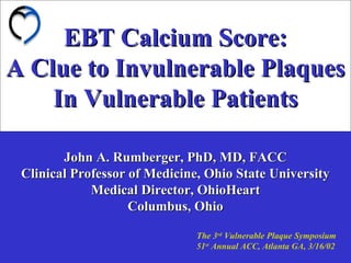
- 1. EBT Calcium Score:EBT Calcium Score: A Clue to Invulnerable PlaquesA Clue to Invulnerable Plaques In Vulnerable PatientsIn Vulnerable Patients John A. Rumberger, PhD, MD, FACCJohn A. Rumberger, PhD, MD, FACC Clinical Professor of Medicine, Ohio State UniversityClinical Professor of Medicine, Ohio State University Medical Director, OhioHeartMedical Director, OhioHeart Columbus, OhioColumbus, Ohio The 3rd Vulnerable Plaque Symposium 51st Annual ACC, Atlanta GA, 3/16/02
- 2. Electron Beam TomographyElectron Beam Tomography [EBT][EBT] Is a unique and established tomographic scanning methodIs a unique and established tomographic scanning method [1[1stst introduced in 1984] using a scanning electron beamintroduced in 1984] using a scanning electron beam rather than a rotating X-ray source [e.g. helical CT]rather than a rotating X-ray source [e.g. helical CT] Rapidly images the patient’s heart [50 or 100 msec/image,Rapidly images the patient’s heart [50 or 100 msec/image, Approximately 10 times faster than helical CT]Approximately 10 times faster than helical CT] Imaging done synchronized at a phase of the cardiacImaging done synchronized at a phase of the cardiac cycle corresponding to the least ballistic coronary motioncycle corresponding to the least ballistic coronary motion
- 3. Electron Beam TomographyElectron Beam Tomography Over the past 18 years there have > 600Over the past 18 years there have > 600 scientificscientific papers published regarding EBT validationpapers published regarding EBT validation andand applications for cardiac imagingapplications for cardiac imaging However, theHowever, the applicationapplication receiving the mostreceiving the most ““notice” (and controversy) has been its abilitynotice” (and controversy) has been its ability
- 4. Coronary Artery CalcificationCoronary Artery Calcification ““Hardening”Hardening” of the coronary arteries has known for 300 years; but over the pastthe past 10 years10 years we havewe have found it is:found it is: An ActiveAn Active ((notnot passive)passive) processprocess CanCan OccurOccur earlyearly in ASO plaque developmentin ASO plaque development AnAn Intimate part ofIntimate part of the fibroproliferative andthe fibroproliferative and inflammatory pathophysiology of CADinflammatory pathophysiology of CAD RegulatedRegulated in a fashion similar to bonein a fashion similar to bone mineralization and repairmineralization and repair
- 5. Coronary Artery CalciumCoronary Artery Calcium and Atherosclerotic Plaqueand Atherosclerotic Plaque Histology X-Ray
- 6. Metabolic Dyslipidemia in Insulin Resistant States Pathobiology & Molecular Mechanisms Khosrow Adeli Ph.D., FCACB, DABCC, NACB Head & Professor, Clinical Biochemistry Laboratory Medicine and Pathobiology Hospital for Sick Children University of Toronto Toronto, CANADA
- 7. Summary of PresentationSummary of Presentation Introduction:Introduction: Insulin Resistance/Metabolic DyslipidemiaInsulin Resistance/Metabolic Dyslipidemia Recent ObservationsRecent Observations Animal Model of Insulin ResistanceAnimal Model of Insulin Resistance (Fructose-Fed Syrian Golden Hamster) • Evidence for Hepatic VLDL Overproduction • Evidence for Hepatic Insulin Resistance • Evidence for Intestinal Lipoprotein Overproduction
- 8. The diverse biological manifestations of the insulin resistant state arise as a consequence of both a blunted insulin action as well as the compensatory hyperinsulinemia per se. Insulin Resistance Insulin resistant peripheral tissues Insulin Increased insulin action in more sensitive tissues or biochemical pathwaysPancreas
- 9. Clinical spectrum of insulin resistant states • Rare (genetic) forms of insulin resistance • Obesity (central, abdominal, visceral, android) • Fasting hyperglycemia/Impaired glucose tolerance • Type 2 diabetes mellitus
- 10. Putative Candidate Gene Mutations inPutative Candidate Gene Mutations in Insulin ResistanceInsulin Resistance •• Glut 1Glut 1 •• Glut 4Glut 4 •• HexokinaseHexokinase IIII •• ISPK-1ISPK-1 •• GSK-3(GSK-3(αα,,ββ)) •• PPIC (PPIC (αα,,ββ,,γγ)) •• PPIGPPIG •• GlycogenGlycogen SynthaseSynthase •• GS-inhibitor-2GS-inhibitor-2 •• GlycogeninGlycogenin •• PhosphofructokinasePhosphofructokinase •• Hormone Sensitive LipaseHormone Sensitive Lipase •• Insulin ReceptorInsulin Receptor •• IRS-1/2IRS-1/2 •• ShcShc •• PI3-PI3-kinasekinase •• ProteinProtein KinaseKinase B (B (αα,,ββ)) •• PPARPPARγγ •• LeptinLeptin •• LeptinLeptin ReceptorReceptor •• b2-b2-adrenergicadrenergic receptorreceptor •• UCP-1UCP-1 •• UCP-2UCP-2 •• NPYNPY •• NPY receptorNPY receptor isoformsisoforms Glucose MetabolismGlucose Metabolism Lipid MetabolismLipid Metabolism Insulin Sensitization/Insulin Sensitization/ desensitizationdesensitization Insulin ActionInsulin Action ObesityObesity
- 11. Disorders associated with insulin resistance • Dyslipidemia • Hypertension • Polycystic ovarian disease • Hyperuricemia • Thrombogenic/fibrinolytic abnormalities • Atherosclerosis
- 12. Features of Metabolic Dyslipidemia •• HypertriglyceridemiaHypertriglyceridemia TG,TG, ApoBApoB VLDL-TG and VLDL-apoB secretionVLDL-TG and VLDL-apoB secretion Small Dense LDLSmall Dense LDL ( LDL particle density)( LDL particle density) •• Reduced HDL-CReduced HDL-C •• Increase FFAIncrease FFA
- 13. FFA FA VLDL DNL Adipose tissue Muscle Liver Intestine TG mobilization by tissue lipases TG, CE ApoB Cytosolic TG stores Oxidation Lipases LPL Mechanisms of VLDL overproductionMechanisms of VLDL overproduction in Insulin Resistancein Insulin Resistance Hepatic Insulin Resistance Adeli K. et al. (2000) J. Biol. Chem. 275: 8416-8425. Adeli K. et al. (2002) J. Biol. Chem. 277:793-803.
- 14. VLDL ApoB mRNA Translation Degradation ER Membrane 5' 3' ApoB mRNA VLDL Assembly Degradation Secretion MTP Proteasome Hepatic Synthesis and Secretion of VLDLHepatic Synthesis and Secretion of VLDL Lipid Poor State Lipid Rich State Lipid Poor State ApoB Gene Expression VLDL Plasma Hepatocy te CE PL TG C
- 15. Mechanisms of VLDL OverproductionMechanisms of VLDL Overproduction in Insulin Resistance (Recent Progress)in Insulin Resistance (Recent Progress) • Development of a Fructose-Fed Hamster Model of Insulin Resistance • Investigations into Mechanisms of Hepatic VLDL Overproduction • Investigations into Mechanisms of Intestinal Lipoprotein Overproduction • Assessment of the Efficacy of hypolipidemic agents and insulin sensitizers in ameliorating metabolic dyslipidemia
- 16. Insulin Resistance Model Fructose-Fed Syrian Golden Hamster • Lipoprotein metabolism closely resembles that in humans • Hamster liver secretes VLDL containing only apoB100 with a density close to that of human VLDL • Hamsters develop hyperTG, hyperCHOL, & atherosclerosis in response to a modest increase in dietary cholesterol & saturated fat • Hamster can be made Obese, Hypertriglyceridemic, Hyperinsulinemic, and Insulin Resistant by carbohydrate feeding (particularly Fructose)
- 17. Male Syrian Golden Hamsters (80-100 grams)Male Syrian Golden Hamsters (80-100 grams) 60% Fructose Diet60% Fructose Diet (2 weeks)(2 weeks) Control HamstersControl Hamsters Control DietControl Diet (2 weeks)(2 weeks) Fructose-fed HamstersFructose-fed Hamsters Plasma Analysis: Glucose, TG, Chol, InsulinPlasma Analysis: Glucose, TG, Chol, Insulin Liver Perfusions >>>>>>Primary HepatocytesLiver Perfusions >>>>>>Primary Hepatocytes Intestinal Fragments >>>>>>Primary EnterocytesIntestinal Fragments >>>>>>Primary Enterocytes Experiments on Hepatic & Intestinal LipoproteinsExperiments on Hepatic & Intestinal Lipoproteins Plasma Glucose, TG, Chol, InsulinPlasma Glucose, TG, Chol, Insulin Insulin Resistance Model Fructose-Fed Syrian Golden Hamster
- 18. Evidence for Development of Insulin Resistance:Evidence for Development of Insulin Resistance: • Increased Plasma Insulin, FFA, TriglycerideIncreased Plasma Insulin, FFA, Triglyceride • Reduced whole body insulin sensitivity (based on Euglycemic-Reduced whole body insulin sensitivity (based on Euglycemic- Hyperinsulinemic Clamp Studies)Hyperinsulinemic Clamp Studies) Adeli K. et al. (2000) J. Biol. Chem. 275: 8416-8425. Evidence for Development of Hepatic VLDL Overproduction:Evidence for Development of Hepatic VLDL Overproduction: • Enhanced hepatic VLDL secretion In Vivo (Triton method)Enhanced hepatic VLDL secretion In Vivo (Triton method) • Enhanced VLDL secretion by primary hamster hepatocytesEnhanced VLDL secretion by primary hamster hepatocytes ex vivoex vivo • Increased intracellular apoB stabilityIncreased intracellular apoB stability • Enhanced MTP expression (mRNA, protein, activity)Enhanced MTP expression (mRNA, protein, activity) Insulin Resistance Model Fructose-Fed Syrian Golden Hamster Hypothesis I:Hypothesis I: Insulin Resistance Induces Hepatic VLDL Overproduction Published Data:
- 19. 0 1 2 Control Fructose-Fed FreeFattyAcids (mmol/L) p=0.0045 0 100 200 300 p=0.0110 PlasmaInsulin (mmol/L) 0.0 2.5 5.0 7.5 p=0.9452 PlasmaGlucose (mmol/L) 0 1 2 3 4 5 p=0.0309 PlasmaTriglyceride (mmol/L) p= 0.0550 0.0 2.5 5.0 7.5 PlasmaCholesterol (mmol/L) A B C D E Increased Plasma Triglyceride, FFA,Increased Plasma Triglyceride, FFA, & Insulin in Fructose-Fed Hamsters& Insulin in Fructose-Fed Hamsters
- 20. Glucose(mmol/l) 0 1 2 3 4 5 6 Control (n=10) Fructose fed (n=9) Insulin(pmol/l) 0 500 1000 1500 2000 2500 3000 Ginf(µmol.kg -1 .min -1 ) 0 10 20 30 40 50 60 SI(10 6 l 2 .kg -1 .min -1 ) 0 1 2 3 4 5 6 p < 0.01 p < 0.01 p = ns p = 0.03 A B C D In Vivo Evidence of Insulin ResistanceIn Vivo Evidence of Insulin Resistance (Euglycemic-hyperinsulinemic Clamp)(Euglycemic-hyperinsulinemic Clamp) Reduced Insulin Sensitivity in Fructose-Fed HamstersReduced Insulin Sensitivity in Fructose-Fed Hamsters
- 21. Enhanced Hepatic VLDL-apoB100 SecretionEnhanced Hepatic VLDL-apoB100 Secretion in Fructose-Fed Hamstersin Fructose-Fed Hamsters (In Vivo Triton WR 1339 Studies) Time (min) 0 20 40 60 80 100 VLDL-apoB(µg/ml) 100 150 200 250 300 350 400 VLDL-apoBsecretion (µg/min) 0 2 4 6 8 10 12 * Control Fructose fed
- 22. 0 100 200 300 400 500 Fructose-FedControl VLDLapoBSecreted (%ofcontrol) ApoB100 * Overproduction of VLDL-apoB byOverproduction of VLDL-apoB by Hepatocytes from Fructose-Fed HamstersHepatocytes from Fructose-Fed Hamsters
- 23. 0 50 100 150 200 250 Control Fructose-Fed MTPActivity (PercentofControl) P=0.042 0 5 10 15 20 Control MTPRNA P<0.02 0 50 100 200 250 MTPProteinMass (percentofcontrol) 150 Fructose-Fed P=0.011 Control Fructose-Fed totalRNA/µgpg)( Protein Mass mRNA Lipid Transfer Activity Evidence for Enhanced Hepatic Microsomal Triglyceride Transfer Protein (MTP) in Fructose-Fed Hamsters
- 24. Insulin Signaling Status in Hepatocytes:Insulin Signaling Status in Hepatocytes: • Ex vivoEx vivo Analysis of Insulin Receptor, IRS-1, PI3-kinase,Analysis of Insulin Receptor, IRS-1, PI3-kinase, PTP-1B in Control and Fructose-Fed Hamster LiversPTP-1B in Control and Fructose-Fed Hamster Livers • In VitroIn Vitro Analysis of Insulin Receptor, IRS-1, PI3-kinase,Analysis of Insulin Receptor, IRS-1, PI3-kinase, PTP-1B in Primary Hepatocytes Exposed to High InsulinPTP-1B in Primary Hepatocytes Exposed to High Insulin Link between Insulin Signaling & VLDL-apoB Secretion:Link between Insulin Signaling & VLDL-apoB Secretion: • In VitroIn Vitro Analysis of ApoB Secretion in Primary HepatocytesAnalysis of ApoB Secretion in Primary Hepatocytes Exposed to High InsulinExposed to High Insulin • Inhibition of Protein Phosphatases byInhibition of Protein Phosphatases by VanadateVanadate and its Impact onand its Impact on VLDL-apoB SecretionVLDL-apoB Secretion (J. Biol. Chem. (2002) 277, 793-803) Hypothesis II:Hypothesis II: VLDL-apoB Overproduction is Linked to Hepatic Insulin ResistanceVLDL-apoB Overproduction is Linked to Hepatic Insulin Resistance Insulin Resistance Model (Fructose-Fed Hamster) Recent Data:
- 25. Y Insulin Signaling Pathway Insulin InsulinReceptorInsulinReceptor αα ββ Y PP IRSIRS ProteinsProteinsSHC Grb2 mSoS Grb2 mSoS RaS p85p85 p110p110 PI 3-KinasePI 3-Kinase PDK1 (PDK2) AktAkt PTP-1B PTP-1B PP PP PTEN aPKCs PDE BAD Anti- apoptosis Anti- lipolysis Glucose transport Gsk3ToRp70rak Glycogen synthesis Protein synthesis RAF MEK MAPK Gene Expression/ mitogenesis 90rak Plasma membrane Protein kinase CK2 Ser/Ther-p CAP CblcrkII Caveolae Glucose & Lipid metabolism Gab 1Shp-2 VanadateVanadate
- 26. CytosolCytosol HepatocyteHepatocyte ApoB DegradationDegradation 5' 3' ApoB mRNAApoB mRNA TranslationTranslation ERMembraneERMembrane Insulin IRS1/IRS2 IR InsulinSignalingPathway InsulinSignalingPathway Plasm a Plasm a VLDLVLDL Insulin Signaling & VLDL OverproductionInsulin Signaling & VLDL Overproduction in Insulin Resistancein Insulin Resistance PI3-Kinase VLDL AssemblyVLDL Assembly PIP3/Phosphorylation CascadePIP3/Phosphorylation Cascade Akt/PKBAkt/PKB PTP-1BPTP-1B 3'
- 27. D InsulinReceptorproteinMass (scanningunits/mgproteinx10-3 ) Control Fructose-fed Control Fructose-fed 0 50 100 150 200 250 IRS-1ProteinMass (scanningunits/mgproteinx10-3 ) Control Fructose-fed Control Fructose-fed 0 40 80 120 E F Control Fructose-fed 0 20 40 60 80 100 120 Control Fructose-fed IRS-2Mass(percentofcontrol) C PhosphorylatedIRS-2 (relativetobasallevelofcontrol) 0 20 40 60 80 100 120 140 160 180 _ + _ + Insulin + + + +Insulin _ _ _ _ Control Fructose-fed Control Fructose-fed Impaired Hepatic Tyrosine Phosphorylation of Insulin Receptor, IRS-1, and IRS-2 in Fructose-Fed Hamsters A PhosphorylatedInsulinReceptorMass (scanningunits/mgproteinx10-3 ) Control Fructose-fed 0 100 200 300 400 500 600 _ + _ + Insulin + + + +Insulin _ _ _ _ * B _ + _ + Insulin PhosphorylatedIRS-1 (scanningunits/mgproteinx10-3 ) Control Fructose-fed 0 20 40 60 80 100 120 Control Fructose-fed + + + +Insulin _ _ _ _ P= 0.04 P= 0.009 P= 0.02 P= 0.03 P= 0.029 * *
- 28. B 0 50 100 150 200 PTP-1BMass (percentofcontrol) Control Fructose-fed Control Fructose-fed C 0 100 200 300 PTP-1BActivity (percentofcontrol) Control Fructose-fed A 0 20 40 60 80 100 120 PI3-KinaseActivity (percentofcontrol) Control Fructose-fed * ** *** D + + + +Insulin 0 20 40 60 80 100 120 140 Insulin-InducedAktSerine Phosphorylation(%ofcontrol) Control Fructose-fed Insulin _ + _ + E Insulin Insulin-InducedAktThreonine Phosphorylation(%ofcontrol) Control Fructose-fed 0 20 40 60 80 100 120 _ + _ + + + + + Insulin F Control Fructose-fed 20 40 60 80 100 120 140 160 AktProteinMass (percentofcontrol) 0 Control Fructose-fed **** *** * Evidence for Reduced PI-3 Kinase Activity, Reduced Akt Phosphorylation, & Enhanced PTP-1B Mass & Activity
- 29. 0 20 40 60 80 100 120 0 20 40 60 80 Vanadate (µM) RecoveredCellularApoB (relativetountreated) B 0 20 40 60 80 100 120 0 20 40 60 80 Vanadate (µM) RecoveredLabeledMediaApoB (relativetountreated) C Vanadate (µM) RecoveredTotalApoB (relativetountreated) 0 20 40 60 80 100 120 0 20 40 60 80 D Vanadate (µM) 0 10 40 80 IR-pY 0 100 200 300 400 500 600 0 20 40 60 80 Vanadate (µM) InsulinReceptorPhosphorylation (relativetountreated) A 0 10 20 40 80Vanadate (µM) Cellular ApoB Secreted ApoB E Inhibition of Cellular Phosphatase Activity with Vanadate Enhances Insulin Signaling and Reduces ApoB Secretion IR-IR-ppYY Cellular ApoBCellular ApoB ApoB StabilityApoB Stability ApoB SecretionApoB Secretion
- 30. Postulated Mechanisms of Insulin ResistancePostulated Mechanisms of Insulin Resistance PTP-1BPTP-1B Mass & ActivityMass & Activity Impaired Phosphorylation ofImpaired Phosphorylation of Insulin Receptor, IRS-1Insulin Receptor, IRS-1 PI-3 Kinase ActivityPI-3 Kinase Activity Attenuated Insulin SignalingAttenuated Insulin Signaling Reduced Phosphorylation of ApoB orReduced Phosphorylation of ApoB or an apoB-chaperonean apoB-chaperone Enhanced Stability and Accelerated Assembly of ApoBEnhanced Stability and Accelerated Assembly of ApoB Overproduction of VLDLOverproduction of VLDL ER-60MTP
- 31. Intestine Contribution of the Intestinal LipoproteinsContribution of the Intestinal Lipoproteins to Metabolic Dyslipidemia in Insulin Resistanceto Metabolic Dyslipidemia in Insulin Resistance Liver ApoB48 ApoB100 Intestinal Lipoprotein Metabolism
- 32. DietaryDietary CholesterolCholesterol DietaryDietary FatFat LuminalLuminal TriglycerideTriglyceride LipasesLipases Bile AcidsBile Acids Fatty AcidsFatty Acids Mocellar CholesterolMocellar Cholesterol Fatty AcidsFatty Acids CholrdyrtolCholrdyrtol ApoB48 + TG + CEApoB48 + TG + CE TGTG CMCM ABCA1ABCA1 ABCG5ABCG5 ABCG8ABCG8 Fatty Acid TransportersFatty Acid Transporters Intestinal Epithelial CellIntestinal Epithelial Cell (Intake)(Intake) (Uptake)(Uptake) (Chylomicron(Chylomicron Assembly)Assembly) (Cholesterol(Cholesterol Excretion)Excretion) Intestinal Lipid Absorption (Trigleride(Trigleride Synthesis)Synthesis)
- 33. Hypothesis III: Fasting and postprandial hyperlipidemia in insulin resistant states may be attributable in part to intestinal oversecretion of apoB-48 containing lipoproteins Experimental Approach: • Dietary induction of an insulin resistant state in the hamster by high fructose feeding • Isolation of adult viable villi from Syrian hamster small intestine. • -In Vivo Studies to assess production rate of intestinal (apoB48- containing) lipoproteins • -Ex Vivo Studies to assess intestinal apoB48 lipoprotein synthesis and secretion, mechanisms of chylomicron assembly, role of de novo lipogenesis in intestinal lipoprotein secretion in the fasting and postprandial states
- 34. Secretion and Regulation of ApoB48 by Primary Hamster Intestinal EnterocytesIntestinal Enterocytes B C LabeledApoB48(%control) LabeledApoB48(%control) 0 20 40 60 80 100 120 Total Cells Media Control MG132 P < 0.05 0 20 40 60 80 100 120 Total Cells Media Control Oleate P = 0.01 Cells Media 0 45 90 45 90 A Chase Time (min)
- 35. Cells Media Fructose-Fed Chow-Fed Chase Time (Min) 0 45 90 45 90 D Chase Time (Min) Chase Time (Min) 100 100 0 10 20 30 40 50 60 70 80 90 0 20 40 60 80 Total apoB 48 LabeledApoB48(%of0Time) LabeledApoB48(%of0Time) p= 0.003 p= 0.001 Secreted apoB 48 0 10 20 30 40 50 60 70 80 90 100 0 20 40 60 80 100 Chow-Fed Fructose-Fed E F Ex vivo evidence for oversecretion of intestinal apoB48 & Enhanced intracellular apoB48 stability in fructose- fed hamster enterocytes
- 36. In Vivo Production of TG & Intestinal ApoB48 in the Fasting State Time (min) 55 60 65 70 75 80 85 Sf>400TGconcentration (mmol/l) 0 1 2 3 4 Sf >400 after Triton WR1339 Time (min) 55 60 65 70 75 80 85 Sf>400ApoB48concentration (µg/ml) 140 160 180 200 220 240 260 280 Time (min) 55 60 65 70 75 80 85 Sf100-400TGconcentration (mmol/l) 0 1 2 3 Sf 100-400 after Triton WR1339 Time (min) 55 60 65 70 75 80 85 Sf100-400ApoB48concentration (µg/ml) 90 100 110 120 130 140 150 160 170 Triglyceridesecretion 0.0 0.2 0.4 0.6 0 1 2 3 ApoB48secretion 0 4 8 12 16 Triglyceridesecretion 0.0 0.1 0.2 0.3 ApoB48secretion 0 2 4 6p = 0.002 p = 0.01 p = 0.14 p = 0.59 A B DC
- 37. In Vivo Production of TG & Intestinal ApoB48 in the Postprandial State Time (min) 55 60 65 70 75 80 85 Triglyceridelevel(mmol/l) 0.0 0.1 0.2 0.3 0.4 0.5 0.6 Time (min) 55 60 65 70 75 80 85 ApoB48level(µg/ml) 80 90 100 110 120 130 140 150 Time (min) 55 60 65 70 75 80 85 Triglyceridelevel(mmol/l) 0.2 0.3 0.4 0.5 0.6 0.7 0.8 0.9 Time (min) 55 60 65 70 75 80 85 ApoB48level(µg/ml) 50 60 70 80 90 100 Triglyceridesecretion(µmol/min) 0.00 0.01 0.02 0.03 0.04 0.05 0.06 0.07 Col 48: 0.07 Col 48: 0.06 Triglyceridesecretion(µmol/min) 0.00 0.02 0.04 0.06 0.08 0.10 Col 50: 0.08 Col 50: 0.09 0 1 2 3 ApoB48secretion(µg/min) 0 1 2 3 4 5 6 7 8 Col 52: 2.65 Col 52: 7.46 ApoB48secretion(µg/min) 0 1 2 3 4 5 6 7 8 Col 54: 3.85 Col 54: 7.26 Sf 100-400 after Triton WR1339 Sf >400 after Triton WR1339 A B C D p = 0.3 p = 0.003 p = 0.8 p = 0.007
- 38. 0 20 40 60 80 100 120 140 160 Fructose-Fed MTPProteinMass (%ofControl) Chow-Fed Fructose-Fed 0 20 40 60 80 100 120 0 0.5 1 1.5 2 2.5 3 3.5 MTP inhibitor (µM) ApoB48Secretion(%ofcontrol) Fructose-fed Chow-fed ∗ ∗ ∗ ∗ Chow-Fed p=0.006D E LabeledCholesterylEster (%ofControl) p= 0.002 0 50 100 150 200 250 Cellular p=0.001 Media B LabeledCholesterol (%ofControl) Chow-Fed Fructose-Fed 0 100 200 300 400 500 600 700 800 Cellular p=0.0018 p=0.0013A Media LabeledTriglyceride (%ofControl) Cellular 0 20 40 60 80 100 120 140 160 180 p=0.04 p=0.003 Media C Ex Vivo Evidence for Intestinal Overproduction of Lipoprotein Lipids and Increase MTP mass/activity
- 39. Fructose-Fed Chow-fed 0 10 20 30 40 50 60 70 80 Large CM Small CM VLDL LDL HDL p<0.05 p= 0.06 p<0.03 %ofTotalLabeledApoB48 A 0 20 40 60 80 100 120 140 CM apoB48 Total-apoB48 LabeledApoB48 (%ofFructose-fed) p<0.0007 p<0.01C 0 10 20 30 40 50 60 70 80 Particles< 1.006 Particles > 1.006 %ofTotalLabeledAPoB48 p<0.024 p<0.026 B 0 40 20 60 80 100 120 CM apoB48 Total apoB48 LabeledApoB48 (%ofFructose-fed) P=0.008 P=0.0001 DFasting Postprandial Density/Size Distribution of Intestinal Lipoproteins Evidence for Increased Number & Size
- 40. Increased De Novo Lipogenesis in FF Enterocytes Evidence that ApoB48 Secretion is Linked to the Rate of De Novo Lipogenesis 0 10 20 30 40 50 60 70 80 90 100 110 0 2 4 6 8 10 12 14 16 Cholesterol Fatty Acid Triglyceride LabeledLipid(%ascontrol) Cerulenin µg/ml 0 50 100 150 200 250 300 350 Chow-fed Fructose-fed LabeledFattyAcid(%ascontrol) 0 10 20 30 40 50 60 70 80 90 100 110 0 2 4 6 8 10 12 14 16 Cerulenin µg/ml LabeledSecretedApoB-48(%ascontrol) A C p= 0.002 Sensitivity of ApoB48 Secretion to Cerulenin Fatty Acid Synthesis Sensitivity of TG & FA Secretion to Cerulenin
- 41. 20 60 80 100 0 40 120 Chow-fed Fructose-fed (2 days) 0 20 40 60 80 100 120 Control Fructose (3mM ) 0 200 400 600 800 1000 enterocytes hepatocytes P < 0.01 LabeledApoB-48(%ofcontrol) LabeledApoB-48(%ofcontrol) Labeledcholesterol 0 50 100 150 200 250 300 350 400 450 enterocytes hepatocytes LabeledTriglyceride P < 0.0001 Acute Fructose Feeding or Exposure Does NOT Affect Intestinal ApoB48 Secretion [14 C] Fructose Incorporation into TG & Cholesterol Two-day Fructose Feeding In Vitro Incubation of Enterocytes with Fructose
- 42. Mechanisms of Intestinal Lipoprotein Overproduction in Insulin Resistant/ Hyperinsulinemic States Apical Surface Basolateral Surface FA monoglyceride FABP-FA FABP TG TG TG ApoB48 PL CE De Novo (FA) Insulin (??) TGdietary SOS Grb-2 IRS PI3-K p110 SHP-2 NCK PTKPTK β β α α IR PKB (Akt) SREBP1c FAS & ACC Glucose + FA MTP ERER GolgiGolgi Intestinal Enterocyte TG ApoB48 Lipoprotein Particles p85Proteasome Degradation
- 43. Acknowledgements Laboratory Group: Changiz Taghibiglou Mehran Haidari Steven Van Iderstine Wei Qui Taryne Chong Farhana Mahboob Biao Chen Leyla Mangaloglu Louisa Pontrelli Fariborz Rashid Rita Kohen Debbie Rudy Collaborators: Gary Lewis- Toronto Andre Carpentier- Sherbrooke Sven Olof Olofsson - Sweden Janet Sparks - Rochester, NY Raphael Cheung – Windsor Michel Tremblay – Montreal Denny Trinh - Toronto Funding: Heart & Stroke Foundation of Ontario Canadian Institutes of Health Research NSERC Hospital for Sick Children Merck-Frosst pfizer GlaxoSmithKline BBDC, University of Toronto
- 44. DNA Microarray Mehran Haidari PhD Application in Vulnerable plaque research Center for Vulnerable Plaque Research University of Texas-Houston & Texas Heart Institute
- 45. Atherosclerosis and the resulting coronary heart disease represent the most common cause of death in industrialized nations. Although certain key risk factors have been identified, the molecular mechanism responsible for this complex disease and its deadly complications remains as a challenge in the years to come. Rupture of atherosclerotic plaque is the predominant underlying process in the pathogenesis of acute coronary syndromes. Although we have gained a great deal of knowledge on underlying pathology involved in plaque vulnerability to rupture, the exact molecular mechanisms underlying the process is still largely unexplored.
- 46. Evolution of genomic and proteomic techniques has opened the door to the world of unknown molecular mechanisms in the body that allowing thorough investigation into susceptibility of certain people / patients to certain outcomes. Investigation of advanced atherosclerosis using the tools for systematic gene and protein expression analysis is a surprisingly neglected area of study and has not been touched widely enough. Only a few numbers of investigators worldwide are actively pursuing this field. (B.C.G Faber, J.A.P Deamen; L.D Adams, Stephen M.Schwartz; M.P. Herman, Uwe Schonbeck; k.J.Haley, Richard T Lee; Timothy A.McCaffrey;L.W.Stanton, R Tyler White;D.Shiffman, Richard M Lawn;Brian K Coombes, ) Deamen Schwartz Lee
- 47. During the last half of the 20th century, the analysis of the regulation and function of genes largely Been driven by step-by-step studies of individual genes and proteins. In the past decade, a paradigm shift has emerged in which we are now able to produce large amounts of data about many genes in a highly parallel and rapidly serialized manner. An important tool in this process has been the development of DNA microarray.
- 48. Low-throughput methods of gene expression Northern Blotting, cumbersome, time-consuming Nuclease protection, at least 10 fold more sensitive Quantitative RT-PCR, state of the art High-throughput Methods of gene expression Serial Analysis of Gene Expression (SAGE) Rapid Analysis of Gene Expression (RAGE) Representational Difference Analysis (RDA) Suppression Subtractive Hybridization (SSH) Differential screening (plus/minus screening) Differential Display (DD) DNA Microarray =400,000 Northern Blotting
- 49. What is DNA Microarray? A large number of genes deposited onto a glass slide (large scale dot blot). The RNA sample is RT with simultaneous incorporation of label, resulting in labeled cDNA. Microarray slides serve as hybridization targets for labeled cDNA. Reverse Northern blotting Patrick O Brown Mark Schena
- 50. Basic Steps in Performing a DNA Microarray Experiments 1- Processing cDNA clones to generate print-ready material 2-Printing cDNA clones (or oligonucleotide) onto a substrate 3-Sample RNA isolation 4-Preparation of the probe (e.g. cDNA synthesis and labeling, RT reaction) 5- Hybridization of labeled probe DNA to the DNA arrayed on the substrate 6-Image acquisition, image analysis and data analysis
- 51. Microarray Fabrication Technologies In Situ Synthesis of Nucleic Acid (Chip ,GeneChip,oligonucleotide array) 15-20 different 25-mer oligonucleotides Exogenous Deposition of cDNA (cDNA, spotted array) Single DNA fragments, greater 0.5 Kb
- 52. Analysis of Gene Expression Monitoring Changes in Genomic DNA Gene Discovery, Sequencing and Pathway Analysis When to use Microarray
- 53. Analysis of Gene Expression 1- Different tissues or different developmental states. 2- Normal or diseased states. 3- Exposure to drugs or different physiological conditions.
- 54. Two basic substrates commonly used for cDNA printing are glass and membrane filters. Chemically treated microscope glass slides are the most widely used support. Microarray, Microscope Slide,80000 Spots. Macroarray, Nylon Membrane, 500,-18000 Spots. Micro or Macro
- 55. RNA Preparation No difference between total RNA or mRNA Type of tissue might have profound effect on extraction process. 10 -20 µg of RNA is needed/slide. Laser captured microdissection (LCM) , incorporation of a PCR step( access to subpopulations cells in vulnerable plaque).
- 56. Sample Labeling Most microarray utilize two fluorophores, Cyanine3(Green emission) and Cyanine5 (Red emission). Fluorophores have different size and different ability for incorporation in Cdna. A single round of transcription is used to generate a labeled cDNA probe (RT-PCR).
- 57. Affymetrix Genechip Biotinylated cRNA is synthesized from cDNA phycoerthrin linked to avidin is used for labeling. Each sample hybridized separately Advantages High density chip Consistent and uniform geometry Single Nucleotide Polymorphisms(SNP) No need for maintaining cDNA clones Disadvantages Sequence data required Oligonucleotid selection rules are not well defined Not best target for hybridization Expensive Hybridization to oligonucleotide is sensitive in detection of single-nucleotide mismatches.
- 58. No consensus on Data Analysis( ANOVA), Clustering (categorizing genes according to their pattern of expression). Normalization First step is during scanning, when sensitivity of detection is adjusted by the laser voltage. Gene expression value can be expressed relative to the expression of housekeeping genes. In the absence of control genes, normalization to the median microarray value is popular.
- 59. Analyzed gene changes are often expressed as a fold increase either greater than twofold or less than 0.5 fold (DeRisi). How Much is Significant??? With a large number of microarrays, small changes can be statistically valid. Elcock et al. detected 1.1 fold changes with 95 % confidence interval when each experimental sample was hybridized to seven microarray slides (with two replicate spots for each gene). Derisi et al.Nat Genet 1996:14:457-60
- 60. Housekeeping genes These are genes that are expressed constitutively and their level of expression is thought to be stable, regardless of the sample used (β Actin, Cyclophilin, GAPDH). DeRisi used 90 housekeeping genes and found that changes that were <0.5 and > 2.4 were acceptable. β Actin is one of the most commonly used housekeeping genes and it has been shown to be downregulated in heat shock experiments. In fact, there is an appreciable amount of literature available to suggest that there is no such thing as housekeeping gene.
- 61. DNA microarray represents a developing technology, there remain substantial obstacles in the design and analysis of these microarray. There are no globally accepted rules or standards for performing controlled microarray experiments. A good experiments include more control component then the real comparison. Accuracy and Precision
- 62. Principles of Q.C in DNA Microarray Replication of each experiments on multiple array. Dual labeling, swapping the dyes for control and treated sample. Using a large number of controls on every array. Rajeevan et al. estimated that 30% of microarray results are false-positive. Microarray findings should be confirmed, at least by one of the low-throughput gene expression methods. Down-Scaling of an experiment makes it generally sensitive to external and internal fluctuation. J.Mol.Diag 2001,3:26-31
- 63. Controls mRNA from genes that are not homologous to the organism understudy (Arabidopsis). cDNA from the organism with high, medium and low expression represented on the array (sensitivity). Cold DNA (e.g., calf thymus DNA, yeast tRNA) is added to block nonspecific annealing. Spots of DNA from another organism whose mRNA is not represented in the sample (Background). Total genomic DNA or cDNA clones of common contaminant such as E.Coli and yeast are represented in the array to monitor for contamination.
- 64. The number of genes encoded by the Human genome has been estimated ∼ 32,000 - 38,000. Between 21,000 - 27,000 genes are expressed in the cardiovascular system Lack of information No cDNA Library for Atherosclerotic plaques Only 5% of total ESTs deposited in GeneBank derived from cardiovascular tissue. ESTs from cardiovascular tissues or cell type or from diseased specimens remain limited.
- 65. Cardiovascular EST data from most model organisms are almost nonexistent. The construction of cardiovascular gene databases at different stages of pathology cast light on the complex genetic mechanisms underlying disease of cardiovascular system. DNA microarray technology is in infancy DNA microarray in atherosclerosis was not born or at least is premature. Premature
- 66. The first study dealing with differential gene expression in whole-mount specimens of rupture plaques using macroarray. Suppression Subtractive Hybridization (SSH) technique isolates low abundant sequence that might not be isolated by use of microarray technology. Mammalian mRNA population 20% Abundant transcript (1000-12000 copies/cell) 25% Medium abundant (100-1000 copies/cell) % 50 small number copies (< 13 copies/cell) Mammalian mRNA encoding proteins that regular cellular behavior are expressed at low abundance. Identification of Gene Potentially Involved in Rupture of Human Atherosclerosis Plaques. Circ Res 2001;89;547554 Deamen
- 67. Perilipin was the known gene that up regulated (confirmed by RT-PCR) , 8 of 10 ruptured plaques expressed perilipin while expression was absent in 10 stable plaque. Perilipin is a protein which present on the surface layer of intracellular lipid droplets in adipocyte and prevent lipolysis. They speculated that the increase in perilipin result in increased lipid retention and plaque destabilization. β actin was down regulated in ruptured plaques. The down regulation of one gene was not confirmed by RT-PCR. A pool of 3 ruptured plaques was compared with a a pool of advanced but stable plaques.
- 68. Prelipin is unlikely to be the sole marker of rupture. The author used only 10% of differentially expressed gene for doing macroarray A large effort at macroarray and then sequencing would have yield more differences. An alternative would be to hybridized the subtractand against a large array. Other alternative is the isolation of cell type-specific genes (LCM) rather than plaque-type-specific genes. (Stephen M.Schwartz et al.Circ Res 2001:89;471-473)
- 69. Richard T Lee et al. Treated cultured Human aortic SMC with TNFα and used DNA microarray with 8600 genes to monitor the gene expression. Marked increase in eotaxin confirmed with northern blotting. Immunohistochemical analysis demonstrated overexpression of eotaxin and its receptor in the Human atheroma (SMC). Circulation;2000:102:2185-2189
- 70. McCaffrey et al. compared transcript profile of fibrous cap vs adjacent media of 13 patients ,using macroarray (membrane 588 known genes). Early growth response gene(Egr-1) was highly expressed in lesion (confirmed by RT-PCR). Many Erg-1 inducible genes including PDGF , TGF-β and ICAM-1 were also strongly elevated in the lesion. Immunocytochemistry indicated that Egr-1 was expressed in SMC. β ACTIN and GAPDH were use as housekeeping gene. J.C.I 2000,105:653-662
- 71. Adams et al. Compared gene expression of media of aorta and vena cava, using cDNA microarray of 4048 known genes. 68 genes had consistent elevation in message expression the aorta. The most differentially gene was Regulator of G Protein Signaling (RGS5). Northern analysis and in situ hybridization were used to confirm the results. Circulation Research 2000.8.623
- 72. R.M Lawn et al. examined the response of macrophages to exposure to oxidized LDL, using microarray containing 10000 Human genes. 268 genes were found to be at least twofold up regulated. Real Time -PCR was used to confirm the results. Orphan nuclear receptors (PPARγ, LXR and RXR) and ABC1 were among genes which unregulated after exposure. J.B.C 2000:275;48, 37324-37332
- 73. L.A Mcintire et al. identified 52 genes with altered expression under shear stress Using DNA microarray in primary human umbilical vein endothelial cells. Significant increases in mRNA levels for 32 and significant decreases in expression for 20 genes were reported. The most enhanced genes were cytocromes P45 1A1 and 1B1 and human prostaglandin transporter. Most dramatically down regulated genes were connective tissue growth factor and endotheline-1. Northern blot analysis confirmed the results obtained on microarray. PNAS2001, 98:8955-8960
- 74. Brian K Coombes et al. used DNA macroarray to study the transcriptional response of Endothelial cells to infection with C.Pneumonia. C.Pneumonia infection up regulated m RNA expression for approximately 8% (20) of the genes studies (268). Genes coding for cytokines (IL-1), Chemkines (MCP-1) and cellular growth factor (PDGF) were the most prominently up regulated genes.
- 75. Proteomic is the study of the proteom or the entire protein complement of a genom It has been readily apparent that examining changes in the proteom offers insight into Understanding cellular and molecular mechanisms that cannot be obtained through genomic analysis. A recent study analyzing human liver samples determined the correlation coefficient between the amount of m RNA present to the corresponding protein abundance to be 0.48 (Anderson and Seilhamer 1997).
- 76. Many genes are expressed constitutively and regulation of their function is at the translational or posttranslational Levels (ApoB ,CFTR, TCR). Several studies have demonstrated selective TnI degradation under Ischemia/reperfusion, partly responsible for contractile dysfunction Observed after myocardial ischemia.( Circ Res.1999;84;9-20) Virtually all known cellular signaling pathways are largely mediated through a complex cascade of reversible protein phosphorylation.
- 77. Acute insults to cells lead to alteration in phenotype through rapid posttranslational Modification of proteins, whereas in chronic disease states cotranslational and Posttranslational protein modification occur in concert with altered gene expression. Most proteomic studies in cardiovascular focused in dilated cardiomyopathy and there is no report of proteomic evaluation in vulnerable plaque. Global proteome analysis provides a better representation of the phenotype than does gene expression analysis.
- 78. Our research group at the vascular biology laboratory of Center for Vulnerable Plaque Research in Texas Heart Institute is conducting a series of genomic and proteomic experiments to shed light on the possible molecular mechanisms involved in the onset and pathogenesis of atherosclerosis. Differential gene and protein expression of morphologically advance, but stable human atherosclerotic lesions and ruptured human atherosclerotic lesions are examined in a large number of patients in the whole-mount specimens.
- 79. Transcript profile of blood monocytes from coronary patients with different presentations and healthy controls will be examined to address the association of gene expression and SNP with coronary risk. Furthermore, Laser Captured Microdissection technology will be employed to evaluate gene and protein expression in different cell populations of atheroma plaques correlated with other markers (such as pH, Temperature, …). We hope these approaches lead to better understanding of the molecular process involved in development and complication of vulnerable plaques.
- 80. The lack of information in genomic and particularly proteomic approaches in vulnerable plaque is apparent and this highlights need for genomic and proteomic evaluation of plaque destabilization
- 82. Coronary Atherosclerosis with Multislice CT: What is beyond coronary atherosclerosis Konstantin Nikolaou Tobias Jakobs Bernd Wintersperger Radiology Alexander Becker Andreas Knez Alexander Leber Cardiology Michael Muders Pathology Christoph R Becker
- 84. CTA Inclusion Criteria • Asymptomatic patients • CV risk factors • Positive calcium scan • Symptomatic patients • No CAD history • Atypical chest pain • Inconsistent stress test < 100 mg CaHA
- 85. Patient Preparation 82 bpm • β-blocker • R/o Contra indications • Informed consent • Metoprolol • 50 - 100 mg orally • 30 - 90 min prior • HR 50 - 60 bpm 65 bpm
- 86. Coronary CTA Parameters • Testbolus 20 ml @ 4 ml/sTestbolus 20 ml @ 4 ml/s • 120 ml (300 mg iodine) @ 3120 ml (300 mg iodine) @ 3 ml/sml/s + NaCl 60 ml+ NaCl 60 ml @ 3 ml/s@ 3 ml/s • 500 ms gantry rotation500 ms gantry rotation • 120 kV, 300 mA120 kV, 300 mA • 4 x 1 mm collimation4 x 1 mm collimation • 3 mm/s table feed3 mm/s table feed • 40 s breath hold40 s breath hold
- 87. ECG Tube Current Modulation Pitch <0,4 250 ms 250 ms250 ms 100% 20% mAs
- 89. Left Coronary Artery (RAO) Coronary Angiography MDCT & VRT
- 90. LAO 60Coronary Angiography MDCT & VRT Right Coronary Artery (LAO)
- 91. Detection of Coronary Stenoses MDCT Coronary Angiography
- 92. Coronary Stenoses CTA & Angiography Author Journal PPV NPV n.a. n Niemann Lancet 2001 81% 97% 30% 35 Achenbach Circulation 2001 59% 98% 32% 64 Mean/sum 70% 98% 31% 99
- 93. CTA Limitations • Artifacts • Cardiac motion • Breathing • Blooming • Poor opacification • Small vessel
- 94. Solutions < Rot. time & β−blocker • Cardiac motion artifacts < Scan times • Breathing artifacts • CM utilization < Slice thickness • Small vessels • Blooming artifact
- 95. 16 Detector Row CT Angiography • 200 ms • 9 Lp/cm • 0.8 mm • 20 s breath hold • 60 ml CM
- 96. Coronary Plaque Imaging MDCT Coronary Angiography
- 97. Non Calcified Plaque Density
- 98. Coronary Atherosclerosis Calcified Nodule Wall changes Occlusion ThrombusFibrocalcified Plaque Stenoses Intimal Thickening Atheroma Healing Hemorrhage Rupture/Erosion
- 99. Atheroma • 38 YOM • Non specific complain • Risk Factors – Cholesterin – Smoker • No calcium 50 HU 50 HU
- 100. Calcified Nodule • 62 YOM • Suspicion of CAD • 12 mg CaHA
- 102. Thrombus • 42 YOM • Epigastric chest pain • Risk Factors – Hypertension – Smoker • No calcium 20 HU
- 103. Thrombus
- 104. Acute Posterior Wall Infarction
- 106. CT Plaque Density Lipid Fibrose50 ± 12 HU 89 ± 31 HU p = 0.018 Lipid Fibrosis
- 107. CTA vs IVUS Schröder Heart 2001;85:576
- 108. Carotid Atherosclerosis Estes 1998 J Cardiovasc Surg 39:527
- 109. Plaque Distribution Leber 2001 Circulation Non- calcifie d 13% Mixe d 33% Calcifie d 54% Myocardial Infarction n = 12 122 Plaque Stable Angina n = 12 135 Plaque Non- calcifie d 6% Mixe d 14% Calcifie d 80%
- 110. Summary • Detection of stenoses – Calcium – Small vessels • Characterization of plaques • Identify atheromas • Follow up under therapy • Acute coronary event • Intracoronary thrombus • Myocardial infarction
- 111. www.CT2002.org
- 112. Coronary Endothelial Shear Stress Profiling In-Vivo to Predict Progression of Atherosclerosis and In-Stent Restenosis in Man Peter H. Stone, M.D. Ahmet U. Coskun, Ph.D. Scott Kinlay, M.D., Ph.D., Maureen E. Clark, M.S. Milan Sonka, Ph.D. Andreas Wahle, Ph.D., Olusegun J. Ilegbusi, Ph.D. Yerem Yeghiazarians, M.D. Jeffrey J. Popma, M.D. Richard E. Kuntz, M.D., M.S. Charles L. Feldman, Sc.D. Cardiovascular Division, Brigham & Women’s Hospital, Harvard Medical School; Department of Mechanical, Industrial and Manufacturing Engineering, Northeastern University; Department of Electrical and Computer Engineering, University of Iowa
- 113. Abstract - 1 The focal and eccentric nature of CAD must be related to local hemodynamic factors. The endothelium is uniquely capable of controlling local arterial responses by transduction of hemodynamic shear stress. Low or reversed shear stress (< ~10 dynes/cm2 ) leads to plaque development and progression. Physiologic shear stress (~10 - 30 dynes/cm2 ) is vasculoprotective, maintaining normal vascular morphology. Increased shear stress (> ~ 30 dynes/cm2 ) promotes outward remodeling and platelet aggregation. Characterization of shear stress along the coronary artery may allow for prediction of progression of atherosclerosis and vascular remodeling.
- 114. Abstract - 2 Current methodologies cannot provide adequate information concerning the micro-environment of the coronary arteries. We developed a unique system using intravascular ultrasound (IVUS), biplane coronary angiography, and measurements of coronary blood flow, to present the artery in accurate 3-D space, and to produce detailed characteristics of intravascular flow, ESS, and arterial wall and plaque morphology. We observed that over 6 mo followup, areas of low ESS demonstrated plaque progression, areas of physiologic ESS remained quiescent, and areas of increased ESS developed outward remodeling. The technology may be invaluable to study the impact of pharmacologic or device interventions on the natural history of coronary disease.
- 115. Fundamental Nature of the Problem • Although all portions of the coronary arterial tree are exposed to the same systemic risk factors, atherosclerosis is focal and eccentric • Each coronary artery has many different obstructions in different “stages” of evolution: – There is not a “wave-front” of vulnerability and consequent rupture.
- 116. Varying Degrees of CAD Lesion Severity in a Single Coronary Artery
- 117. Fundamental Nature of the Problem • Coronary atherosclerotic obstructions behave differently based on the degree of luminal obstruction and morphology: – Lesions > 50-75% obstruction Angina Pectoris – Lesions < 50% obstruction Rupture,superimposed thrombus, MI, death These small, potentially lethal lesions are,These small, potentially lethal lesions are, therefore, “clinically silent” until they rupture.therefore, “clinically silent” until they rupture. • It would be of enormous value to identify minorIt would be of enormous value to identify minor obstructions which were progressing and/orobstructions which were progressing and/or evolving towards “vulnerability” since they could beevolving towards “vulnerability” since they could be treated before rupture occurred, thereby avertingtreated before rupture occurred, thereby averting an acute coronary syndrome.an acute coronary syndrome.
- 118. Nature of Progression of Atherosclerosis • The only truly local phenomena which could lead to varying local vascular responses are endothelial shear stresses (ESS) • Local ESS variations are critical: – Low ESS and disturbed flow (< 6-10 dynes/cm2 ) • Causes atheroma; pro-thrombotic, pro-migration, pro-apoptosis – Physiologic shear stress and laminar flow (10-30 dynes/cm2 ) • Vasculoprotective, anti-thrombotic, anti-migration, pro-survival – High shear stress and turbulent flow (> 30 dynes/cm2 ) • Promotes platelet activation, thrombus formation, and probably plaque rupture • Until now,Until now, in vivoin vivo determination of intracoronary flow velocitydetermination of intracoronary flow velocity and endothelial shear stress has not been possible.and endothelial shear stress has not been possible.
- 119. The Detrimental Effect of Low Shear Stress on Endothelial Structure and Function Low shear stresses and disturbed local flow (< ~ 6 dynes/cm2 ) are atherogenic: (Malek, et al. JAMA 1999; 282:2035) • Cell proliferation, migration • Expression of vascular adhesion molecules, cytokines, mitogens • Monocyte recruitment and activation • Procoagulant and prothrombotic state • Local oxidation Promotes:
- 120. The Effect of Physiologic Shear Stress on Endothelial Structure and Function Physiologic shear stress (~15-50 dynes/cm2 ) is vasculoprotective: (Malek, et al. JAMA 1999; 282:2035) • Enhances endothelial quiescence - decreases proliferation • Enhances vasodilation • Enhances anti-oxidant status • Enhances anti-coagulant and anti-thrombotic status
- 121. Overview of Intracoronary Flow Profiling System Patient • Coronary angiography • Intracoronary ultrasound • Coronary flow (TIMI Frame Count) Acquire image data 3D reconstruction of lumen, EEL, Plaque Generation of grid for Computational Fluid Dynamics Numerical computation Determination of local velocity vectors and shear stress Application of vascular data to patient care Prediction of restenosis Prediction of CAD progression
- 122. Intracoronary Flow Profiling Methods • The intracoronary ultrasound (ICUS) “core” is positioned in the relevant section of the artery and a biplane angiogram is recorded using dilute contrast. • ICUS is performed with controlled pull-back at 0.5 mm/sec with biplane angiography. ECG is simultaneously recorded for “gating.” • A dynamic programming technique extracts the lumen and EEL outline from the ICUS at end-diastolic frames and re-aligns them. • The ICUS frames are realigned in 3-D space perpendicular to the ICUS core image. • The reconstructed lumen is divided into computational control volumes comprising 0.3 mm thick slices along the segment, 40 equal intervals around the circumference, and 16 intervals in the radial direction. • Dividing the blood into small “cubes” on the grid, the Navier-Stokes equations of fluid flow are solved numerically using an iterative procedure (Computational Fluid Dynamics). • Shear stress at the wall is obtained by multiplying viscosity by the velocity gradient at the wall.
- 123. Selected ICUS frames Total number of frames ≈ 100-200/arterial segment
- 124. Measurements of Lumen, Outer Vessel Wall, and Plaque by IVUS (DeFranco. AJC 2001; 88 [Suppl]: 7M) • Lumen • Outer Vessel Wall = Area within EEM • Plaque = Intimal-Medial Thickness
- 125. Stacking of ICUS frames
- 127. Creation of Computational Mesh 640 Cells per cross-section 3mm
- 128. Representative Example of 3-D Reconstruction of Coronary Artery RAO projection LAO projection
- 129. Example of 3-D Reconstruction of Coronary Artery Solid line passing through the centroid of the lumen defines a pathline Perpendicular distance between pathline and lumen border defines local lumen radius, perpendicular distance between EEL border and pathline defines the local EEL radius Difference between local EEL and lumen radii defines local plaque thickness
- 130. Original angiogram of a portion of an artery studied Composite reconstruction of portion of the arterial segment, consisting of outer arterial wall, plaque, and lumen: Isolated view of reconstructed outer arterial wall: Isolated view of reconstructed lumen: Isolated view of reconstructed atherosclerotic plaque: Example of 3-D Reconstruction of Arterial Segment
- 131. Velocity Field Presented As A Longitudinal Section
- 132. Coronary Endothelial Shear Stress w y u WSS ∂ ∂ µ= dynes/cm2 [Artery is displayed as if it were cut and opened longitudinally, as a pathologist would view it.]
- 133. Reproducibility Studies of Intra-coronary Flow Profiling Measurements Cardiac catheterization and coronary angiography – Patients studied completely with ICUS pullback and biplane angiography (“Test A”) – All catheters removed, and after a few minutes, entire procedure repeated (“Test B”): • catheters reinserted • angle, skew, table height reproduced to mimic the initial procedure – All calculations performed to measure lumen, outer vessel, plaque morphology, and endothelial shear stress
- 134. Reproducibility of 3-D Coronary Artery Reconstruction “Test A” and “Test B” Performed Separately Lumen Radius [mm] EEL Radius [mm] Plaque Thickness [mm] Endothelial SS [dynes/cm2 ] r = 0.96 r = 0.95 r = 0.91 r = 0.88 Grid divided into 2,560-10,640 areas/artery (average 5,900/artery) Each p < 0.0001 (Coskun, et al. JACC 2002, 39; 44A) ArterialSegmentLength(mm)
- 135. In-Vivo Determination of the Natural History of Restenosis and Atherosclerosis • First pilot study of its kind in the world • Complete intra-coronary flow profiling at index catheterization and repeated at 6-month followup • 10 patients enrolled: – Followup catheterization completed in 8 patients • one refused recath; one had clinical event prior to recath
- 136. Pilot Study of Natural History of Progression of Coronary Atherosclerosis and In-Stent Restenosis Effect of Candesartan vs. Felodipine ConsentandRandomize Identification of appropriate CAD substrate: -PTCA/stent -obstruction < 50% in adj artery, not revascularized Cath # 1 Cath # 2 Enter BWH System Candesartan active Felodipine placebo Candesartan placebo Felodipine active Titration to BP < 140/90 mmHg (Outpatient visits) Time Line: Hours Time 0 Mo 1 Mo 2 Mo 3 Mo 6 Preliminary identification of hypertensive patient Inclusion Criteria: • Hypertension • CAD requiring stent • Additional minor CAD
- 137. Pilot Study of Natural History of Progression of Coronary Atherosclerosis and In-Stent Restenosis Followup Status: One patient refused repeat catheterization One patient developed acute coronary syndrome and required urgent cath and restenting Serial Study Cohort: 8 patients Native CAD Endpoints: 6 patients with serial studies 5 Felodipine and 1 patient Candesartan Restenosis Endpoints: 6 patients with serial studies 3 Candesartan and 3 Felodipine
- 138. Pilot Study of Candesartan to Reduce Coronary In-Stent Restenosis and Progression of Atherosclerosis Patient Population: 10 patients 9 men; 1 woman Mean age: 60.8 years (range 37-83 years) Concomitant medications: B-blockers, statins, and aspirin (all patients) Mean fasting lipids: Total cholesterol: 156 mg/dl LDL cholesterol: 95 mg/dl HDL: 36 mg/dl Triglycerides: 150 mg/dl Blood Pressure:Baseline: 156/89 mmHg Followup: 137/78 mmHg
- 139. Example of Coronary Atherosclerosis Progression Over 6-Month Period (Stone, et al. JACC 2002, 39: 217A) Plaque Thickness [mm] Lumen Radius [mm] EEL Radius [mm] ESS [dynes/cm 2 ] Arterylength[mm] Plaque Thickness Increases in Areas of Low ESS Lumen Radius Decreases in Areas of Increased Plaque Thickness EEL Radius Increases in Distal Areas ESS Increases in Areas of Plaque Increase and Decreases in Distal Areas
- 140. Example of Coronary Artery “Outward Remodeling” Over 6-Month Period Lumen Radius [mm] EEL Radius [mm] Plaque Thickness [mm] Endothelial SS [dynes/cm2 ] Lumen radius enlarges Outer vessel radius enlarges Plaque thickness does not change ESS returns to normal values (Stone, et al. JACC 2002, 39: 217A) ArterySegmentLength(mm)
- 141. Example of Instent Restenosis Over 6-Month Period Lumen Radius [mm] EEL Radius [mm] Plaque Thickness [mm] Endothelial SS [dynes/cm2 ] Lumen radius smaller within stent, larger outside of stent Outer vessel radius enlarges Plaque thickens within stent, no change outside stent Endothelial shear stress increases within stent, normalizes outside stent (Kinlay, et al. JACC 2002, 39: 5A) ArterySegmentLength(mm)
- 142. Example of No Change in Stented Segment Over 6-Month Period Lumen Radius [mm] EEL Radius [mm] Plaque Thickness [mm] ESS [dynes/cm 2 ] ArterySegmentLength(mm) (Kinlay, et al. JACC 2002, 39: 5A)
- 143. Conclusions • This methodology allows for the first time in man the systematic and serial in vivo investigation of the natural history of CAD and consequent vascular responses. • There are different and rapidly changing behaviors of different areas within a coronary artery in response to different ESS environments. • The methodology can evaluate in detail the ESS that are responsible for the development and progression of CAD, as well as the remodeling that occurs in response to CAD. • The technology may be invaluable to study the impact of pharmacologic or device interventions on these natural histories
- 144. References • Asakura T, Karino T. Flow patterns and spatial distribution of atherosclerotic lesions in human coronary arteries. Circ 1990; 66: 1045-66. • Nosovitsky VA, et al. Effects of curvature and stenosis-like narrowing on wall shear stress in a coronary artery model with phasic flow. Computer and Biomed Res 1997; 9: 575-580. • Malek A, et al. Hemodynamic shear stress and its role in atherosclerosis. JAMA 1999; 282: 2035-42. • Ward M, et al. Arterial remodeling. Mechanisms and clinical implications. Circ 2000; 102: 1186-91. • Ilegbusi O, et al. Determination of blood flow and endothelial shear stress in human coronary artery in vivo. J Invas Cardiol 1999; 11: 667-74. • Feldman CL, et al. Determination of in vivo velocity and endothelial shear stress patterns with phasic flow in human coronary arteries: A methodology to predict progression of coronary atherosclerosis. Am Heart J 2002; 143: (in press). • Feldman CL, Stone PH. Intravascular hemodynamic factors responsible for progression of coronary atherosclerosis and development of vulnerable plaque. Curr Opin in Cardiol 2000; 15: 430-40.
- 145. References • Coskun AU, et al. Reproducibility of 3-D lumen, plaque and outer vessel reconstructions and of endothelial shear stress measurements in vivo to determine progression of atherosclerosis. JACC 2002; 39: 44A. • Stone PH, et al. Prediction of sites of progression of native coronary disease in vivo based on identification of sites of low endothelial shear stress. JACC 2002; 39: 217A. • Kinlay S, et al. Endothelial shear stress identified in vivo within the stent is related to in-stent restenosis and remodeling of stented coronary arteries. JACC 2002; 39: 5A. • Feldman CL, et al. In-vivo prediction of outward remodeling in native portions of stented coronary arteries associated with sites of high endothelial shear stress at the time of deployment. JACC 2002; 39: 247A.
- 147. Multi-slice fast CT and Electron Beam Tomography: the first screening step in imaging coronary atherosclerosis? Stephan Achenbach, MD Department of Cardiology, University of Erlangen, Germany
- 148. Coronary artery disease events such as myocardial infarction or coronary death frequently occur in previously healthy individuals without prior symptoms. Tests which permit identification of individuals at increased risk may thereore be beneficial. Since coronary events are in most cases caused by plaque rupture, imaging methods which permit the identification and quantification of coronary atherosclerotic plaque are potentially useful for risk stratification. Most non-invasive imaging techniques, however, lack the combination of high temporal and spatial resolution which is necessary to reliably visualize the coronary arteries. Electron beam tomography (EBT) and, more recently, multi-slice spiral CT have been shown to permit visualization and quantification of coronary calcium in a non- invasive fashion. Coronary calcium is always caused by coronary atherosclerosis and the amount of coronary calcification correlates to the overall atherosclerotic plaque burden. Numerous clinical studies conducted by electron beam tomography have proven the method´s potential to identify individuals at increased risk for coronary events through detection and quantification of coronary calcifications. Most (but not all) studies have demonstrated a higher predictive value of coronary calcifications as compared to traditional risk factors. Even though some smaller studies have shown that mulit-slice CT (MSCT) in conjunction with ECG-gated reconstruction techniques permits the detection and quantification of coronary calcium with accuracies similar to electron beam tomography, no clinical outcome data have so far been published using MSCT. In conclusion, the detection and quantification of coronary calcification my be a useful tool for the identification of individuals at increased risk for coronary events. Abstract:
- 149. Coronary events - such as myocardial infarction - are usually caused by plaque rupture and frequently occur in previously asymptomatic individuals Introduction
- 150. Traditional risk factors frequently do not permit satisfactory identification of individuals who are at increased risk for coronary artery events Introduction
- 151. Imaging techniques for the non- invasive detection of atherosclerotic plaque in the coronary arteries may be helpful to identify individuals at increased coronary event risk. However, both high temporal and high spatial resolution are necessary to visualize the corornary arteries in a non-invasive fashion. Introduction
- 152. Electron beam tomography is a cross-sectional x- ray imaging technique with a temporal resolution of 100 ms. Introduction
- 153. Electron beam tomography permits the sensitive detection and quantification of coronary artery calcification. Calcium in LAD & LCX Calcium in RCA Introduction
- 154. Aquisition protocols and methods for quantification of coronary calcium by EBT are standardized and large reference data bases are available1, 2 . Severe calcification in LAD Abscence of coronary Introduction
- 155. Recent pre-clinical work has shown that multi-slice spiral CT using the last hardware generation and sophisticated ECG-correlated image reconstruction software also permits coronary calcium detection4,5 . LAD calcifications in retrospectively ECG-gated multislice CT Introduction
- 156. Introduction However, care has to be taken in order to avoid motion artifacts which may be more frequent due to the longer acquisition window as compared to EBT Same patient: prospectively triggered (left) and retrosplectively triggered MSCT
- 157. What is the rationale behind the detection of coronary artery calcification? Discussion
- 158. Why detect coronary calcium? Coronary calcification is always caused by artherosclerosis 6 Discussion
- 159. Why detect coronary calcium? The amount of calcium correlates to overall plaque burden 7,8 However: no close relationship between calcium in a vessel segment and degree of luminal stenosis. Discussion
- 160. Even though calcium does not permit to specifically detect vulnerable plaque, it is wrong to assume that calcified plaques are stable or more frequently stable than non-calcified plaques9 . erosion stable vulnerable healed rupture acute rupture Presence of Calcium Discussion
- 161. Discussion Coronary calcium does not permit to detect the „vulnerable plaque“, but it permits to detect the patient with high coronary atherosclerotic plaque burden in an asymptomatic stage.
- 162. A number of clinical trials have evaluated the predictive value of coronary calcium detection by electron beam tomography in symptomatic and asymptomatic individuals. Discussion
- 163. Raggi et al10 : 632 asymptomatic patients 32 +/- 7 months follow-up myocardial infarction and death Annual event rate: 0.1% for calcium score of 0 2.1% for calcium score 1-99 4.1% for calcium score 100-400 4.8% for calcium score > 400 Raggi et al, Circulation 2000 Discussion
- 164. Arad et al11 : 1173 asymptomatic patients 1 year and 3.5 year follow-up Risk ratio for coronary events: 23 for calcium score > 160 Discussion
- 165. Meta analysis by O´Malley et al12 : Calcium score above median: All events: RR 8.6 „Hard“ events: RR 4.2 Discussion
- 166. Discussion In most studies, coronary calcium by EBT was more predictive than conventional risk Arad et al 1996: ROC 0.91 for calcium, 0.74 for RF 1173 asymptomatic subjects (mean age: 53 years) Raggi et al 2000: OR 22 for calcium, 7.0 for RF 632 asymptomatic subjects (mean age: 52 years) Detrano et al 1999: ROC 0.65 for calcium, 0.67 for RF 1196 asymptomatic high-risk subjctes (mean age: 67 years)
- 167. Keelan et al13 : 288 patients with CAD who underwent coronary angiography. Follow-up 6.9 years. Event-free survival was significantly higher for patients with calcium score < 100 than for those with scores > 100. Discussion
- 168. In summary, a number of studies have proven the prognostic value of coronary calcium detection by electron beam tomography in asymptomatic and symptomatic populations. Study results are not completely unanimous concerning the superiority of coronary calcium over traditional risk factors, but most studies found coronary calcium to have a higher predictive value. No clinical outcome studies have so far been performed using multi-slice CT. Discussion
- 169. What is the potential clinical role of coronary calcium detection? AHA/ACC statement14 : „A positive EBCT confirms the presence of coronary atherosclerotic plaque.“ „Total amount of calcium correlates ... total amount of atherosclerotic plaque.“ „A negative EBCT test makes the presence of atherosclerotic plaque, including unstable plaque, very unlikely.“ „A high calcium score may be consistent with a moderate to high cardiovascular event risk within 2-5 years.“ „A negative test ... low risk of a cardiovascular event in the next 2 to 5 years.“ Discussion
- 170. What is the potential clinical role of coronary calcium detection? In clinical practice, cleary low-risk and clearly high-risk individuals probably do not need further testing for risk stratification. Intermediate risk patients, however, might profit: ACC/AHA14 : „selected use of coronary calcium scores when a physician is faced with the patient with intermediate coronary artery disease risk may be appropriate“ Discussion
- 171. What is the potential clinical role of coronary calcium detection? Discussion
- 172. Role of EBT and MSCT in risk stratification? Coronary calcium, even though it does not permit to detect the „vulnerable plaque“, permits to identify the patient with high plaque burden. The detection of coronary calcium therefore permits identification of patients at increased risk for coronary artery events. It may be beneficially applied in patients who seem to be at „intermediate“ risk. Conclusion
- 173. Initial results have shown that EBT and especially MSCT - after i.v. injection of contrast agent - also permit visualization of non-calcified plaque: Partly calcified plaque in the proximal right coronary artery visualized by multi- slice CT MSCT Conclusion
- 174. Non-calcified plaque in EBT: EBT EBT Conclusion
- 175. Non-calcified plaque in MSCT: Conclusion
- 176. Some authors have compared plaque morphology in MSCT to intravascular ultrasound15 , but the clinical implications and the exact meaning of non-calcified plaque in MSCT or EBBT currently are not clear. Conclusion
- 177. SUMMARY: EBT and MSCT have sufficient spatial and temporal resolution for coronary artery visualization. Clinical studies have shown a high prognostic value of coronary calcium for identification of asymptomatic individuals at increased coronary artery disease risk. The meaning of non-calcified plques which can also be detected (after injection of contrast agent) is not yet clear. Conclusion
- 178. SUMMARY: Future clinical studies, some are currenty being conducted, will help to define the role of coronary calcium detection in the clinical work-up of patients ín whom risk stratification for coronary artery events may be beneficial. Conclusion
- 179. References 1. Hoff JA, et al: Age and gender distributions of coronary artery calcium detected by electron beam tomography in 35246 adults. Am J Cardiol 2001;87:1335-1339 2. Raggi P: Introduction. Am J Cardiol 2001:88(2A);1E-3E. 4. Carr JJ, et al: Coronary artery calcium quantification with retrospectively gated helical CT: protocols and techniques. Int J Card Imaging 2001;17:213-220 5. Becker CR, et al: Coronary artery calcium measurement: agreement of multirow detector and electron beam CT. Am J Roentgenol 2001;176:1295-1298 6. Blankenhorn DH: Coronary arterial calcification. Am J Med Sci 1961; 41-50 7. Rumberger JA, et al: Coronary artery calcium area by electron-beam computed tomography and coronary atherosclerotic plaque area. A histopathologic correlative study. Circulation 1995:92:2157-2162.
- 180. References 8. Sangiorgi G, et al: Arterial calcification and not lumen stenosis is highly correlated with atherosclerotic plaque burden in humans: a histologic study of 723 coronary artery segments using nondecalcifying methodology. J AM Coll Cardiol 1998;31:126-133 9.Burke et al: Coronary calcification: insights from sudden coronary death victims. Z Kardiol 2000;89(Suppl. 2):49-53 10. Raggi P et al: Identification of patients at increased risk of first unheralded acute myocardial infarction by electron-beam computed tomography. Circulation 2000;101:850-855 11. Arad Y et al: Prediction of coronary events with electron beam computed tomography. J Am Coll Cardiol 200:36:1253-1260 12. O´Malley et al: Prognostic value of coronary electron-beam computed tomography for coronary heart disease events in asymptomatic populations. Am J Cardiol 2000;85:945-948 13. Keelan PC et al: Long-term prognostíc value of coronary calcification detected by electron beam computed tomography in patients undergoing coronary angiography. Circulation 2001;104:412-417 14. ACC/AHA expert consensus document on electron-beam computed tomography for the diagnosis and prognosis of coronary artery disease. Circulation 2000;102:126- 140 15. Kopp AF et al: Non-invasive characterization of coronary lesion morphology and composition by multislice CT: first results in comparison with intracoronary ultrasound. Eur Radiol 2001:1607-1611
- 181. Vascular InterventionsVascular Interventions Ergin Atalar, Ph.D.Ergin Atalar, Ph.D. Johns Hopkins UniversityJohns Hopkins University Departments of Radiology and Biomedical EngineeringDepartments of Radiology and Biomedical Engineering DISCLOSURE:DISCLOSURE: E. Atalar is a founder and stock holder of Surgi-Vision, Inc.E. Atalar is a founder and stock holder of Surgi-Vision, Inc.
- 182. Johns Hopkins University OverviewOverview – Intravascular MRI (first human experiments)Intravascular MRI (first human experiments) – Balloon AngioplastyBalloon Angioplasty – Stent PlacementStent Placement – MR-guided Gene TherapyMR-guided Gene Therapy
- 183. Johns Hopkins University Intravascular MRIIntravascular MRI
- 184. MR Imaging Guidewire Surgi-Vision, Inc.
- 185. Johns Hopkins University mm B FSE, 1200/13-msec TR/TE, Double IR blood suppression, 16 ETL, 4-cm FOV, 32 NEX, 256x256 matrix, 10 min 14 sec acquisition time ACM • Resolution: 150 µm J. M. Serfaty et al. Watanabe rabbit with a 0.032” MRI-GuidewireWatanabe rabbit with a 0.032” MRI-Guidewire Aortic wall imagingAortic wall imaging
- 186. Johns Hopkins University Post stent: human iliac IVMRIPost stent: human iliac IVMRI L. Hofmann, D. Bluemke Rt common iliac art. Guidewire (venous) Fibrous cap Lipid core T1 - pre gad T1 - post gad
- 187. Johns Hopkins University L. Hofmann, D. Bluemke Fibrous cap Lipid core Plaque CharacterizationPlaque Characterization
- 188. Johns Hopkins University Restenosis - s/p renal stentRestenosis - s/p renal stent 5 mm L. Hofmann, D. Bluemke Rt renal artery Guidewire in IVC restenosis
- 189. In-Vivo Human Iliac Artery:In-Vivo Human Iliac Artery: Concentric AtherosclerosisConcentric Atherosclerosis Angiography 20 MHz IVUS IVMRI No Abnormality Concentric Atherosclerosis 5 mm 5 mm Vein K. Yucel, et. al. Brigham and Women’s Hospital
- 190. Johns Hopkins University MR-guided Balloon AngioplastyMR-guided Balloon Angioplasty Technical Challenges:Technical Challenges: – MR compatible/visible Balloon AngioplastyMR compatible/visible Balloon Angioplasty CatheterCatheter – Methods for Monitoring Balloon AngioplastyMethods for Monitoring Balloon Angioplasty ProcedureProcedure
- 193. Johns Hopkins University Scan RoomScan Room J. Serfaty et. al.
- 194. Johns Hopkins University MRI-guided PTCAMRI-guided PTCA 90° 10° 90°10° slice selection projection J. Serfaty et. al.
- 195. DilatationDilatation of the Pulmonary Arteryof the Pulmonary Artery C. Rickers, 2001
- 196. Dilatation with Gd filled balloonDilatation with Gd filled balloon C. Rickers, 2001
- 197. Dilatation with air filled balloonDilatation with air filled balloon C. Rickers, 2001
- 198. Johns Hopkins University MR-guided Stent PlacementMR-guided Stent Placement Technical ChallengesTechnical Challenges – MR compatible and visible stent deploymentMR compatible and visible stent deployment devicedevice – MR compatible stentMR compatible stent – Methods of monitoring stent placementMethods of monitoring stent placement procedureprocedure
- 199. Johns Hopkins University MR Guidewire Tracking/PlacementMR Guidewire Tracking/Placement sheath FGREFGRE 256x162256x162 20 mm slice20 mm slice 4 element4 element cardiac coilcardiac coil A. Lardo et. al.
- 200. Johns Hopkins University MRI Guided Stent Positioning and DeploymentMRI Guided Stent Positioning and Deployment stent liver stomach Ao SPGRSPGR 256x162256x162 TR/TE=4.4/1.2TR/TE=4.4/1.2 20 mm slice20 mm slice 4 element cardiac4 element cardiac coilcoil A. Lardo et. al.
- 201. Johns Hopkins University Intravascular Wire Stent CrossingIntravascular Wire Stent Crossing SPGRSPGR 256x162256x162 TR/TETR/TE 20 mm slice20 mm slice 3 element3 element cardiac coilcardiac coil + 1 element+ 1 element internal coilinternal coil stent 1 stent 2 MRI guidewire A. Lardo et. al.
- 202. Johns Hopkins University High Resolution Aortic ImagingHigh Resolution Aortic Imaging stent MRIG (Imaging Guidewire) SPGR, 256x256, FOV=4 cm Guidewire element only stented wall 156µm A. Lardo et. al.
- 203. Johns Hopkins University MR-guided Gene Therapy
- 204. A Remedy gene delivery balloon catheter Gene delivery channel Angioplasty balloon channel Guidewire channel 0.014” MRIG Tuning box X. Yang, et al. Circulation 2001
- 205. Johns Hopkins University Design Plaque Plaque Vessel 0.014” MRIG Balloon inflation with 3% Gd Gd/blue-dye medium or Gd/GFP-lentivirus medium X. Yang, et al. Circulation 2001
- 206. Johns Hopkins University BA X Clinical significance? X. Yang, et al. Circulation 2001
- 207. Johns Hopkins University ConclusionConclusion MRI has potential to guide new andMRI has potential to guide new and conventional vascular interventionsconventional vascular interventions
- 208. Content and GraphicsContent and Graphics Zorina Galis, Ph.D.Zorina Galis, Ph.D. ““Macrophage-induced proteolysis:Macrophage-induced proteolysis: how many MMPs and non-MMPs arehow many MMPs and non-MMPs are involved?”involved?”?? Zorina S. Galis,Zorina S. Galis, Ph.D.Ph.D.Division of Cardiology , EmoryDivision of Cardiology , Emory University School of MedicineUniversity School of Medicine Department of BiomedicalDepartment of Biomedical Engineering Emory/Georgia Tech,Engineering Emory/Georgia Tech, Atlanta GAAtlanta GA March 16, 2002March 16, 2002 33rdrd Vulnerable Plaque SymposiumVulnerable Plaque Symposium March 16, 2002March 16, 2002 33rdrd Vulnerable Plaque SymposiumVulnerable Plaque Symposium
- 209. Content and GraphicsContent and Graphics Zorina Galis, Ph.D.Zorina Galis, Ph.D. LipiLipi dd corcor ee ThrombusThrombus Natural history of humanNatural history of human atherosclerosisatherosclerosis Natural history of humanNatural history of human atherosclerosisatherosclerosis M. Davies,M. Davies, 19981998 Acute cardiovascular events representAcute cardiovascular events represent a late stage of arterial remodelinga late stage of arterial remodeling Acute cardiovascular events representAcute cardiovascular events represent a late stage of arterial remodelinga late stage of arterial remodeling adaptatioadaptatio nn sustainedsustained adaptatioadaptatio n andn and repairrepair destructiondestruction Culprit = ruptureCulprit = ruptureCulprit = ruptureCulprit = rupture
- 210. Content and GraphicsContent and Graphics Zorina Galis, Ph.D.Zorina Galis, Ph.D. SelectedSelected MMPMMP Selected substratesSelected substrates StromelysinStromelysin (SL / MMP-3)(SL / MMP-3) StromelysinStromelysin (SL / MMP-3)(SL / MMP-3) Proteoglycans, fibronectin, lamininProteoglycans, fibronectin, laminin pro-MMP-1, pro-MMP-9pro-MMP-1, pro-MMP-9 Proteoglycans, fibronectin, lamininProteoglycans, fibronectin, laminin pro-MMP-1, pro-MMP-9pro-MMP-1, pro-MMP-9 Gelatinases (GL)Gelatinases (GL) 72 kD GL, GL a (MMP-2)72 kD GL, GL a (MMP-2) 92 kD GL, GL b (MMP-9)92 kD GL, GL b (MMP-9) Gelatinases (GL)Gelatinases (GL) 72 kD GL, GL a (MMP-2)72 kD GL, GL a (MMP-2) 92 kD GL, GL b (MMP-9)92 kD GL, GL b (MMP-9) Collagen type IV / VCollagen type IV / V degraded collagen, elastindegraded collagen, elastin Collagen type IV / VCollagen type IV / V degraded collagen, elastindegraded collagen, elastin Interstitial collagenaseInterstitial collagenase (CL / MMP-1)(CL / MMP-1) Interstitial collagenaseInterstitial collagenase (CL / MMP-1)(CL / MMP-1) Fibrillar collagenFibrillar collagenFibrillar collagenFibrillar collagen membrane-type MMP-1membrane-type MMP-1 (MT-MMP)(MT-MMP) membrane-type MMP-1membrane-type MMP-1 (MT-MMP)(MT-MMP) pro-MMP-2, pro-MMP-13,pro-MMP-2, pro-MMP-13, collagen, fibronectin, laminincollagen, fibronectin, laminin pro-MMP-2, pro-MMP-13,pro-MMP-2, pro-MMP-13, collagen, fibronectin, laminincollagen, fibronectin, laminin The matrix metalloproteinase (MMP)The matrix metalloproteinase (MMP) family of enzymes can break-downfamily of enzymes can break-down matrix componentsmatrix components The matrix metalloproteinase (MMP)The matrix metalloproteinase (MMP) family of enzymes can break-downfamily of enzymes can break-down matrix componentsmatrix components MatrilysinMatrilysin (MMP-7)(MMP-7) MatrilysinMatrilysin (MMP-7)(MMP-7) fibronectin, collagen type IV,fibronectin, collagen type IV, laminin, elastinlaminin, elastin fibronectin, collagen type IV,fibronectin, collagen type IV, laminin, elastinlaminin, elastin Could MMPs beCould MMPs be responsible for theresponsible for the weakening ofweakening of atheroscleroticatherosclerotic plaques?plaques?
- 211. Content and GraphicsContent and Graphics Zorina Galis, Ph.D.Zorina Galis, Ph.D. Galis et al. 1994, JCIGalis et al. 1994, JCI Normal coronaryNormal coronary arteryartery Coronary atheromaCoronary atheroma Immunohistochemistry:Immunohistochemistry: MMP-3MMP-3 Immuhistochemistry:Immuhistochemistry: MMP-3MMP-3 LumenLumen fibrous capfibrous cap In situIn situ zymography (activity assay)zymography (activity assay)In situIn situ zymography (activity assay)zymography (activity assay) Lysis ofLysis of fluorescentfluorescent substratesubstrate First…First… are MMPs expressed in humanare MMPs expressed in human atheroma?atheroma? First…First… are MMPs expressed in humanare MMPs expressed in human atheroma?atheroma? The shoulders of human atherosclerotic plaques contain active MMPs MMP proteins are overexpressed in the vulnerable shoulders, but are they enzymaticaly active ? MMP proteins are overexpressed in the vulnerable shoulders, but are they enzymaticaly active ?
- 212. Content and GraphicsContent and Graphics Zorina Galis, Ph.D.Zorina Galis, Ph.D. Vulnerable plaques have a highVulnerable plaques have a high percentage of macrophage-foam cellspercentage of macrophage-foam cells Farb &VirmaniFarb &VirmaniFarb &VirmaniFarb &Virmani LumenLumenMacrophagMacrophag e foame foam cellscells
- 213. Content and GraphicsContent and Graphics Zorina Galis, Ph.D.Zorina Galis, Ph.D. Macrophage (MMacrophage (MΦΦ) foam cells in the) foam cells in the shoulders of human atheroma expressshoulders of human atheroma express MMPsMMPs Immunohistology: Detection of MMP-3Immunohistology: Detection of MMP-3 Double immunohistology: Detection of MMP-1 and MΦ Double immunohistology: Detection of MMP-1 and MΦ (Galis et al., 1994, J Clin Invest)(Galis et al., 1994, J Clin Invest)
- 214. Content and GraphicsContent and Graphics Zorina Galis, Ph.D.Zorina Galis, Ph.D. Other MMP sightings in theOther MMP sightings in the atheroma…atheroma… Other MMP sightings in theOther MMP sightings in the atheroma…atheroma… • Messenger RNA for MMP-3 colocalizes withMessenger RNA for MMP-3 colocalizes with macrophage foam cells (Henney et al., 1991)macrophage foam cells (Henney et al., 1991) • Active MMP-9 synthesis in atherectomyActive MMP-9 synthesis in atherectomy specimens from patients with unstable angina andspecimens from patients with unstable angina and MMP-13 colocalize with degraded collagen (BrownMMP-13 colocalize with degraded collagen (Brown et al, 1995)et al, 1995) • MMP-7 is expressed by macrophage foam cells atMMP-7 is expressed by macrophage foam cells at sites of potential plaque rupture (Halpert et al.sites of potential plaque rupture (Halpert et al. 1996)1996) • Macrophages can also express the elastolyticMacrophages can also express the elastolytic MMP-8 (Herman, 2001)MMP-8 (Herman, 2001) Macrophage foam cells are associated with increased MMP expression and activity Macrophage foam cells are associated with increased MMP expression and activity
- 215. Content and GraphicsContent and Graphics Zorina Galis, Ph.D.Zorina Galis, Ph.D. Do MMPs degrade the collagenDo MMPs degrade the collagen of the fibrous cap?of the fibrous cap? Do MMPs degrade the collagenDo MMPs degrade the collagen of the fibrous cap?of the fibrous cap? • Macrophage MMP-2 and MMP-9 degrade exMacrophage MMP-2 and MMP-9 degrade ex vivo the collagen of the plaque’s fibrousvivo the collagen of the plaque’s fibrous cap (Shah et al, 1995)cap (Shah et al, 1995) • MMP-1 and MMP-13 colocalize in theMMP-1 and MMP-13 colocalize in the plaque with epitopes expressed byplaque with epitopes expressed by degraded collagendegraded collagen (immunohistochemistry, Sukhova et al,(immunohistochemistry, Sukhova et al, 1999)1999) • MMP-8 colocalizes with epitopesMMP-8 colocalizes with epitopes expressed by cleaved type I collagen in theexpressed by cleaved type I collagen in the shoulders of human plaqueshoulders of human plaque (immunohistochemistry, Herman et al.,(immunohistochemistry, Herman et al., 2001)2001)
- 216. Content and GraphicsContent and Graphics Zorina Galis, Ph.D.Zorina Galis, Ph.D. Overexpression ofOverexpression of interstitial collagenaseinterstitial collagenase (MMP-1 ) coincides with the(MMP-1 ) coincides with the places subjected to theplaces subjected to the highest tensile stress withinhighest tensile stress within the vulnerable shouldersthe vulnerable shoulders (Lee(Lee et alet al. 1996). 1996) Bad luck?!?Bad luck?!?Bad luck?!?Bad luck?!?
- 217. Content and GraphicsContent and Graphics Zorina Galis, Ph.D.Zorina Galis, Ph.D. Determinants of atherosclerotic plaqueDeterminants of atherosclerotic plaque stabilitystability TissueTissue characteristicscharacteristics MechanicalMechanical stressstress Active degradation of matrixActive degradation of matrix scaffold in the vulnerablescaffold in the vulnerable shoulders by MMPsshoulders by MMPs (Galis(Galis et alet al 1994, Galis1994, Galis et al.et al. 1995)1995) Tissue characteristicsTissue characteristics Mechanical stressMechanical stress Rupture of atherosclerotic plaqueRupture of atherosclerotic plaqueRupture of atherosclerotic plaqueRupture of atherosclerotic plaque thin fibrous capthin fibrous cap large lipid corelarge lipid core ((Cheng et al. 1993)Cheng et al. 1993) inflammationinflammation ((LendonLendon et al.et al. 1991, van der Wal1991, van der Wal et alet al., 1994)., 1994) mechanical “hotmechanical “hot spots”coincide withspots”coincide with the weak pointsthe weak points ((Lee et al. 1996)Lee et al. 1996)
- 218. Content and GraphicsContent and Graphics Zorina Galis, Ph.D.Zorina Galis, Ph.D. • Cytokine stimulation: human EC (Hanemaaijeret al. 1993), human SMC (Galis et al. 1994) • mechanical stretch: shoulders (Lee et al, 1996) • engagement of cell surface receptors: VCAM-1 (Romanic & Madri 1994), CD40 (Malik 1996, Schonbeck, Mach et al 1997), ICAM-1 (Aoudjit et al 1998) • modified lipoproteins: vascular cells (Rajavashist et al. 1999), macrophages (Xu et al 1999) • proteases: thrombin (Galis et al. 1995), plasmin/uPA (Carmeliet et al. 1997), cathepsins (Sukhova et al 1997), MT-MMP (Wang et al.,1998) • oxidative stress: • increased by superoxide, hydrogen peroxide, peroxynitrite (Rajagopalan et al. 1996), • Inhibited by N-acetyl cysteine (Galis et al. 1998), nitric oxide (Gurjar et al. 1999) • matrix composition: collagen I increases macrophage MMP(Wesley et al. 1997) • Infections: Chlamydia (Kol et al, 1998)Kol et al, 1998) Potential modulators of MMP expression and activity in atheroma Expression of pro- MMPs Activation of MMP enzymatic activity
- 219. Content and GraphicsContent and Graphics Zorina Galis, Ph.D.Zorina Galis, Ph.D. Experimental modelExperimental model for investigation offor investigation of macrophage foam cellmacrophage foam cell MMPsMMPs Subcutaneous granulomaSubcutaneous granuloma Balloon angioplastyBalloon angioplasty HypercholesterolemiHypercholesterolemi c dietc diet MacrophageMacrophage (anti-RAM 11)(anti-RAM 11) Intracellul ar lipid (Nile red)
- 220. Content and GraphicsContent and Graphics Zorina Galis, Ph.D.Zorina Galis, Ph.D. 0 200 400 600 MΦ FC x103 counts/min/106 cells PMA - + - + Macrophage (MMacrophage (MΦΦ)-derived foam)-derived foam cells (FC) producecells (FC) produce reactive oxygen speciesreactive oxygen species (Rajagopalan et al. 1996 JCI)(Rajagopalan et al. 1996 JCI) Macrophage-derivedMacrophage-derived FC activate theFC activate the zymogen of MMP-9zymogen of MMP-9 in vitroin vitro (Galis et al., 1998 Circulation)(Galis et al., 1998 Circulation) Macrophage-derivedMacrophage-derived FC activate theFC activate the zymogen of MMP-9zymogen of MMP-9 in vitroin vitro (Galis et al., 1998 Circulation)(Galis et al., 1998 Circulation) SuperoxideSuperoxide PeroxidesPeroxides MΦMΦ FCFC 66 -66 - 97 -97 - 46 -46 - pro-MMP-9pro-MMP-9 MMP-9MMP-9 MMΦΦ FCFC MMP-9MMP-9
- 221. Content and GraphicsContent and Graphics Zorina Galis, Ph.D.Zorina Galis, Ph.D. Reactive oxygen species activate latentReactive oxygen species activate latent MMPs produced by human vascular smoothMMPs produced by human vascular smooth muscle cellsmuscle cells ++ Pro-MMP-2 Pro-MMP-9 MMP-9 MMP-2 Pro-MMP-2 MMP-2 In vivo?In vivo?In vivo?In vivo? + Xanthine/ Xanthine Oxidase + Xanthine/ Xanthine Oxidase (Rajagopalan et al. 1996, JCI)(Rajagopalan et al. 1996, JCI)
- 222. Content and GraphicsContent and Graphics Zorina Galis, Ph.D.Zorina Galis, Ph.D. N-acetyl cysteine (NAC) treatment decreasesN-acetyl cysteine (NAC) treatment decreases inin situsitu MMP-9 expression and activity inMMP-9 expression and activity in experimental rabbit atheromaexperimental rabbit atheroma MMP- 9 Macrophag es MMP-9 + NAC Macrophage s + NAC100100 µµmm IELIEL Pro-MMP-2 - MWM (kDa) Abdomina l aorta Thoracic aorta + NAC + NAC0 0 MMP-2 - Pro-MMP-9 - MMP-9 - (Galis et al., 1998 Circulation)(Galis et al., 1998 Circulation)
- 223. Content and GraphicsContent and Graphics Zorina Galis, Ph.D.Zorina Galis, Ph.D. Lipid lowering therapy mayLipid lowering therapy may increase plaque stability byincrease plaque stability by decreasing the oxidative stressdecreasing the oxidative stress •improvement ofimprovement of endothelial functionendothelial function •decreased plaque lipiddecreased plaque lipid •decreased MMPdecreased MMP productionproduction increased plaqueincreased plaque stabilitystability •improvement ofimprovement of endothelial functionendothelial function •decreased MMPdecreased MMP productionproduction increased plaqueincreased plaque stabilitystability OxidativOxidativ e stresse stress LipidLipid herapeutic interventions and plaque stabiliherapeutic interventions and plaque stabilitherapeutic interventions and plaque stabilitherapeutic interventions and plaque stabilit
- 224. Content and GraphicsContent and Graphics Zorina Galis, Ph.D.Zorina Galis, Ph.D. Experimental macrophage-rich arterialExperimental macrophage-rich arterial lesions (ApoE KO mouse carotid arterylesions (ApoE KO mouse carotid artery ligation)ligation) Macrophage-Macrophage- rich neointimarich neointima Normal carotidNormal carotid arteryartery Normal carotidNormal carotid arteryartery Atherosclerotic carotidAtherosclerotic carotid arteryartery Atherosclerotic carotidAtherosclerotic carotid arteryartery (Lessner et al.,(Lessner et al., unpublished)unpublished)
- 225. Content and GraphicsContent and Graphics Zorina Galis, Ph.D.Zorina Galis, Ph.D. Our studies indicate that arteries withOur studies indicate that arteries with macrophage-rich lesions undergomacrophage-rich lesions undergo enhanced positive remodelingenhanced positive remodeling Our studies indicate that arteries withOur studies indicate that arteries with macrophage-rich lesions undergomacrophage-rich lesions undergo enhanced positive remodelingenhanced positive remodeling 55 1010 11 55 2020 2525 303055 1010 11 55 2020 2525 3030 Macrophage areaMacrophage area 10001000 20002000 Outer perimeterOuter perimeter Time (days) Time (days) 1010 2020 3030 4040 AreaArea ((µµmm22 )) LengtLengt h (h (µµm)m) - - Pro MMP-9 Pro MMP-2 98 kDa 72 kDa - - Days 0 14 2 8 WT W T WTKO KO KOS T Arteries with macrophage-rich lesions have enhanced MMP activity Arteries with macrophage-rich lesions have enhanced MMP activity
- 226. Content and GraphicsContent and Graphics Zorina Galis, Ph.D.Zorina Galis, Ph.D. Positively remodeling isPositively remodeling is associated with plaqueassociated with plaque instabilityinstability Positively remodeling isPositively remodeling is associated with plaqueassociated with plaque instabilityinstability Schoenhagen et al., 2000 Circulation 101Schoenhagen et al., 2000 Circulation 101Schoenhagen et al., 2000 Circulation 101Schoenhagen et al., 2000 Circulation 101 UnstableUnstable StableStable PositivePositive NegativeNegativeAbsentAbsent 2020 3030 4040 5050 1010 RemodelingRemodeling %cohort%cohort
- 227. Content and GraphicsContent and Graphics Zorina Galis, Ph.D.Zorina Galis, Ph.D. FoamFoam cell/macrophagcell/macrophag e-drivene-driven SmoothSmooth muscle cell-muscle cell- drivendriven UnstablUnstabl ee plaqueplaque StableStable plaqueplaque Constrictive arterial remodelingConstrictive arterial remodeling Outward arterial remodelingOutward arterial remodeling(Galis and(Galis and Khatri,Khatri, 2002)2002) Normal arteryNormal artery
- 228. Content and GraphicsContent and Graphics Zorina Galis, Ph.D.Zorina Galis, Ph.D. Macrophage-foam cells are key regulators of MMP-dependent degradation of vascular matrix Macrophage-foam cells are key regulators of MMP-dependent degradation of vascular matrix ROSROS TFTF ThrombinThrombin ModulateModulate MMPMMP activityactivity ExpressExpress pro-pro- MMPsMMPs CytokinesCytokines UpregulateUpregulate vascularvascular cell MMPscell MMPs Macrophage foam cellsMacrophage foam cells
- 229. Content and GraphicsContent and Graphics Zorina Galis, Ph.D.Zorina Galis, Ph.D. Macrophage non-MMPMacrophage non-MMP proteases?proteases? Macrophage non-MMPMacrophage non-MMP proteases?proteases? • CathepsinsCathepsins K and S – have elastolytic activityhave elastolytic activity – Expressed by macrophages in atheromaExpressed by macrophages in atheroma (Sukhova et al. 1998)(Sukhova et al. 1998) • ThrombinThrombin – generated at sites of disruption, can begenerated at sites of disruption, can be generated in the atherosclerotic plaques viagenerated in the atherosclerotic plaques via tissue factor expressed by macrophage foamtissue factor expressed by macrophage foam cells (Wilcox et al. 1989)cells (Wilcox et al. 1989) – can activate latent MMP-2 (Galis et al., 1997)can activate latent MMP-2 (Galis et al., 1997)
- 230. Content and GraphicsContent and Graphics Zorina Galis, Ph.D.Zorina Galis, Ph.D. Potential protease networksPotential protease networksPotential protease networksPotential protease networks • Macrophage MMPs may increase the activity ofMacrophage MMPs may increase the activity of other proteases through:other proteases through: – Activation of MMP zymogens - e.g., MMP-3 can activateActivation of MMP zymogens - e.g., MMP-3 can activate pro-MMP-1pro-MMP-1 – inactivation of inhibitors - e.g., MMP-1, MMP-3, and MMP-inactivation of inhibitors - e.g., MMP-1, MMP-3, and MMP- 9can inactivate alpha 1-antitrypsin, the primary9can inactivate alpha 1-antitrypsin, the primary physiologic inhibitor of human leukocyte elastase (Siresphysiologic inhibitor of human leukocyte elastase (Sires et al. 1994et al. 1994); MMP-1, MMP-7, MMP-9, and MMP-12 cleave); MMP-1, MMP-7, MMP-9, and MMP-12 cleave tissue factor pathway inhibitor (Belaaouaj et al, 2000)tissue factor pathway inhibitor (Belaaouaj et al, 2000) • Macrophage non-MMPs may increase theMacrophage non-MMPs may increase the activity of other proteases through:activity of other proteases through: – Activation of MMP zymogens -- e.g., uPA can activate pro-Activation of MMP zymogens -- e.g., uPA can activate pro- MMP-3, thrombin can activate pro-MMP-2MMP-3, thrombin can activate pro-MMP-2 – inactivation of inhibitors ?inactivation of inhibitors ?
- 231. Content and GraphicsContent and Graphics Zorina Galis, Ph.D.Zorina Galis, Ph.D. RupturedRuptured plaqueplaque The mutual stimulation of generation ofThe mutual stimulation of generation of active MMPs and thrombin may the basisactive MMPs and thrombin may the basis of sustained plaque instability, withof sustained plaque instability, with recurring episodes of plaque disruptionrecurring episodes of plaque disruption and thrombosisand thrombosis MMPsMMPs ThrombinThrombin MMPsMMPs ThrombinThrombin ThrombinThrombin activatesactivates latent MMPslatent MMPs (Galis et al.,(Galis et al., 1997)1997) ActiveActive MMPsMMPs stimulatestimulate generationgeneration of thrombinof thrombin (Sawicki et(Sawicki et al., 1997)al., 1997) roteases and the unstable atherosclerotic plaquroteases and the unstable atherosclerotic plaquroteases and the unstable atherosclerotic plaquroteases and the unstable atherosclerotic plaqu
- 232. Content and GraphicsContent and Graphics Zorina Galis, Ph.D.Zorina Galis, Ph.D. Are these proteasesAre these proteases redundant?redundant? What are the potential actionsWhat are the potential actions of all these in relation toof all these in relation to plaque rupture?plaque rupture? Are these proteasesAre these proteases redundant?redundant? What are the potential actionsWhat are the potential actions of all these in relation toof all these in relation to plaque rupture?plaque rupture?• MMPs can degrade all the components of theMMPs can degrade all the components of the extracellular matrixextracellular matrix • uPAuPA activatesactivates MMP zymogens (MMP zymogens (Carmeliet et al. 1997) • Thrombin can activate MMP zymogens (Galis et al.Thrombin can activate MMP zymogens (Galis et al. 1997)1997) Do MMPs provide a common pathway forDo MMPs provide a common pathway for plaque weakening by other proteases?!?plaque weakening by other proteases?!? Do MMPs provide a common pathway forDo MMPs provide a common pathway for plaque weakening by other proteases?!?plaque weakening by other proteases?!?