Microscopy - Magnification, Resolving power, Principles, Types and Applications
•
7 likes•11,298 views

Magnification, Resolving power, Principles and Applications of Simple, Compound, Stereozoom, Phase contrast, Fluorescent and Electron microscopes (TEM & SEM). Microscopy is the technical field that uses microscopes to observe samples which are not in the resolution range of the normal-unaided eye.

Recommended
More Related Content
What's hot
What's hot (20)
Similar to Microscopy - Magnification, Resolving power, Principles, Types and Applications
Similar to Microscopy - Magnification, Resolving power, Principles, Types and Applications (20)
More from Nethravathi Siri
More from Nethravathi Siri (20)
Recently uploaded
Recently uploaded (20)
Microscopy - Magnification, Resolving power, Principles, Types and Applications
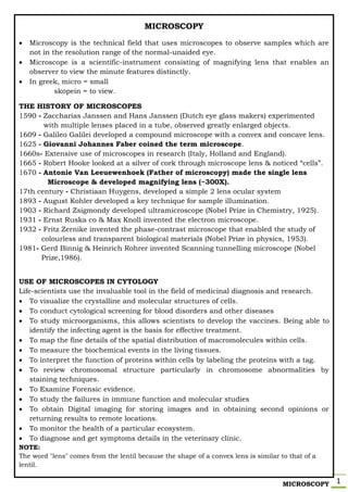
- 1. MICROSCOPY 1 MICROSCOPY Microscopy is the technical field that uses microscopes to observe samples which are not in the resolution range of the normal-unaided eye. Microscope is a scientific-instrument consisting of magnifying lens that enables an observer to view the minute features distinctly. In greek, micro = small skopein = to view. THE HISTORY OF MICROSCOPES 1590 - Zaccharias Janssen and Hans Janssen (Dutch eye glass makers) experimented with multiple lenses placed in a tube, observed greatly enlarged objects. 1609 - Galileo Galilei developed a compound microscope with a convex and concave lens. 1625 - Giovanni Johannes Faber coined the term microscope. 1660s- Extensive use of microscopes in research (Italy, Holland and England). 1665 - Robert Hooke looked at a silver of cork through microscope lens & noticed “cells”. 1670 - Antonie Van Leeuewenhoek (Father of microscopy) made the single lens Microscope & developed magnifying lens (~300X). 17th century - Christiaan Huygens, developed a simple 2 lens ocular system 1893 - August Kohler developed a key technique for sample illumination. 1903 - Richard Zsigmondy developed ultramicroscope (Nobel Prize in Chemistry, 1925). 1931 - Ernst Ruska co & Max Knoll invented the electron microscope. 1932 - Fritz Zernike invented the phase-contrast microscope that enabled the study of colourless and transparent biological materials (Nobel Prize in physics, 1953). 1981- Gerd Binnig & Heinrich Rohrer invented Scanning tunnelling microscope (Nobel Prize,1986). USE OF MICROSCOPES IN CYTOLOGY Life-scientists use the invaluable tool in the field of medicinal diagnosis and research. To visualize the crystalline and molecular structures of cells. To conduct cytological screening for blood disorders and other diseases To study microorganisms, this allows scientists to develop the vaccines. Being able to identify the infecting agent is the basis for effective treatment. To map the fine details of the spatial distribution of macromolecules within cells. To measure the biochemical events in the living tissues. To interpret the function of proteins within cells by labeling the proteins with a tag. To review chromosomal structure particularly in chromosome abnormalities by staining techniques. To Examine Forensic evidence. To study the failures in immune function and molecular studies To obtain Digital imaging for storing images and in obtaining second opinions or returning results to remote locations. To monitor the health of a particular ecosystem. To diagnose and get symptoms details in the veterinary clinic. NOTE: The word "lens" comes from the lentil because the shape of a convex lens is similar to that of a lentil.
- 2. MICROSCOPY 2 PRINCIPLE: MAGNIFICATION AND RESOLVING POWER MAGNIFICATON Magnification is defined as “The degree of enlargement of an object provided by the microscope for detailed analysis of sample”. The magnification by microscope is the product of individual magnifying powers of ocular lens (eye piece) and objective lens. Magnification = Magnifying Power of ocular lens X Magnifying Power of objective Lens For example: If ocular lens is 10X and objective is 40X. Then, Magnification = Magnifying Power of ocular lens X Magnifying Power of objective lens = 10 X 40 = 400X The Magnifying Power of Microscope is defined as “The ratio of the final image observed through the microscope to the size of sample observed via naked eye”. Magnifying Power = The ratio of the final image observed through the microscope The size of sample observed via naked eye Magnification has no limit, but beyond certain point the view becomes blur or unclear. This is termed as EMPTY MAGNIFICATION. Therefore, magnification alone does not provide quality information of the sample. Thus, Resolution plays a crucial role. MICROSCOPE OPTICAL MICROSCOPE SIMPLE MICROSCOPE COMPOUND MICROSCOPE STEREOZOOM MICROSCOPE PHASE CONTRAST MICROSCOPE FLUORESCENT MICROSCOPE ELECTRON MICROSCOPE TRANSMISSION ELECTRON MICROSCOPE SCANNING ELECTRON MICROSCOPE
- 3. MICROSCOPY 3 RESOLVING POWER Resolving Power is defined as “the performance capacity or ability of the microscope to distinguish between two very closely associated particles”. For example; Human eye has resolving power of 0.25nm. Resolving Power of the microscope is the reciprocal of limit of resolution. Limit of Resolution is the shortest distance between the two objects when they can be distinguished as two separate entities. Limit of Resolution (d) = 0.61 X λ n Sinθ Where, λ → wavelength of light n → refractive index of the medium between specimen and objective θ → half angle formed between the specimen and lens As, Resolving Power of the microscope = 1 _ Limit of Resolution Therefore, Resolving Power of the microscope = n Sinθ 0.61 X λ Where, n → refractive index of the medium between specimen and objective θ → half angle formed between the specimen and lens λ → wavelength of light Since, Resolving Power of the microscope ∝ n Sinθ λ Resolving power can be increased by following 3 steps: 1. by increasing Refractive Index; n immersion oil = 1.5 n air = 1 2. by increasing Sinθ 3. by decreasing wavelength of light; λ blue light = 400nm λ red light = 600nm NOTE: Numerical Aperture (NA) of the objective is defined as the property of lens that decides the quantity of light that enters into objective. Numerical Aperture = n Sinθ
- 4. MICROSCOPY 4 GENERIC CONSTRUCTION OF MICROSCOPE Any microscope is constructed based on mechanical system and optical adjustments. Henceforth, could be separated into; 1. MECHANICAL PARTS provides physical support to the optical parts and help in focusing the sample. 2. OPTICAL PARTS confers required adjustments for magnification and optical pathway. LIST OF MECHANICAL PARTS LIST OF OPTICAL PARTS 1. Base / Metal stand / Foot 2. Pillar 3. Inclination joint 4. Curved arm 5. Stage 6. Stage knobs 7. Stage clips 8. Revolving nosepiece 9. Coarse adjustment 10. Fine adjustment 11. Body tube 12. Draw tube 1. Light source / illuminator 2. Diaphragm 3. Sub stage - Condenser 4. Objective lens 5. Ocular lens / eye piece NOTE: Optical pathway is the light path from source of illumination passing through the optical parts (condenser, specimen, objective lens, ocular lens) and finally creates a magnified virtual image in eyes of an observer).
- 5. MICROSCOPY 5 SIMPLE MICROSCOPE A simple microscope works on the principle that when a tiny object is placed within its focus, a virtual, erect and magnified image of the object is formed at the least distance of distinct vision from the eye held close to the lens. Refer… Magnification*** and Construction - mechanical and optical parts Working Principle: Light from a light source (mirror) passes through a glass stage with slide containing a thin transparent specimen. A biconvex - ocular lens based on its capacity magnifies the size of the object, resulting in an enlarged virtual image. APPLICATIONS OF SIMPLE MICROSCOPE 1. Simple microscope is used to obtain small magnifications such as morphology. 2. Simple microscope is usually used for study of microscopic algae, fungi and biological specimen. 3. Simple microscope is used by skin specialists to scan for various skin disorders. 4. Simple microscope is used to see the magnified view of different particles present in diverse soil forms. COMPOUND MICROSCOPE A compound microscope is an optical instrument used to observe the magnified images of small objects on a glass slide. Compound microscopes are so called because they are designed with a compound lens system. The objective lens provides the primary magnification which is compounded (multiplied) by the ocular lens (eyepiece). It provides higher magnification and overcomes the limited clarity of image observed by stereo or other low power microscopes and reduces chromatic aberration. It facilitates detailed study of specimen in a two-dimensional spatial lane. High-quality Compound Microscopes are available in Monocular, Binocular, and Trinocular configurations. It has a series of two lenses; (i) the objective lens ((4x, 10x, or 100x)close to the object to be observed and (ii) the ocular lens or eyepiece (5x-30x), through which the image is viewed by eye.
- 6. MICROSCOPY 6 Compound microscopy classified based on the field observed; 1. Bright-field microscopes 2. Dark-field microscopes 1. BRIGHTFIELD MICROSCOPES The bright-field microscope is the simplest optical microscope and is popularly employed. The object to be inspected is normally placed on a clear glass slide, and light is transmitted though the object. This makes the object appear against a bright background, hence the term Bright-field. WORKING PRINCIPLE Light from the illumination (light) source from the base of the Microscope stand is aimed at sub-stage condenser lens. The sub-stage condenser lens focuses light through slit in the stage onto the sample. The sample absorbs some amount of light based on stain, pigmentation or thickness. The projected light from the sample is collected by objective lens and is magnified according to its capacity, creating a primary image. The primary image is magnified by ocular lens (eye piece), which also act as magnifying glass by allowing the observer to view virtual and magnified image of the sample. APPLICATIONS Widely used for stained or naturally pigmented or highly contrasted specimens mounted on a glass microscope slide. Used in biology classrooms (mitosis & meiosis, etc.) and clinical laboratories. Used in pathology to view fixed tissue sections or cell smears / smears.
- 7. MICROSCOPY 7 2. DARKFIELD MICROSCOPES Used to observe unstained – transparent specimens. Samples having very close refractive indices value as that of surroundings are difficult to observe with conventional bright-field microscopes, such samples are ideal for observation with dark background. Example: small aquatic organisms, oocytes and other thin-transparent materials with Refractive Index from 1.2 to 1.4 WORKING PRINCIPLE Light from the illumination (light) source from the base of the Microscope stand is aimed at dark-field ring. Dark-field ring is an opaque disk blocks the central rays of the light. The marginal/peripheral light rays are directed to sub-stage dark-field condenser lens. The specimen on the stage is illuminated only with the peripheral oblique rays. As a result of this, the field appears dark. The scattered ray from bright specimen is collected by objective lens and is magnified according to its capacity, creating a primary image. The primary image is magnified by ocular lens (eye piece), which also act as magnifying glass by allowing the observer to view virtual and magnified image of the sample. APPLICATIONS Used for examination of live sample. Unstained or lightly stained specimen or fluids could also be observed. Useful for diagnosis of disease. The bacterial motility can be studied. Precious stones are viewed.
- 8. MICROSCOPY 8 STEREO MICROSCOPE The stereo microscope, also called a Dissecting microscope, as it allows the operator to manipulate/dissect the specimen while it is being observed through the microscope. It provides relatively lower magnification usually below 100x. They provide a close-up, 3-Dimensional view of objects surface textures. Stereo microscopes are used for large biological samples ( insects, leaf, tissues…) and medical science applications as well as in the electronics industry, such as by those who make circuit boards or watches. WORKING PRINCIPLE The Optical binocular stereo microscope consists of two objective lens and two ocular lens. Two spatially separated optical path focuses sample on the same point from slightly different angles. The laterally correct, upright-erect image is obtained. ADVANTAGES They can have a single fixed magnification, several discrete magnifications, or a zoom magnification system. Many stereo microscopes are modular in design. It does not require a slide preparation. It enables to switch from bright-field to dark-field and vice-versa.
- 9. MICROSCOPY 9 PHASE-CONTRAST MICROSCOPE The first phase contrast microscope was developed by FRITZ ZERNIKE (FREDRICK ZERNIKE) in 1933, hence also referred as ZERNIKE MICROSCOPE. The phase contrast microscope enables to differentiate transparent, unstained, living (without killing or altering the living component) structures. Phase contrast is an illumination technique provides greater degree differentiation inside the cells by phase contrast optics. WORKING PRINCIPLE Light from the illumination (light) source from the base of the Microscope stand is aimed at annular diaphragm stop. The annular-diaphragm-stop allows only the hollow cone of light rays to pass through sub-stage condenser lens. The sub-stage condenser lens focuses light through slit in the stage onto the sample. The projected light from the sample is collected by special set of objective lens with phase plate and phase rings which are placed in the back/rare focal plane of the objective. The direct rays (unaltered amplitude and phase, but retarded by ¼ wavelength ) from transparent sample converge on the phase ring within the objective and produce phase shift. The most diffracted rays (altered rays due to difference in density) pass through plate plate by missing phase ring. The convergence of diffracted and direct rays on the image plane results in image. ADVANTAGES Specimens which have a refractive index similar to their surroundings can be invisible in Brightfield, but are well defined in Phase Contrast. Phase Contrast is normally used to examine unstained biological specimens. Living microorganisms and their minute details such as Cilia, flagella can be observed.
- 10. MICROSCOPY 10 FLUORESCENT MICROSCOPE When a substance absorbs light, the electrons present at the outermost orbit absorbs energy and get excited; on the way back to the ground state, it emits a part of the energy absorbed. This phenomenon is termed as fluorescence. Fluorescent microscope involves staining of specimens with special fluorescent dyes (fluorescein, acridine orange, etc). When a fluorescent dye is applied to a substance, it absorbs a wavelength of light (excitation wavelength) and emits light of different wavelength (emission wavelength). WORKING PRINCIPLE Illumination (light) is provided by a bright mercury vapor lamp (very expensive + harmful), produces light range of 200-400nm and generates considerable amount of heat. The heat filter absorbs heat, allows UV rays and visible rays by blocking infrared rays. The exciter filter ensures high energy - short wavelength - monochromatic light towards dichroic mirror. Dichroic mirror (beam splitter) eliminates visible light and reflects excited UV light to the dark-field condenser, which provides high contrast for fluorescence and also deflects majority of UV light. The excitation light is focused on to the fluorochrome specimen. The fluorescent labeled specimen absorbs light and emits excitatory light along with florescent light, which reaches objective lens. As per the capacity of objective lens, the specimen would be magnified and are directed towards barrier filter. The additional barrier filter permits only the fluorescent wavelength and rejects excitation light. The fluorescent light passing through ocular lens creates the magnified image, which can also be detected by detector to give a photographic image. APPLICATIONS Imaging the genetic material (DNA & RNA) and other structural components. Monitoring the environment for microbial contamination. Certain micro-organism can be detected and identified only by this microscopy.
- 11. MICROSCOPY 11 ELECTRON MICROSCOPE Electron beam is the source of illumination. Image is produced by magnetic field. Contrasting features between light microscope and electron microscope are construction, working principle, specimen preparation, cost-expenses and designed room (vacuum chamber). ELECTRON Electrons are sub-atomic particles around the nucleus with negative charge. Electrons have high velocity and shorter wavelength about 0.05 A0 [105K times shorter than wavelength of visible light- 5500 A0] Shorter the wavelength, higher is the resolution. Electrons are sensitive to magnetic field. In 1924, BROGLIE proposed Dual nature of electrons (wave and particular) PRINCIPLE OF ELECTRON MICROSCOPE A vacuum chamber with heating metal filament such as tungsten [at about ~6000volts] generates electron rays. Multiple electro-magnetic lenses i.e., the copper wires coiled around hallow cylindrical tube induces electromagnetic field during current flow and converts electron rays into electron beam. Electron beam is similar to light rays, but have shorter wavelength. Electron beam on interaction with atoms of the biological sample produces image and is displayed on fluorescent screen. Faster the electron moves, shorter the wavelength and greater is the image quality. TYPES OF ELECTRON MICROSCOPE 1. Transmission Electron Microscope [TEM] 2. Scanning Electron Microscope [SEM] TRANSMISSION ELECTRON MICROSCOPE [TEM] The Transmission Electron Microscope [TEM] was first type of Electron Microscope. TEM was developed by MAX RUSKA in 1931 and was awarded Nobel Prize for Physics in 1986. WORKING PRINCIPLE Electron generator is the source of illumination with a tungsten filament. When heated by electric current, it emits a stream of electrons. The stream of electrons is directed through anode aperture into a condenser lens system. The condenser lens system (1st electromagnetic coils) adjusts the beam and guides the beam towards the specimen. As the electron beam passes through the specimen placed below the condenser, electron beam is scattered depending on the varying refractive index of the specimen. From the specimen, the beam of electrons passes through objective/intermediary lens (2nd set of electromagnetic coils) forming an intermediary image. The projection lens (3rd set of electromagnetic coils) produces final image and is projected on a fluorescent screen/ photographic plate.
- 12. MICROSCOPY 12 PREPARATION OF THE SPECIMEN FOR TEM 1. DEHYDRATION Specimen is dehydrated i.e., water molecules are removed, in order to avoid shrinkage of specimen under high temperature and preserve the structural integrity. 2. FIXATION The specimen is mounted in proper orientation and fixed in a required angle. This minimizes any disturbance in the specimen observation. Cryo-fixation could also be used. 3. ULTRA-SECTIONING Very thin section Specimen is necessary to visualize their internal structures. Ultra- sectioning is done with the help of ultra-microtome, which uses a mechanical instrument to move specimen (embedded in renin) slowly across a knife surface (made up of glass/diamond) to create thin slices. 4. STAINING Staining is used to improve the contrast between the specimen and the background. The stains used in TEM contain electron dense heavy metal salts. There are two types of staining; Positive staining and negative staining. In Positive staining, the cell components are combined with metals of high atomic weight (lead-Pb207, U238) and the specimen appears dark in light background. In Negative staining, electron opaque materials (phospho-tungsic acid) are deposited which does not combine with cell components but make background appear dark and specimen appears light.
- 13. MICROSCOPY 13 TEM ADVANTAGES TEM provides most powerful magnification. TEM offers detailed and high quality image. They are easy to operate with proper training. TEM is ideal for a number of different fields such as life-sciences, nanotechnology, medical, biological and material research, forensic analysis, gemology and metallurgy. TEM provides topographical, morphological, compositional and crystalline information. TEM DISADVANTAGES TEMs are large and very expensive. Dehydration may alter morphological features dealing to mis-interpretation. Requires large, special housing and maintenance. They are expensive and as laborious sample preparation Images are black and white. Operation and analysis requires special training. SCANNING ELECTRON MICROSCOPE [SEM] Scanning Electron Microscope [SEM] was developed by DENNIS MC MULLAN (PhD student - England) and CHARLES OUTLAY (Engineer) in 1948. SEM generates an image by scanning the specimens with a beam of electrons and enables topographical study of the specimen surface. NOTE: The path of the electron beam within SEM differs from that of the TEM. WORKING PRINCIPLE Electron gun is the source of illumination in a vacuum chamber that produces a stream of electrons and is directed into a condenser lens, thus generating the narrow electron beam. Rapidly moving electron beam passes through the beam deflector, enters the objective lens and primary electron beam is created. The primary electron beam strikes the specimen, the surface atoms discharge shower of second electrons and are called as Secondary electrons. The secondary electrons are collected by a Scintillator detector (composed of scintillator and photomultiplier) which generates an electronic signal. These signals help in the formation of the final image on a CRT/Video screen. The secondary electrons emitted from each point on the specimen are characteristic of the surface. The image on the screen thus reflects the composition and topography of the specimen surface. This image gives a three-dimensional appearance. PREPARATION OF THE SPECIMEN FOR SEM 1. DEHYDRATION SEM allows observing the surface topography. So, dehydration is achieved by critical point drying which minimizes artifact formation (disturbance in surface configuration). In critical point drying, at a particular temperature and pressure the liquid changes to gas without any surface tension damage to the specimen. The specimen is first immersed in ethanol or acetone to remove water and then in pressurized liquid of CO2. Simultaneously, rising the temperature above 320C (the critical point of CO2). At this temperature, the liquid vaporizes without surface tension leaving the specimen perfectly dry.
- 14. MICROSCOPY 14 2. SHADOW CASTING In this technique, the specimen is coated with an extremely thin layer of gold, gold palladium or platinum at an oblique angle, so that the specimen produces a shadow on the uncoated side. The shadow casting technique results in three dimensional topographic image of the specimen. Coating is done with a device called sputter coater. 3. SURFACE REPLICA In this technique a thin layer of a coherent material is coated on to the specimen evenly. The coated specimen is then floated on to a water surface, from where it is transferred to a strong acid or alkali. This dissolves the specimen without damaging the replica. This replica is then dried and kept on the mental grid for viewing. SEM ADVANTAGES SEM provides detailed three-dimensional and topographical imaging. Easy to operate with proper training, associated with user-friendly software. SEM is used as research tool and as got various application in the industrial fields. SEM samples require relatively minimal preparation than TEM. SEM DISADVANTAGES SEM is expensive and occupies large space. Special training is mandatory. Additional cooling and system maintenance is required. SEMs are limited to solid, inorganic samples. Sample size must be small enough to fit inside the chamber.